Review
. 2019 Jan 24:14:497-516.
doi: 10.1146/annurev-pathmechdis-012418-013109. Epub 2018 Oct 24.

Cellular and Molecular Mechanisms of Prion Disease

Affiliations
Review

Cellular and Molecular Mechanisms of Prion Disease

Christina J Sigurdson et al. Annu Rev Pathol. .

Abstract

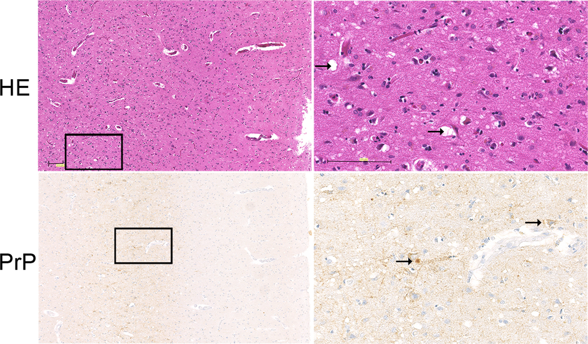
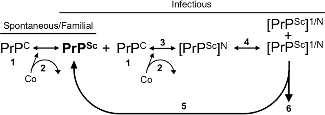
Prion diseases are rapidly progressive, incurable neurodegenerative disorders caused by misfolded, aggregated proteins known as prions, which are uniquely infectious. Remarkably, these infectious proteins have been responsible for widespread disease epidemics, including kuru in humans, bovine spongiform encephalopathy in cattle, and chronic wasting disease in cervids, the latter of which has spread across North America and recently appeared in Norway and Finland. The hallmark histopathological features include widespread spongiform encephalopathy, neuronal loss, gliosis, and deposits of variably sized aggregated prion protein, ranging from small, soluble oligomers to long, thin, unbranched fibrils, depending on the disease. Here, we explore recent advances in prion disease research, from the function of the cellular prion protein to the dysfunction triggering neurotoxicity, as well as mechanisms underlying prion spread between cells. We also highlight key findings that have revealed new therapeutic targets and consider unanswered questions for future research.

Keywords: amyloid; neurodegeneration; neurotoxicity; prion transmission; strains.


Figures

Figure 1.
Figure 1.
Hematoxylin and eosin and PrP immunostain of brain (frontal cortex) from a sCJD patient. Spongiosis is visible in the deep layers of the cortex (HE) (arrow indicates intaneuronal and parenchymal spongiform change, arrow) and synaptic, plaque-like, and perineuronal deposits of pathological prion protein (arrow indicates plaque-like and perineuronal deposits). The synaptic deposits of pathological prion protein are pronounced in the deep layers of the cortex. Scale bar = 100 μm.
Figure 2.
Figure 2.
Possible pathways of prion spread from cell-to-cell. Prion aggregates may spread through transport in tunneling nanotubes (1), GPI painting, by which GPI-anchored proteins transfer from one cell surface to a neighboring cell surface (2), trafficking within exosomes (3), or from membrane budding and transport within vesicles (4).
Figure 3.
Figure 3.
Strain-specific factors in prion formation. Prion formation is dependent on the presence of PrPC (1). For the conversion of PrPC to PrPSc in spontaneous, familial, or infectious etiologies, cofactors (Co) may participate in the formation of PrPSc, although it is unknown if they are incorporated into the growing polymer or simply used as a structural scaffold (2). The rate of PrPSc formation (3) is dictated by the incoming prion strain (PrPSc), the level of PrPC (1), and the cofactors present (2). PrPSc fragmentation can result in newly fragmented PrPSc serving as a seed for conversion (5) or PrPSc clearance from the cell (6). The rate of prion formation (3) must be greater than the rate of clearance (6) to establish a productive infection. Strain-specific PrPSc conformations may utilize specific subpopulations of PrPC, cofactors, and clearance mechanisms that may all contribute to strain-specific cellular and tissue tropism.

References

    1. Bolton DC, McKinley MP, Prusiner SB. 1982. Identification of a protein that purifies with the scrapie prion. Science 218: 1309–11 - PubMed
    1. Prusiner SB. 1982. Novel proteinaceous infectious particles cause scrapie. Science 216: 136–44 - PubMed
    1. Prusiner SB, Scott MR, DeArmond SJ, Cohen FE. 1998. Prion protein biology. Cell 93: 337–48 - PubMed
    1. Basler K, Oesch B, Scott M, Westaway D, Walchli M, Groth DF, McKinley MP, Prusiner SB, Weissmann C. 1986. Scrapie and cellular PrP isoforms are encoded by the same chromosomal gene. Cell 46: 417–28 - PubMed
    1. DeArmond SJ, Prusiner SB. 1995. Etiology and pathogenesis of prion diseases. Am J Pathol 146: 785–811 - PMC - PubMed

Publication types