Review
. 2019 Jan 24:14:421-447.
doi: 10.1146/annurev-pathol-020117-043711. Epub 2018 Oct 24.

Pathology and Pathogenesis of Chagas Heart Disease

Affiliations
Review

Pathology and Pathogenesis of Chagas Heart Disease

Kevin M Bonney et al. Annu Rev Pathol. .

Abstract

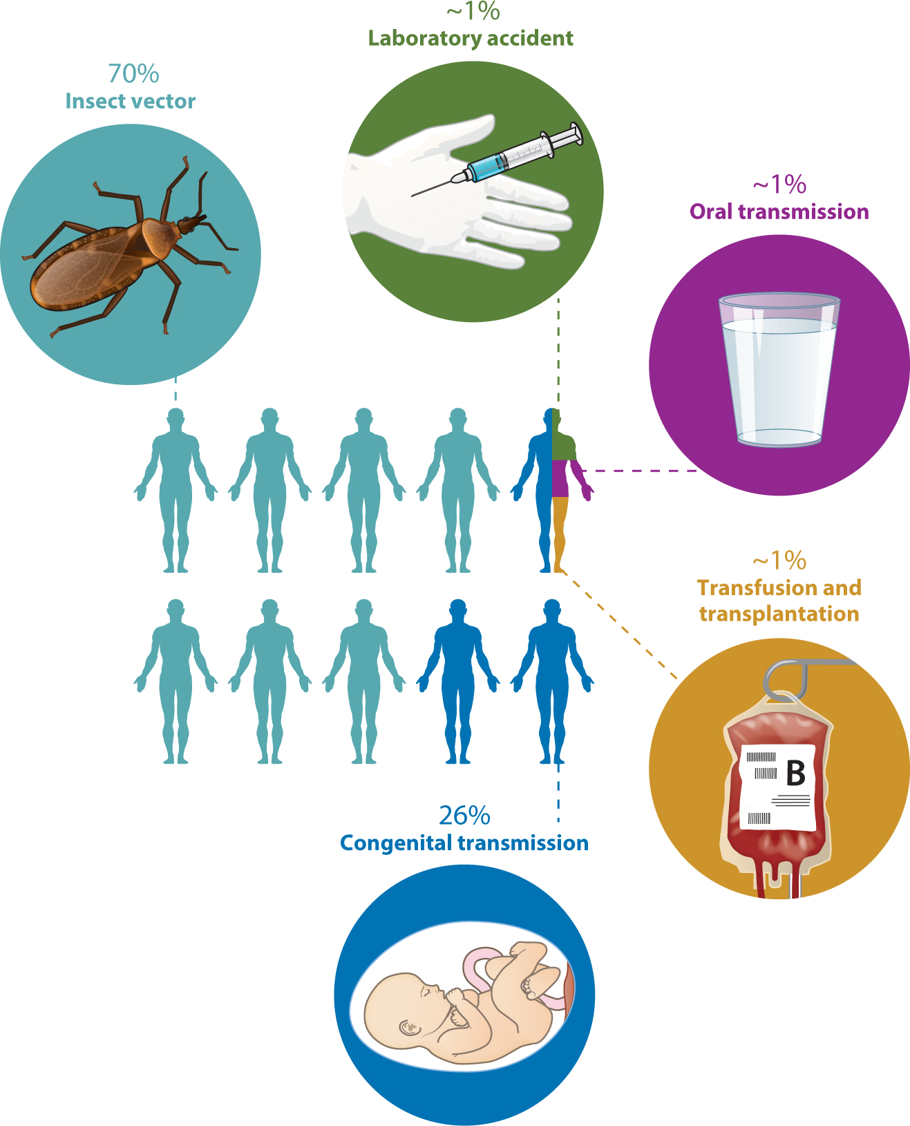
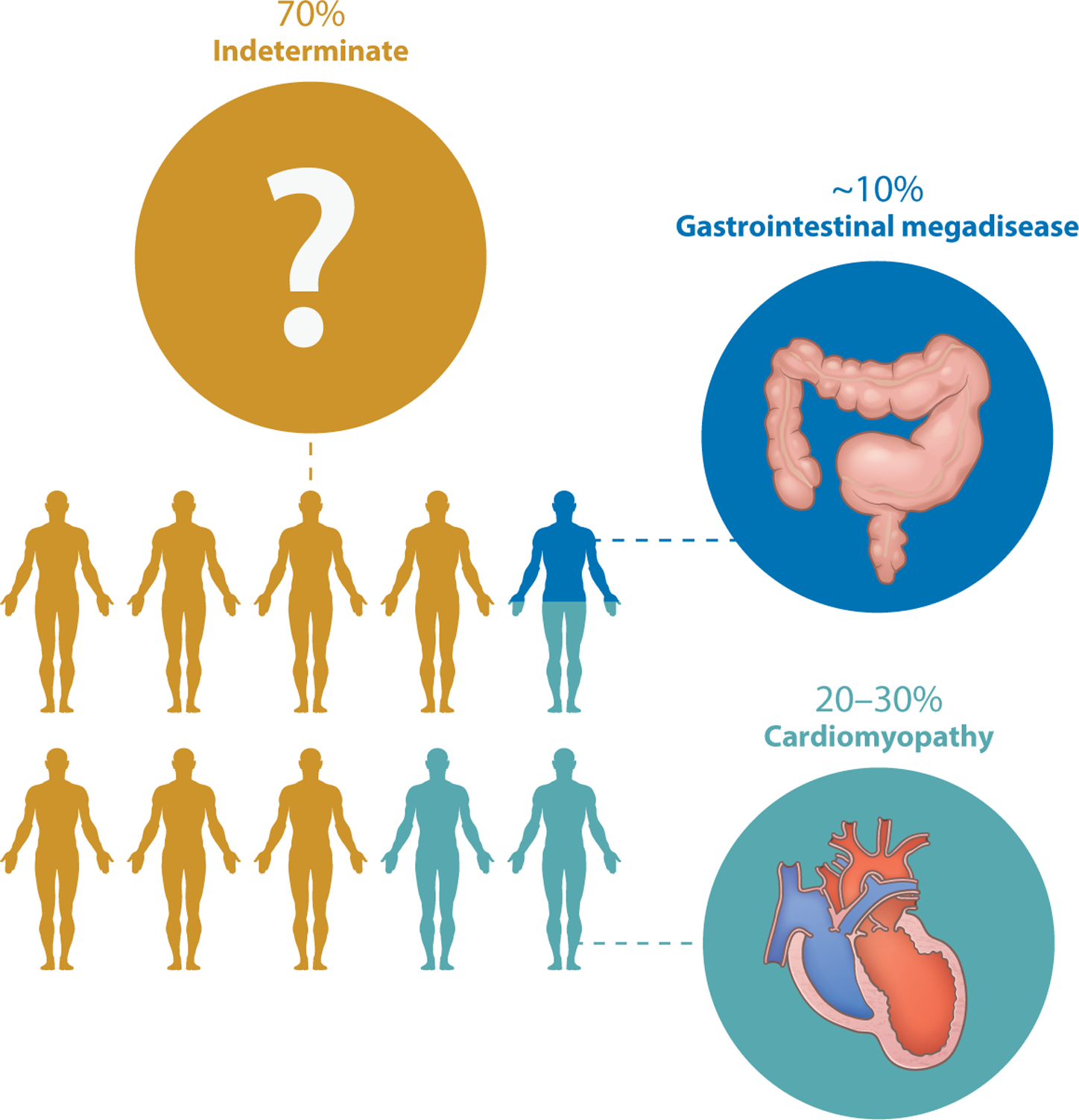
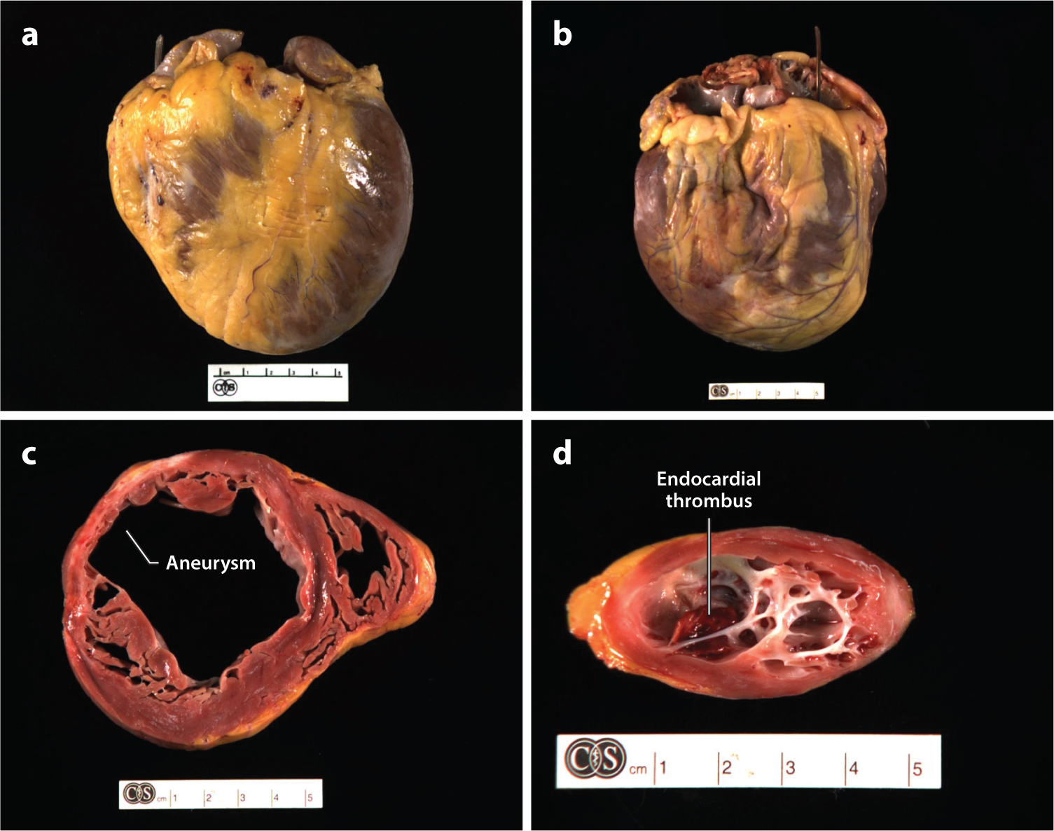
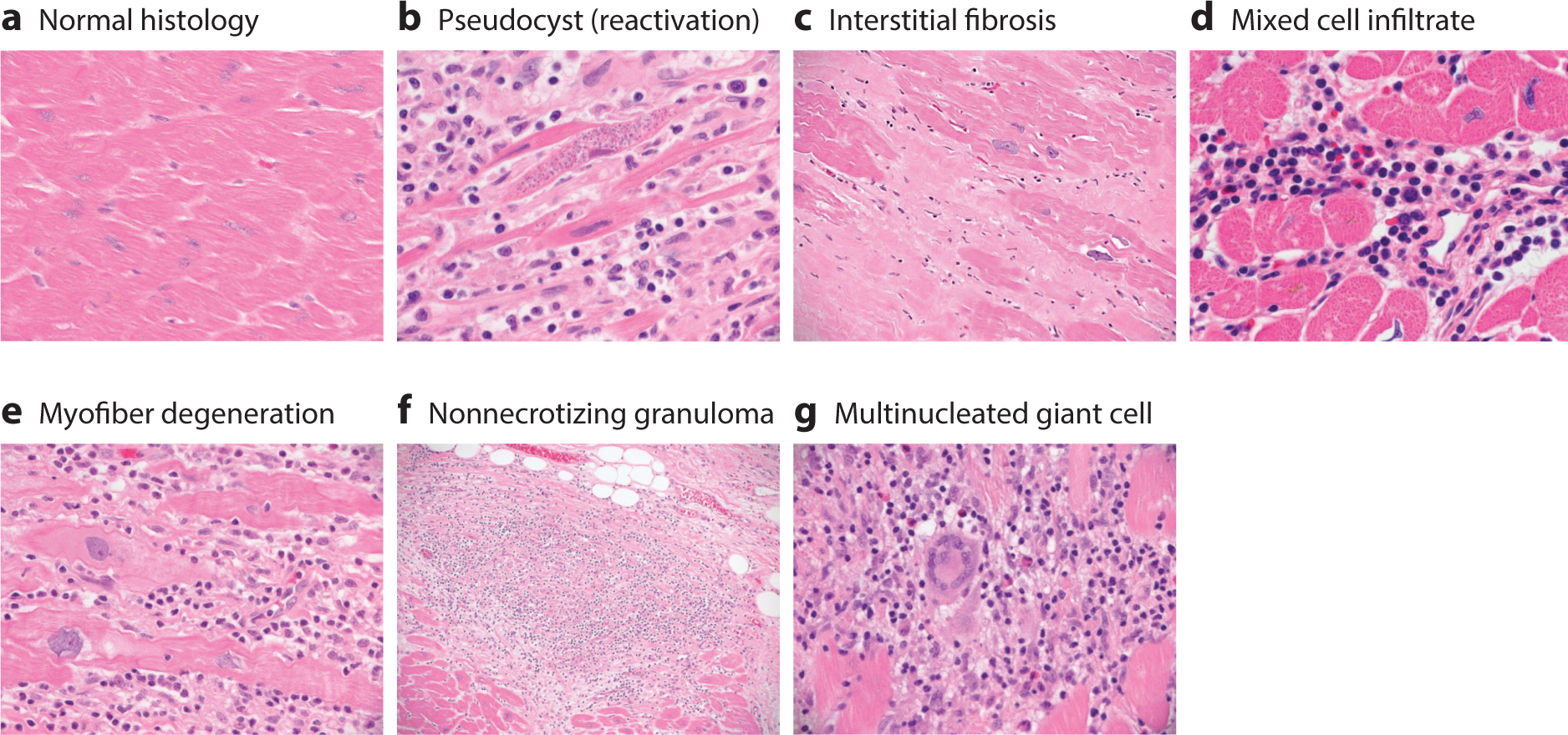
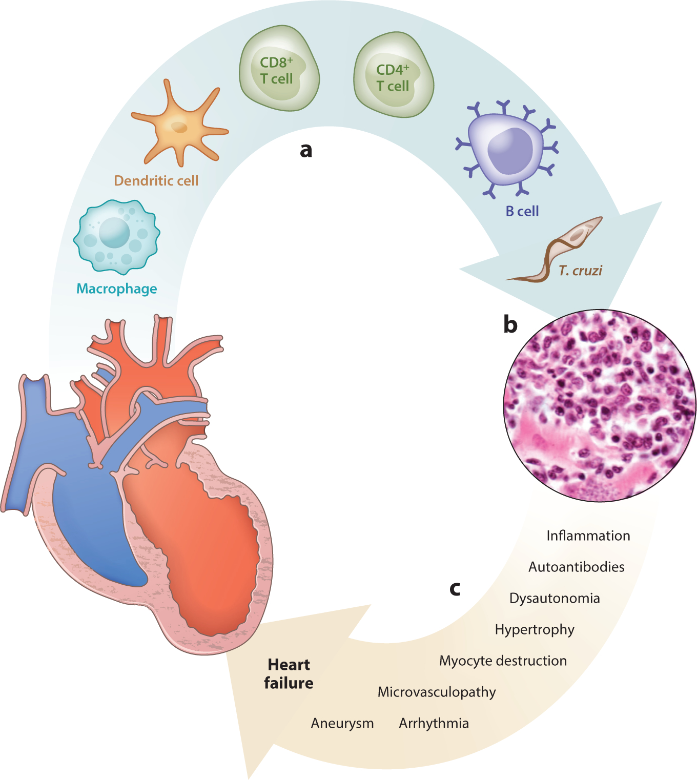
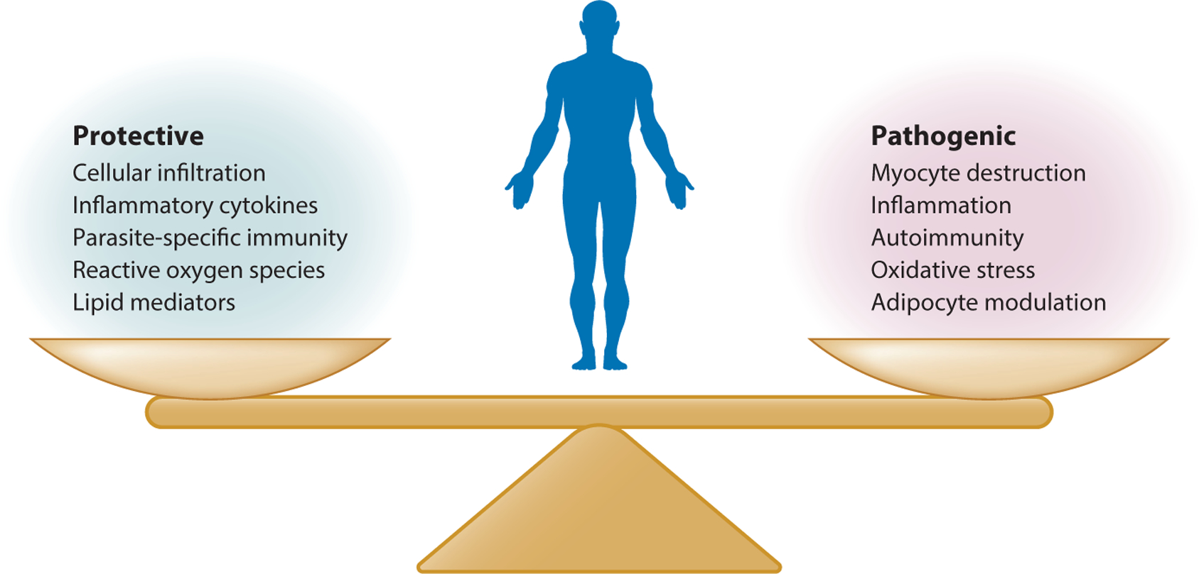
Chagas heart disease is an inflammatory cardiomyopathy that develops in approximately one-third of people infected with the protozoan parasite Trypanosoma cruzi. One way T. cruzi is transmitted to people is through contact with infected kissing bugs, which are found in much of the Western Hemisphere, including in vast areas of the United States. The epidemiology of T. cruzi and Chagas heart disease and the varied mechanisms leading to myocyte destruction, mononuclear cell infiltration, fibrosis, and edema in the heart have been extensively studied by hundreds of scientists for more than 100 years. Despite this wealth of knowledge, it is still impossible to predict what will happen in an individual infected with T. cruzi because of the tremendous variability in clonal parasite virulence and human susceptibility to infection and the lack of definitive molecular predictors of outcome from either side of the host-parasite equation. Further, while several distinct mechanisms of pathogenesis have been studied in isolation, it is certain that multiple coincident mechanisms combine to determine the ultimate outcome. For these reasons, Chagas disease is best considered a collection of related but distinct illnesses. This review highlights the pathology and pathogenesis of the most common adverse sequela of T. cruzi infection-Chagas heart disease-and concludes with a discussion of key unanswered questions and a view to the future.

Keywords: Chagas; cardiomyopathy; heart; myocarditis; pathogenesis; trypanosome.


Figures

Figure 1
Figure 1
Modes of transmission of Trypanosoma cruzi. Approximately 70% of infections occur when infected reduviid bugs feed on or otherwise come into contact with people. The bug’s feces contain infective metacyclic trypomastigotes, which can enter the body through any break in the skin, for example the bite wound, or through any mucous membrane. Approximately 26% of infections are due to maternal–fetal transmission during pregnancy. A few infections occur as a result of laboratory accidents, the consumption of contaminated food or beverages, or blood transfusion or the transplantation of tissue from an infected donor. Oral transmission can occur when fresh juice is prepared by crushing plants such as sugar cane. If there are infected reduviids in the plants, trypomastigotes end up in the juice when the plants (and bugs) are crushed. Transmission by blood transfusion was a major problem before blood suppliers in many countries, including the United States, began screening for T. cruzi.
Figure 2
Figure 2
Outcomes of chronic Trypanosoma cruzi infection. Approximately 70% of T. cruzi–infected people live normal lives without developing any adverse sequelae of infection. Approximately 10% develop megaesophagus or megacolon, and 20–30% develop cardiomyopathy. A tiny number of people die during acute infection from fatal cardiac dysrhythmias (not shown), and a few others develop additional sequelae involving other systems.
Figure 3
Figure 3
Gross pathology of Chagas heart disease. Examples of explanted hearts from patients with chronic Chagas cardiomyopathy highlight the characteristic features of (a,b) cardiomegaly, multichamber dilatation, and epicardial scarring. Cross-sections through (c) the ventricles and (d ) left apex reveal patchy myocardial, endocardial, and epicardial fibrosis. Aneurysms are frequently present in different stages of development and are visible in the 10 o’clock position in panel c and viewed from above in panel d. In the aneurysm in panel d, the thinning of the ventricular wall can be appreciated, and an apical endocardial thrombus (deep red ) is visible in the aneurysm cavity. All explants are from Cedars-Sinai Medical Center in Los Angeles.
Figure 4
Figure 4
Microscopic pathology of Chagas heart disease. Essentially, every possible histologic picture of Trypanosoma cruzi infection can be observed by examining the hearts of patients with acute, chronic, or chronic reactivation (e.g., upon immunosuppression) disease. (a) Normal cardiac histology. (b) Heart section with mononuclear cell infiltration, myofibrillar degeneration, fibrosis, and an amastigote pseudocyst, commonly seen in reactivation disease. (c) Massive interstitial fibrosis replacing depleted myocytes. (d ) A mixed cell infiltrate comprising histiocytes, lymphocytes, eosinophils, and mast cells. (e) In addition to the chronic inflammation described in the earlier panels, myocardial fibers show evidence of degeneration, with expanded basophilic cytoplasm, corrugated cell membranes, and nuclei with degenerative features, including open and clumped chromatin and irregular nuclear borders. ( f ) Nonnecrotizing granulomas are occasionally observed. ( g) Multinucleated giant cells are commonly found. We emphasize that T. cruzi amastigotes are rarely observed, except in the case of reactivation (b).
Figure 5
Figure 5
Pathogenesis of Chagas heart disease. (a) A variety of cells, including Trypanosoma cruzi parasites, infiltrate the myocardium, which develops (b) histopathology characterized by mononuclear cell infiltration, edema, myocyte destruction, and fibrosis. Parasites are typically not observed in chronic infection. (c) Mechanisms are listed that may lead to progressive cardiac dysfunction that progresses to heart failure during T. cruzi infection.
Figure 6
Figure 6
Protective and pathogenic responses to Trypanosoma cruzi infection. The various protective and pathogenic responses are shown. Importantly, many protective processes can also have deleterious effects, which is a general challenge in mounting innate and adaptive responses to pathogens.

Similar articles

Cited by

References

    1. Brener Z 1973. Biology of Trypanosoma cruzi. Annu. Rev. Microbiol 27:347–82 - PubMed
    1. WHO (World Health Organ.). 2002. Control of Chagas Disease: Second Report of the WHO Expert Committee. Geneva, Switzerland: WHO
    1. Shikanai-Yasuda MA, Carvalho NB. 2012. Oral transmission of Chagas disease. Clin. Infect. Dis 54:845–52 - PubMed
    1. Tanowitz HB, Weiss LM, Montgomery SP. 2011. Chagas disease has now gone global. PLOS Negl. Trop. Dis 5:e1136. - PMC - PubMed
    1. PAHO (Pan Am. Health Organ.). 2018. Chagas disease portal. Pan Am. Health Organ http://www.paho.org/hq/index.php?option=com_topics&view=article&id=10&It...

Publication types

LinkOut - more resources