. 2018 Aug 31;123(6):660-672.
doi: 10.1161/CIRCRESAHA.118.313105.

Role of Thrombospondin-1 in Mechanotransduction and Development of Thoracic Aortic Aneurysm in Mouse and Humans

Affiliations

Role of Thrombospondin-1 in Mechanotransduction and Development of Thoracic Aortic Aneurysm in Mouse and Humans

Yoshito Yamashiro et al. Circ Res. .

Erratum in

Abstract

Rationale: Abnormal mechanosensing of smooth muscle cells (SMCs) resulting from the defective elastin-contractile units has been suggested to drive the formation of thoracic aortic aneurysms; however, the precise molecular mechanism has not been elucidated.

Objective: The aim of this study was to identify the crucial mediator(s) involved in abnormal mechanosensing and propagation of biochemical signals during the aneurysm formation and to establish a basis for a novel therapeutic strategy.

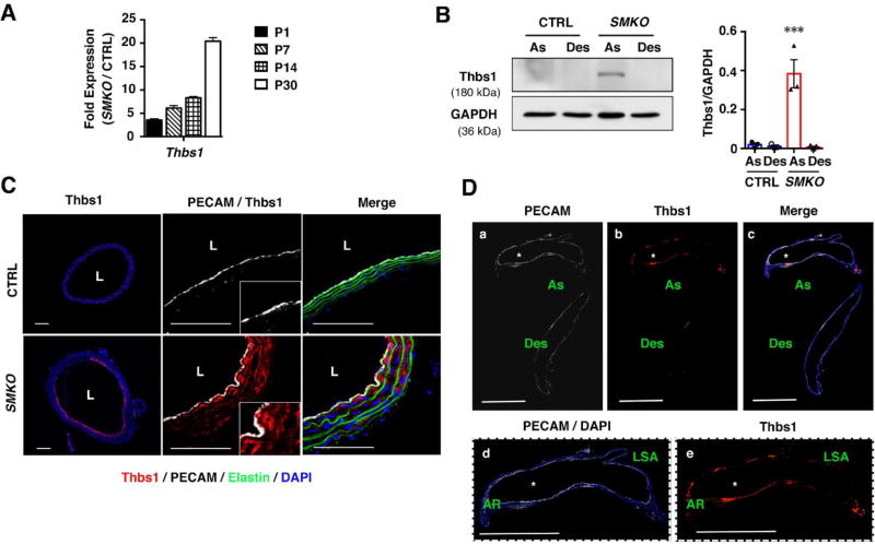
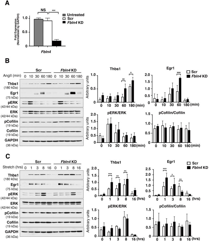
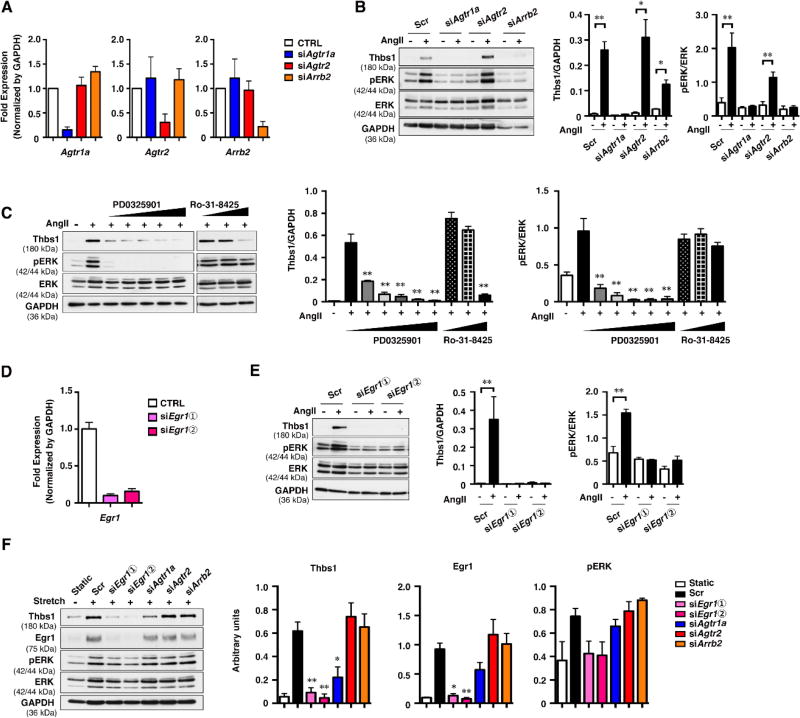
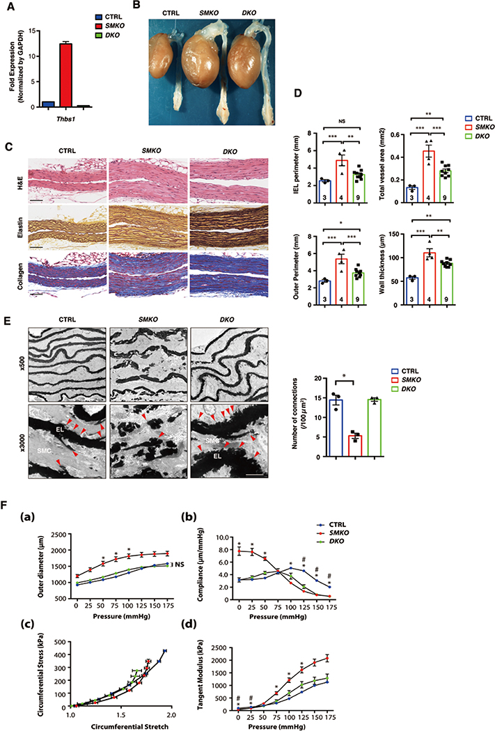
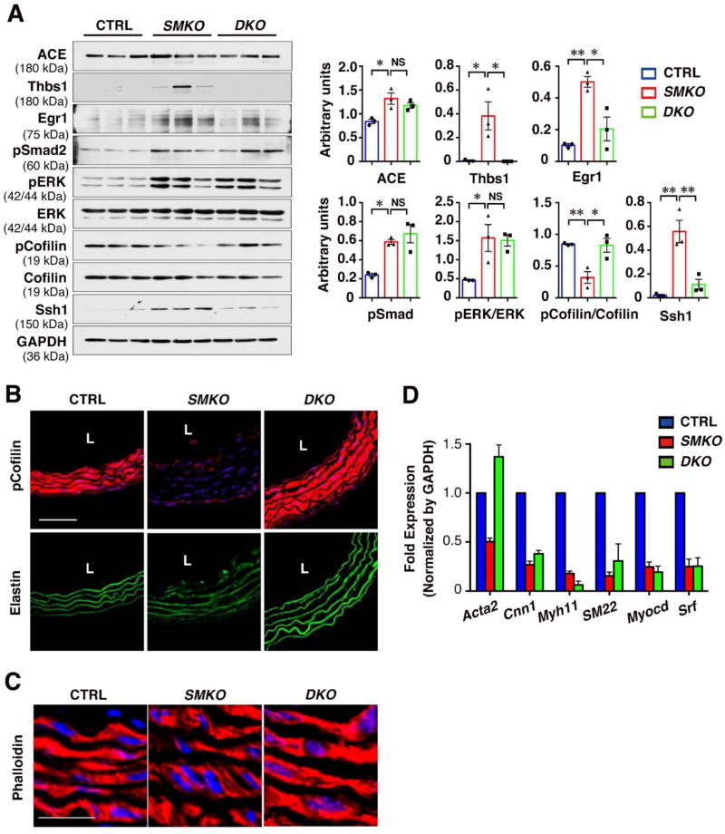
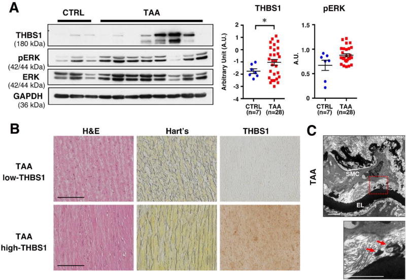
Methods and results: We used a mouse model of postnatal ascending aortic aneurysms ( Fbln4SMKO; termed SMKO [SMC-specific knockout]), in which deletion of Fbln4 (fibulin-4) leads to disruption of the elastin-contractile units caused by a loss of elastic lamina-SMC connections. In this mouse, upregulation of Egr1 (early growth response 1) and angiotensin-converting enzyme leads to activation of Ang II (angiotensin II) signaling. Here, we showed that the matricellular protein, Thbs1 (thrombospondin-1), was highly upregulated in SMKO ascending aortas and in human thoracic aortic aneurysms. Thbs1 was induced by mechanical stretch and Ang II in SMCs, for which Egr1 was required, and reduction of Fbln4 sensitized the cells to these stimuli and led to higher expression of Egr1 and Thbs1. Deletion of Thbs1 in SMKO mice prevented the aneurysm formation in ≈80% of DKO (SMKO;Thbs1 knockout) animals and suppressed Ssh1 (slingshot-1) and cofilin dephosphorylation, leading to the formation of normal actin filaments. Furthermore, elastic lamina-SMC connections were restored in DKO aortas, and mechanical testing showed that structural and material properties of DKO aortas were markedly improved.

Conclusions: Thbs1 is a critical component of mechanotransduction, as well as a modulator of elastic fiber organization. Maladaptive upregulation of Thbs1 results in disruption of elastin-contractile units and dysregulation of actin cytoskeletal remodeling, contributing to the development of ascending aortic aneurysms in vivo. Thbs1 may serve as a potential therapeutic target for treating thoracic aortic aneurysms.

Keywords: angiotensin II; aortic aneurysm, thoracic; elastic tissue; extracellular matrix; humans.


Figures

Figure 1
Figure 1. Thbs1 is highly expressed in aneurysmal wall
(A) qPCR analysis of Thbs1 from ascending aortas of CTRL (pooled P1, n=12; P7, n=18; P14, n=9; and P30, n=12) and Fbln4SMKO (SMKO, pooled P1, n=12; P7, n=18; P14, n=8; and P30, n=11) mice performed in technical triplicate. (B) Representative Western blots of ascending (As) and descending (Des) aortas of CTRL and SMKO at P30. n=3, Bar are means ± SEM. ***P < 0.001, one-way ANOVA. (C) Cross sections of the ascending aorta from CTRL and SMKO at P30 immunostained with Thbs1 (red), DAPI (blue), PECAM (white). Elastic fibers are green (autofluorescence). n=5. Scale bars are 50 µm. (D) Longitudinal sections of the aorta from SMKO mice (a–c) immunostained with PECAM (white), Thbs1 (red) and merged image with DAPI (blue). As (ascending aorta), Des (descending aorta). Expanded images of ascending aorta (d and e). AR (aortic root), LSA (left subclavian artery). Scale bars are 1 cm. n=5.
Figure 2
Figure 2. Fbln4 deficiency enhances the response to mechanical stress and Ang II in rat SMCs
(A) qPCR analysis confirming knockdown of Fbln4 in rat SMCs. Total RNA from siRNA-transfected cells were extracted 3 days after transfection. The mRNA levels relative to untreated siRNA are shown. n=4, Bars are means ± SEM. *** P < 0.001, one-way ANOVA. (B) Rat SMCs treated with scramble siRNA (Scr) or Fbln4 siRNA (Fbln4 KD) cultured in serum-free media for 21 hr, then stimulated with Ang II (1 nM) for 10, 30, 60 or 180 min. Cell lysates were analyzed by Western blotting with indicated antibodies. Representative blots and quantification graphs are shown from quadruplicate experiments. Bars are means ± SEM. *P < 0.05, **P < 0.01, ***P < 0.001. Two-way ANOVA. (C) Scr- or Fbln4 KD cells were subjected to cyclic stretch (20 % strain, 1.0 Hz) by FlexCell system for 1, 3, 8 or 16 hr. Representative Western blots showing expression of Thbs1, Egr1, pERK, total ERK, pCofilin, total cofilin and GAPDH. Quantification graph are shown from triplicate experiments. Bars are means ± SEM. *P < 0.05, **P < 0.01, ***P < 0.001. Two-way ANOVA.
Figure 3
Figure 3. Upregulation of Thbs1 is mediated by Egr1
(A and D) qPCR analysis confirming the knockdown of Agtr1a, Agtr2, and Arrb2 (A) and Egr1 (D) in rat SMCs. Total RNA from siRNA-treated cells were extracted 3 days after transfection. The mRNA levels relative to scramble siRNA (Scr) are shown. Bars indicate technical triplicate. (B and E) Scr- or siRNA-treated rat SMCs were cultured in serum-free media for 21 hr and stimulated with or without Ang II (100 nM) for 3 hr. Representative Western blots and quantification graphs are shown. (C) Rat SMCs were cultured in serum-free media for 21 hr, then pretreated with or without MEK inhibitor PD0325901 (1 nM, 10 nM, 100 nM, 1 µM, 10 µM) or PKC inhibitor Ro-31-8425 (100 nM, 1 µM, 10 µM) for 1 hr followed by 3 hr of stimulation with Ang II (100 nM). Representative Western blots and quantification graphs are shown. Bar are means ± SEM, **P < 0.01, two-way ANOVA. (F) Scr- or siRNA-treated rat SMCs were subjected to uniaxial cyclic stretch (20 % strain, 1.0 Hz) in the presence of 20% FBS for 8 hr. Representative Western blots showing Thbs1, Egr1, pERK, total ERK and GAPDH. Quantification graphs are shown from triplicate experiments. Bars are means ± SEM. *P < 0.05, **P < 0.01. One-way ANOVA.
Figure 4
Figure 4. Deletion of Thbs1 attenuates aneurysm formation in SMKO aortas
(A) qPCR analysis of Thbs1 on aortas harvested from P30 CTRL (pooled n=12), SMKO (pooled n=11) and DKO (pooled n=3) mice in technical triplicate. (B) Gross photos of CTRL, SMKO and SMKO crossed with Thbs1 null mice (SMKO; Thbs1−/−, DKO) at 3 months of age. (C) Histological images of cross sections of the ascending aorta from P30 CTRL, SMKO and DKO stained with hematoxylin and eosin (H&E), Hart’s (elastin) and Masson trichrome (collagen). Scale bars, 20 µm. (D) Morphometric analysis showing IEL perimeter, outer perimeter, total vessel area and wall thickness. Bars are means ± SEM. *P < 0.05, **P < 0.01, ***P < 0.001, one-way ANOVA. Number of animals are indicated in each bar. (E) Electron microscopy (EM) images from P30 CTRL (n=3), SMKO (n=3) and DKO (n=3) ascending aortas. Elastic lamina (EL) -SMC connections seen in CTRL (red arrowheads) were significantly decreased in SMKO and restored in DKO. Scale bars, 2 µm. Quantification of the connection between EL and SMC by Kruskal-Wallis test with Dunn’s multiple comparisons is shown. (F) Mechanical testing using CTRL (n=7), SMKO (n=7) and DKO (n=3) aortas at P30. (a) Aortic pressure-outer diameter curves, (b) Aortic pressure-compliance curves, (c) circumferential stress-stretch plots, and (d) tangent modulus. Bars are means ± SEM. Symbols indicate significant difference (P < 0.05) between CTRL and SMKO (*), and CTRL and DKO (#). One-way ANOVA with Tukey’s post-hoc test.
Figure 5
Figure 5. Thbs1 is upstream of Ssh1-cofilin pathway leading to aneurysm formation
(A) Western blots showing downregulation of Ssh1 expression and upregulation of pCofilin levels in P30 DKO aortas. Quantification of Western blots are shown in right graphs. Bars are means ± SEM. *P < 0.05, **P < 0.01, one-way ANOVA. (B and C) Cross sections of the ascending aorta from P30 CTRL, SMKO and DKO immunostained with pCofilin (red in B), Phalloidin (red in C) and DAPI (blue). Elastic fibers are green (autofluorescence). n=3. Scale bars are 50 µm in (B), 100 µm in (C). (D) qPCR analysis of SMC-specific genes from ascending aortas of CTRL (pooled n=12), SMKO (pooled n=11) and DKO (pooled n=3) mice performed in technical triplicate.
Figure 6
Figure 6. THBS1 expression is significantly increased in human TAA
(A) Representative Western blots showing the comparison of THBS1, GAPDH and pERK levels in aortic tissues of control (n=7, pooled 2–3 tissues per group) and TAA samples (n=28). Log-converted value of THBS1/GAPDH was compared using unpaired t-test with Welch correction. pERK/GAPDH value was compared by unpaired t-test. * P < 0.05. (B) Representative histological images of cross sections of the ascending aorta from human TAA tissues from high THBS1 group (n=6) and low THBS1 group (n=4) stained with hematoxylin and eosin (H&E), Hart’s (elastin) and anti-THBS1. Bars are 50 µm. (C) EM images of the ascending aortas from human TAA tissues. The lower panel is an enlargement of the area shown in red box. Arrows indicate loss of connections between elastic lamina (EL) and SMCs. n=1. Scale bar, 2 µm.

Similar articles

Cited by

References

    1. Neptune ER, Frischmeyer PA, Arking DE, Myers L, Bunton TE, Gayraud B, Ramirez F, Sakai LY, Dietz HC. Dysregulation of tgf-beta activation contributes to pathogenesis in marfan syndrome. Nat Genet. 2003;33:407–411. - PubMed
    1. Mizuguchi T, Collod-Beroud G, Akiyama T, Abifadel M, Harada N, Morisaki T, Allard D, Varret M, Claustres M, Morisaki H, Ihara M, Kinoshita A, Yoshiura K, Junien C, Kajii T, Jondeau G, Ohta T, Kishino T, Furukawa Y, Nakamura Y, Niikawa N, Boileau C, Matsumoto N. Heterozygous tgfbr2 mutations in marfan syndrome. Nat Genet. 2004;36:855–860. - PMC - PubMed
    1. Loeys BL, Chen J, Neptune ER, Judge DP, Podowski M, Holm T, Meyers J, Leitch CC, Katsanis N, Sharifi N, Xu FL, Myers LA, Spevak PJ, Cameron DE, De Backer J, Hellemans J, Chen Y, Davis EC, Webb CL, Kress W, Coucke P, Rifkin DB, De Paepe AM, Dietz HC. A syndrome of altered cardiovascular, craniofacial, neurocognitive and skeletal development caused by mutations in tgfbr1 or tgfbr2. Nat Genet. 2005;37:275–281. - PubMed
    1. Maki JM, Rasanen J, Tikkanen H, Sormunen R, Makikallio K, Kivirikko KI, Soininen R. Inactivation of the lysyl oxidase gene lox leads to aortic aneurysms, cardiovascular dysfunction, and perinatal death in mice. Circulation. 2002;106:2503–2509. - PubMed
    1. Dasouki M, Markova D, Garola R, Sasaki T, Charbonneau NL, Sakai LY, Chu ML. Compound heterozygous mutations in fibulin-4 causing neonatal lethal pulmonary artery occlusion, aortic aneurysm, arachnodactyly, and mild cutis laxa. Am J Med Genet A. 2007;143A:2635–2641. - PubMed

Publication types

MeSH terms