. 2018 Aug 31;123(6):673-685.
doi: 10.1161/CIRCRESAHA.118.312751.

miR-146a Suppresses SUMO1 Expression and Induces Cardiac Dysfunction in Maladaptive Hypertrophy

Affiliations

miR-146a Suppresses SUMO1 Expression and Induces Cardiac Dysfunction in Maladaptive Hypertrophy

Jae Gyun Oh et al. Circ Res. .

Abstract

Rationale: Abnormal SUMOylation has emerged as a characteristic of heart failure (HF) pathology. Previously, we found reduced SUMO1 (small ubiquitin-like modifier 1) expression and SERCA2a (sarcoplasmic reticulum Ca2+-ATPase) SUMOylation in human and animal HF models. SUMO1 gene delivery or small molecule activation of SUMOylation restored SERCA2a SUMOylation and cardiac function in HF models. Despite the critical role of SUMO1 in HF, the regulatory mechanisms underlying SUMO1 expression are largely unknown.

Objective: To examine miR-146a-mediated SUMO1 regulation and its consequent effects on cardiac morphology and function.

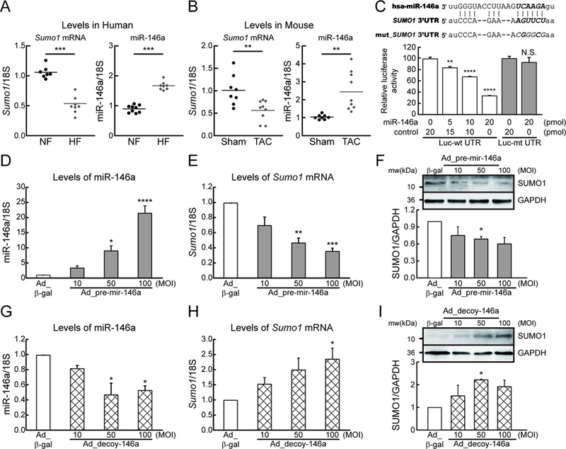
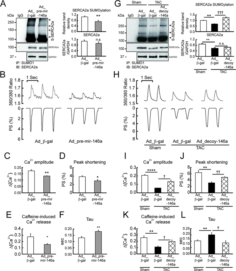
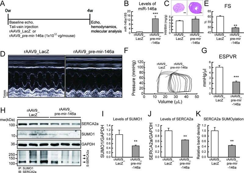
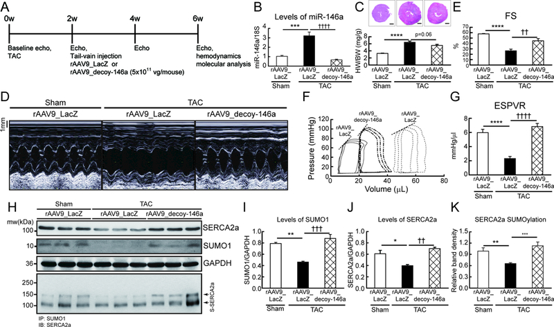
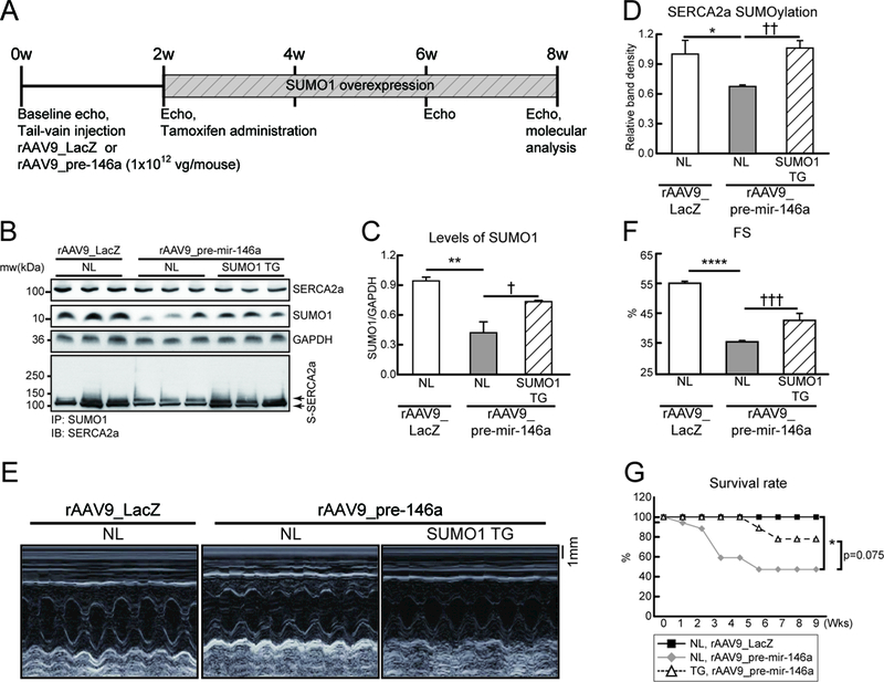
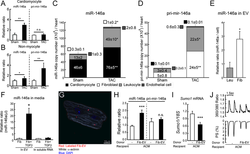
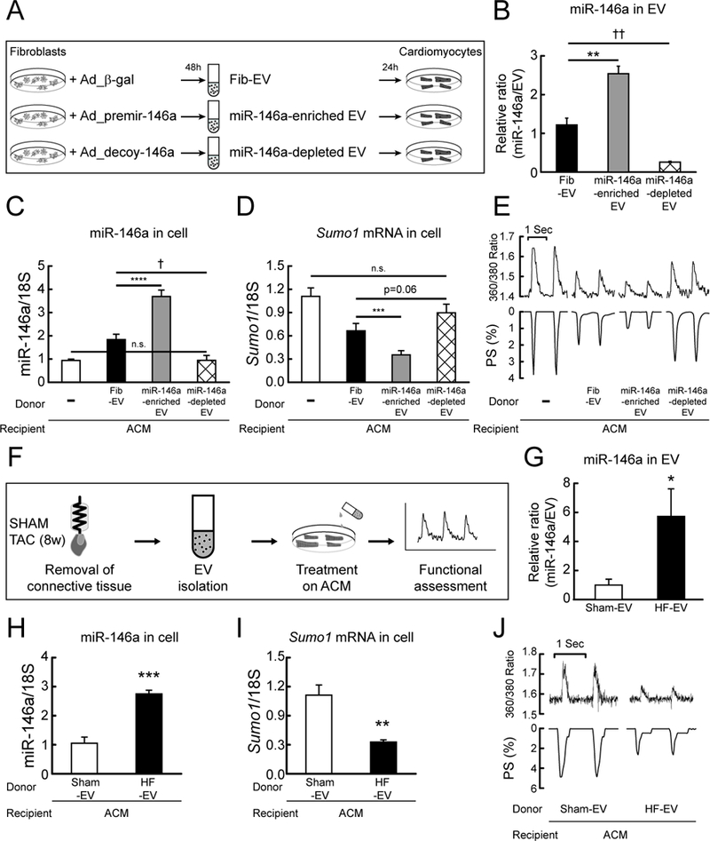
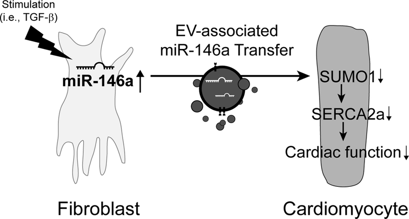
Methods and results: In this study, miR-146a was identified as a SUMO1-targeting microRNA in the heart. A strong correlation was observed between miR-146a and SUMO1 expression in failing mouse and human hearts. miR-146a was manipulated in cardiomyocytes through AAV9 (adeno-associated virus serotype 9)-mediated gene delivery, and cardiac morphology and function were analyzed by echocardiography and hemodynamics. Overexpression of miR-146a reduced SUMO1 expression, SERCA2a SUMOylation, and cardiac contractility in vitro and in vivo. The effects of miR-146a inhibition on HF pathophysiology were examined by transducing a tough decoy of miR-146a into mice subjected to transverse aortic constriction. miR-146a inhibition improved cardiac contractile function and normalized SUMO1 expression. The regulatory mechanisms of miR-146a upregulation were elucidated by examining the major miR-146a-producing cell types and transfer mechanisms. Notably, transdifferentiation of fibroblasts triggered miR-146a overexpression and secretion through extracellular vesicles, and the extracellular vesicle-associated miR-146a transfer was identified as the causative mechanism of miR-146a upregulation in failing cardiomyocytes. Finally, extracellular vesicles isolated from failing hearts were shown to contain high levels of miR-146a and exerted negative effects on the SUMO1/SERCA2a signaling axis and hence cardiomyocyte contractility.

Conclusions: Taken together, our results show that miR-146a is a novel regulator of the SUMOylation machinery in the heart, which can be targeted for therapeutic intervention.

Keywords: extracellular vesicle; heart failure; mice; microRNA; sarcoplasmic reticulum.


Figures

Figure 1:
Figure 1:. MicroRNA-146a (miR-146a) inhibits SUMO1 expression in cardiomyocytes.
A, Changes in SUMO1 mRNA and miR-146a levels in human hearts of non-failing (NF) and heart failure (HF) patients (n=7–9). B, Changes in SUMO1 mRNA and miR-146a levels in mouse hearts of sham and TAC operated mice (6 weeks after TAC) (n=8–9). C, Sequence alignment of SUMO1 wild-type and mutant 3′ UTRs with miR-146a (top) and luciferase reporter assay (bottom). Luciferase reporters with a putative miR-146a–binding site (Luc-wt UTR) or mutant miR-146a-binding site (Luc-mt UTR) were co-transfected into H9C2 cells with miR-146a mimic or control (cel-miR-39) for 24h, and luciferase activity determined. Values are presented as relative luciferase activity ± s.e.m. (n=3). D and E, The expression of miR-146a (D) and SUMO1 mRNA (E) in isolated adult mouse cardiomyocytes which were infected with an adenovirus encoding pre-mir-146a (Ad_pre-mir-146a) for 48h (n=3). F, Representative blot (top) and quantitative analysis (bottom) showing the expression of SUMO1 protein. G and H, The expression of miR-146a (G) and SUMO1 mRNA (H) in isolated adult mouse cardiomyocytes which were infected with an adenovirus encoding mir-146a decoy (Ad_decoy-146a) for 48h (n=3). I, Representative blot (top) and quantitative analysis (bottom) showing the expression of SUMO1 protein. *, p<0.05, **, p<0.01, ***, p<0.001, ****, p<0.001 versus the respective control, as determined by One-way ANOVA. Data are presented as mean ± s.e.m.
Figure 2:
Figure 2:. miR-146a inhibits cardiac contractility via suppression of SERCA2a SUMOylation.
Isolated mouse cardiomyocytes infected with Ad_pre-mir-146a (MOI=50) for 48h. A, Representative blot (left) and quantitative analysis (right) showing the levels of SERCA2a SUMOylation and SERCA2a expression (n=6). B, Representative trace of calcium transients (top) and cardiac contractility (bottom). C and D, Averaged data of calcium transient amplitude (C) and sarcomere length shortening (D) (n=100–150 cells/4 hearts). E, Caffeine-induced Ca2+ transient amplitude at 10 mmol/L Caffeine (n=10 cells/3 hearts). F, Rate constant for the decay of the Ca2+ transient (n=100–150 cells/4 hearts). *, p<0.05, **, p<0.01 versus cardiomyocytes infected with Ad_β-gal, as determined by Student’s t-test. Isolated failing cardiomyocytes from mice subjected to TAC for 6 weeks infected with Ad_decoy-146a (MOI=50) for 48h. G, Representative blot (left) and quantitative analysis (right) showing the levels of SERCA2a SUMOylation and SERCA2a expression (n=3). H, Representative trace of calcium transients (top) and cardiac contractility (bottom). I and J, Averaged data of calcium transient amplitude (I) and sarcomere length shortening (J) (n=100–150 cells/4 hearts). K, Caffeine-induced Ca2+ transient amplitude at 10 mmol/L Caffeine (n=10 cells/3 hearts). L, Rate constant for the decay of the Ca2+ transient (n=100–150 cells/4 hearts). *, p<0.05, **, p<0.01, ****, p<0.0001 versus the sham mouse and , p<0.05, ††, p<0.01 versus the failing cardiomyocyte treated with Ad_β-gal, as determined by one-way ANOVA. Data are represented as mean ± s.e.m. in all panels.
Figure 3:
Figure 3:. Overexpression of miR-146a promotes cardiac dysfunction in vivo.
A, Protocol for AAV9-mediated pre-mir-146a gene transfer into normal mice. Six week-old male B6C3/F1 mice received AAV9 carrying pre-mir-146a (rAAV9_pre-mir-146a, 1×10¹² vg/mouse) or control virus (rAAV9_LacZ) via tail-vein injection. After 4 weeks of gene delivery, cardiac function was measured by echocardiography and then, further analyzed by hemodynamic measurements. B, levels of miR-146a. C, Cross-section images of a mouse heart (top) and quantification of heart weight/body weight ratio (bottom). Scale bars, 1 mm D and E, Representative LV M-mode images (D) and Factional shortening (FS) (E) (n=8). F and G, Representative pressure-volume loops (F) and ESPVR (G) (n=6). G, Representative blot showing SERCA2a, SUMO1 and total SUMOylation levels after infection with rAAV9_pre-mir-146a. I-K, The protein quantification of SUMO1 (I), SERCA2a (J) and SERCA2a SUMOylation (K) (n=6). *, p<0.05, **, p<0.01, ***, p<0.001 versus mouse infected with rAAV9_LacZ, as determined by Student’s t-test. Data are presented as mean ± s.e.m. in all panels.
Figure 4:
Figure 4:. Inhibition of miR-146a prevents cardiac dysfunction under pressure overload in vivo.
A, Protocol for AAV9-decoy-146a gene transfer in mouse model of pressure overload. Six week-old male B6C3/F1 mice were subjected to TAC and received AAV9 carrying miR-146a decoy (rAAV9_decoy-146a, 5×10¹¹ vg/mouse) or LacZ (rAAV9_LacZ) via tail-vein injection 2 weeks post-TAC surgery. Cardiac function was measured by echocardiography and analyzed by hemodynamic measurements 4 weeks after gene delivery. B, levels of miR-146a. C, Cross-section images of a mouse heart (top) and quantification of heart weight/body weight ratio (bottom). Scale bars, 1 mm D and E, Representative LV M-mode images (D) and Factional shortening (FS) (E) (n=8–10). F and G, Representative pressure-volume loops (F) and ESPVR (G) (n=6–8). H, Representative blot showing SERCA2a, SUMO1 and total SUMOylation after infection of rAAV9_decoy-146a. I-K, Protein quantification of SUMO1 (I), SERCA2a (J) and SERCA2a SUMOylation (K) (n=6). *, p<0.05, **, p<0.01, ***, p<0.001 versus sham operated mice and ††, p<0.01, †††, p<0.001 versus the TAC operated mice infected with rAAV9_LacZ, as determined by one-way ANOVA. Data are represented as mean ± s.e.m. in all panels.
Figure 5:
Figure 5:. Complementation of SUMO1 expression attenuates miR-146a-induced cardiac dysfunction in vivo.
A, Protocol for SUMO1 re-expression in mouse model injected with rAAV9_pre-mir-146a. Six week-old male B6C3/F1 mice were received AAV9 carrying pre-miR-146a (rAAV9_pre-mir-146a, 1×10¹² vg/mouse) or LacZ (rAAV9_LacZ) via tail-vein injection. Tamoxifen was administered to induce SUMO1 expression in the SUMO1 TG mice at 2 weeks post- rAAV9_pre-mir-146a injection. Cardiac function was measured by echocardiography and analyzed by hemodynamic measurements 8 weeks after gene delivery. B, Representative blot showing SERCA2a, SUMO1 and total SUMOylation after infection of rAAV9_decoy-146a. C and D, The protein quantification of SUMO1 (C) and SERCA2a SUMOylation (D) protein levels (n=6). E and F, Representative LV M-mode images (E) and Factional shortening (FS) (F) (n=5–8). G, Survival of animals after rAAV9_pre-mir-146a in negative littermate (NL) or SUMO1 TG. The Kaplan-Meier method was used to analyze animal lifespan (NL with rAAV9_LacZ, n=10; NL with rAAV9_pre-mir-146a, n=17; TG with rAAV9_pre-mir-146a, n=9). *, p<0.05, **, p<0.01, ***, p<0.001 versus NL mice injected with rAAV9_LacZ and ††, p<0.01, †††, p<0.001 versus NL mice injected with rAAV9_pre-mir-146a, as determined by one-way ANOVA. Data are represented as mean ± s.e.m. in all panels.
Figure 6:
Figure 6:. miR-146a is transferred from fibroblasts into cardiomyocytes through an EV-mediated mechanism.
A and B, Changes in miR-146a and pri-mir-146a levels in isolated cardiomyocyte (A) or non-myocyte (B) fractions from mouse hearts of sham and TAC operated mice (TAC, 6 weeks after TAC) (n=3). The RNA levels were normalized to 18S rRNA. C and D, Copy number of miR-146a (C) and pri-mir-146a (D) in sorted cardiac cells from sham and TAC operated mice (6 weeks after TAC) (n=5). E, Levels of miR-146a in isolated EVs from leukocyte (Leu) or fibroblast (Fib)-cultured media. F, Levels of miR-146a in isolated EVs from fibroblast-cultured media with/without TGF-β. G, Representative confocal microscopic image of isolated cardiomyocytes treated with labeled EVs for 24h. H and I, Levels of miR-146a, pri-mir-146a (H) and SUMO1 mRNA (I) in isolated cardiomyocytes treated with/without fibroblast-derived EVs for 24h (n=3). J, Representative trace of calcium transient (top) and cardiac contractility (bottom) (n=60–80 cells/3 hearts). *, p<0.05, **, p<0.01, ***, p<0.001 versus the indicated control, as determined by Student’s t-test. Data are represented as mean ± s.e.m. in all panels.
Figure 7:
Figure 7:. EV-mediated miR-146a transfer affects cardiac function.
A, Scheme depicting a protocol used for the production of miR-146a enriched or depleted EVs. B, Levels of miR-146a in fibroblast-derived (Fib EV), miR-146a-enriched fibroblast-derived (miR-146a enriched EV), and miR-146a-depleted fibroblast-derived (miR-146a depleted EV) EVs. C and D, Levels of miR-146a (C) and SUMO1 mRNA (D) in isolated cardiomyocytes non-treated or treated with Fib-EV, miR-146a-enriched EV or miR-146a-depleted EV for 24h (n=4). E, Representative trace of calcium transients (top) and cardiac contractility (bottom) (n=60–80 cells/3 hearts). F, Scheme depicting a protocol used for the isolation of EVs from normal and failing hearts. G, Levels of miR-146a in EVs from normal (Sham-EV) and failing mouse hearts (HF-EV) (6 weeks after TAC, n=3). H and I, Levels of miR-146a (H) and SUMO1 mRNA (I) in isolated cardiomyocytes treated with Sham-EVs or HF-EVs for 24h (n=3). J, Representative trace of calcium transient (top) and cardiac contractility (bottom) (n=60–80 cells/3 hearts). *, p<0.05, **, p<0.01, ***, p<0.001, , p<0.05, ††, p<0.01 versus the indicated control, as determined by Student’s t-test. Data are represented as mean ± s.e.m. in all panels.
Figure 8:
Figure 8:. Schematic model depicting miR-146a regulation of SUMOylation in heart failure. During heart failure, miR-146a is expressed and processed in fibroblasts. The mature miR-146a is secreted as an EV-associated form from the activated fibroblasts and then transferred into cardiomyocytes. The fibroblast-derived miR-146a targets SUMO1 and attenuates SERCA2a SUMOylation, thus reducing cardiomyocyte function.

References

    1. Marks AR. Calcium cycling proteins and heart failure: mechanisms and therapeutics. J Clin Invest. 2012;123(1):46–52. - PMC - PubMed
    1. Luo M, Anderson ME. Mechanisms of altered Ca(2)(+) handling in heart failure. Circ Res. 2013;113(6):690–708. - PMC - PubMed
    1. Mozaffarian D, Benjamin EJ, Go AS, Arnett DK, Blaha MJ, Cushman M, Das SR, de Ferranti S, Despres JP, Fullerton HJ, Howard VJ, Huffman MD, Isasi CR, Jimenez MC, Judd SE, Kissela BM, Lichtman JH, Lisabeth LD, Liu S, Mackey RH, Magid DJ, McGuire DK, Mohler ER, 3rd, Moy CS, Muntner P, Mussolino ME, Nasir K, Neumar RW, Nichol G, Palaniappan L, Pandey DK, Reeves MJ, Rodriguez CJ, Rosamond W, Sorlie PD, Stein J, Towfighi A, Turan TN, Virani SS, Woo D, Yeh RW, Turner MB. Heart Disease and Stroke Statistics-2016 Update: A Report From the American Heart Association. Circulation. 2016;133(4):e38–360. - PubMed
    1. Miyamoto MI, del Monte F, Schmidt U, DiSalvo TS, Kang ZB, Matsui T, Guerrero JL, Gwathmey JK, Rosenzweig A, Hajjar RJ. Adenoviral gene transfer of SERCA2a improves left-ventricular function in aortic-banded rats in transition to heart failure. Proc Natl Acad Sci U S A. 2000;97(2):793–798. - PMC - PubMed
    1. del Monte F, Hajjar RJ, Harding SE. Overwhelming evidence of the beneficial effects of SERCA gene transfer in heart failure. Circ Res. 2001;88(11):E66–67. - PubMed

Publication types

MeSH terms