. 2018 Oct 23;25(4):1040-1050.e5.
doi: 10.1016/j.celrep.2018.09.081.

Jumonji Inhibitors Overcome Radioresistance in Cancer through Changes in H3K4 Methylation at Double-Strand Breaks

Affiliations

Jumonji Inhibitors Overcome Radioresistance in Cancer through Changes in H3K4 Methylation at Double-Strand Breaks

Juan Bayo et al. Cell Rep. .

Abstract

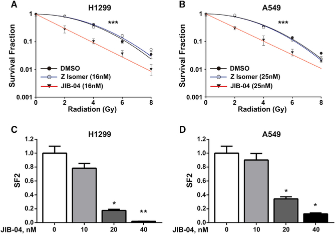
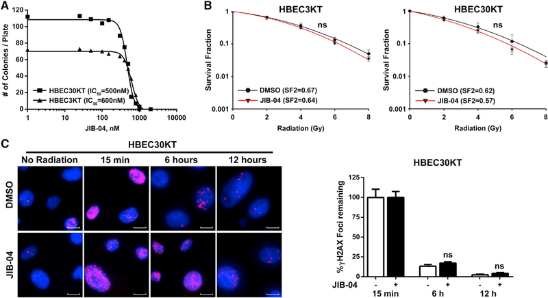
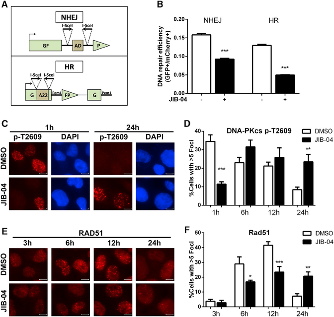
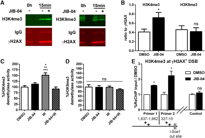
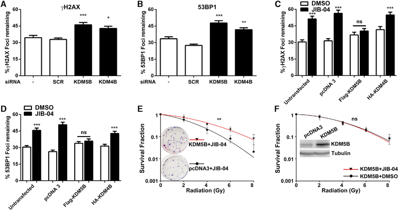
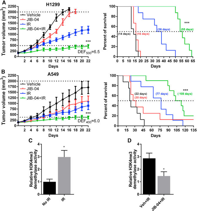
We have uncovered a role for Jumonji inhibitors in overcoming radioresistance through KDM5B inhibition. Pharmacological blockade of Jumonji demethylases with JIB-04 leads to specific accumulation of H3K4me3 at sites marked by γH2AX and impaired recruitment of DNA repair factors, preventing resolution of damage and resulting in robust sensitization to radiation therapy. In DNA-repair-proficient cancer cells, knockdown of the H3K4me3 demethylase KDM5B, but not other Jumonji enzymes, mimics pharmacological inhibition, and KDM5B overexpression rescues this phenotype and increases radioresistance. The H3K4me3 demethylase inhibitor PBIT also sensitizes cancer cells to radiation, while an H3K27me3 demethylase inhibitor does not. In vivo co-administration of radiation with JIB-04 significantly prolongs the survival of mice with tumors even long after cessation of treatment. In human patients, lung squamous cell carcinomas highly expressing KDM5B respond poorly to radiation. Thus, we propose the use of Jumonji KDM inhibitors as potent radiosensitizers.

Keywords: DNA repair; H3K4me3; JARID; JIB-04; Jumonji KDM; KDM5B; lung cancer; radiation therapy; radioresistance; radiosensitization.


Conflict of interest statement

DECLARATION OF INTERESTS

The authors declare no competing interests.

Figures

Figure 1.
Figure 1.. JIB-04 Robustly Sensitizes Radioresistant NSCLC to IR
(A and B) Clonogenic survival of H1299 (A) or A549 (B) cells treated with 16 nM or 25 nM JIB-04, respectively, and then irradiated as indicated (0–8 Gy). The survival of cells treated with JIB-04 alone was set to 1. Graphs show one of two independent experiments, each done in triplicate. Error bars represent SD across triplicates. ***p < 0.001, DMSO or Z-isomer versus JIB-04 (A) (2-way ANOVA); ***p < 0.001, DMSO or Z-isomer versus JIB-04 (2 way ANOVA). (C and D) Clonogenic survival of H1299 (C) and A549 (D) cells treated with increasing doses of JIB04 (0–40 nM) and 2 Gy IR. The number of colonies was normalized to the colonies formed by cells treated with the corresponding dose of JIB-04 alone (without IR). Graph represents one of two experiments (n = 3). Error bars show SD. *p < 0.05; **p < 0.01 versus vehicle (0 nM) (Kruskal-Wallis). See also Figure S1 and Table S1.
Figure 2.
Figure 2.. Defective Resolution of γH2AX and 53BP1 Foci in JIB-04-Treated Cells
(A and B) DNA DSB repair kinetics after IR in H1299 (A) and A549 (B) cells. Cells were incubated with DMSO vehicle or JIB-04 for 4 hr (16 nM for H1299 and 25 nM for A549), irradiated (2 Gy), fixed at the indicated time points, and then immunostained for γH2AX (red) and 53BP1 (green). Scale bars, 10 μm. Foci per nucleus were counted (> 100 nuclei per treatment). Representative images are shown in the left panels for 15 min and 12 hr. Repair kinetics are plotted as the percentage + SEM of remaining foci against time in the right bar graphs for measured time points. ***p < 0.001; **p < 0.01 versus control (ANOVA). Representative data from one of three independent experiments are shown. See also Figures S2 and S3 and Table S2.
Figure 3.
Figure 3.. The Radiosensitizing Effects and DNA Repair Defects Induced by JIB-04 Are Cancer Selective
(A) JIB-04 IC50 determined by liquid colony formation in immortalized non-transformed human bronchial epithelial cells HBEC3KT and HBEC30KT. Graph represents one experiment done in triplicate. (B) Clonogenic survival of HBEC3KT and HBEC30KT cells treated with 600 nM or 500 nM JIB-04, respectively, and then irradiated as indicated. Curves werederived as in Figure 1B. Graph represents one of three experiments done in triplicate. Values represent the average survival fraction ± SD of triplicate samples. ns indicates that no significant differences in radiosensitization curves across treatments were observed (p ≥ 0.1, 2-way ANOVA). (C) DNA DSB repair kinetics in HBEC30KT cells. Cells were incubated with vehicle or 500 nM JIB-04 for 4 hr, irradiated (2 Gy), fixed, immunostained for γH2AX, and then quantified as in Figure 2. Representative images are shown on the left panel. Scale bars, 10 μm. Repair kinetics are plotted in the right bar graphs as percentage + SEM of remaining foci over time. ns indicates that no significant differences were observed between vehicle and treatment groups at any time point (p > 0.8, ANOVA). Data are from >50 nuclei per treatment for one of two experiments. See also Figure S3.
Figure 4.
Figure 4.. JIB-04 Inhibits Both NHEJ and HR Repair
(A) Schematic of the reporter constructs used inHR and NHEJ repair assays. (B) H1299 cells stably containing the NHEJ or theHR constructs were treated with JIB-04 or DMSO for 4 hr and then transfected with the pCMV3×nls-I-SceI (functional endonuclease) and a pN1-mCherry plasmid as transfection control in the continuous presence of treatment (300 nM JIB-04). Cells were analyzed by flow cytometry for GFP and mCherry expression 24 hr after transfection. 20,000 cells were analyzed in each sample and NHEJ or HR repair frequency calculated (%GFP+ cells/%mCherry+ cells). Average + SEM values of triplicates for one of three representative experiments are shown. ***p < 0.001 versus vehicle control (Kruskal-Wallis). (C–F) DNA-PKcs p-T2609 (C and D) and RAD51 (E and F) foci kinetics in H1299 cells. Cells were incubated with vehicle or 16 nM JIB-04 for 4 hr, irradiated (10 Gy), fixed, and immunostained, and then the number of foci per nucleus in >100 cells was counted for each time point. (C) and (E) show representative immunofluorescence images (scale bars, 10 μm), and (D) and (F) show foci formation and resolution kinetics obtained by plotting the percentage + SEM of cells with more than 5 foci per nucleus over time (***p < 0.001; **p < 0.01; *p < 0.05 versus DMSO control per time point [ANOVA]). Data are representative of one of two independent experiments. See also Figure S5.
Figure 5.
Figure 5.. Jumonji Inhibition by JIB-04 Results in H3K4me3 Enrichment at DSBs
(A) Cells were incubated with vehicle or 300 nM JIB-04 for 4 hr, irradiated (20 Gy), and lysed, and nucleosomes were solubilized. γH2AX was immunoprecipitated from 2 mg soluble nucleosome extract and immune-precipitates blotted for γH2AX and H3K4me3 (left) or H3K9me3 (right). (B) The immunoblot data from three independent experiments were quantified and expressed as the average ratio H3K4me3 signal/γH2AX signal or H3K9me3 signal/γH2AX signal + SEM. *p < 0.05; ns, not significant (p = 0.74); DMSO versus JIB-04(t test). (C and D) H1299 cells were pretreated with JIB04 (16 nM) for 4 hr followed by IR (8 Gy) and collected at 15 min after radiation. Cellular extracts were prepared and H3K4me3 (C) and H3K9me3 (D) activity measured. Values in (C) and (D) are expressed as percentage + SEM ofDMSO-treated activity across three independent experiments. *p < 0.05 IR versus JIB-04; **p < 0.001 IR versus DMSO; ***p < 0.001 IR versus JIB-04+IR; ns, not significant (p > 0.8) (Kruskal-Wallis). (E) H1299-NHEJ stable cells pretreated with DMSO or 300 nM JIB-04 were transfected with I-Sce1 plasmid to induce DSBs, and 20 hr later, ChIP/re-ChIP experiments were carried out by immunoprecipitating the product of γH2AX ChIP with H3K4me3 antibodies. Levels of associated DNA were measured by qPCR. *p = 0.012 (DMSO versus JIB-04 for primer 1); *p = 0.011 (DMSO versus JIB-04 for primer 2); ns, p = 0.3 (DMSO versus JIB-04 for control region) (onetailed paired t test). n = 2–4 replicates of one experiment; average percentage of re-ChIP input relative to DMSO ± SEM is shown. See also Figure S6.
Figure 6.
Figure 6.. Jumonji Enzyme Knockdown Phenocopies the JIB-04 Repair Defect, and Overexpression of KDM5B Rescues the JIB-04-Induced DNA Repair Defects and Radiosensitization of Cancer Cells
(A and B) DNA DSB repair kinetics in H1299 cells after knockdown of KDM5B or KDM4B. Cells were irradiated 48 hr after transfection (2 Gy) and immunostained for γH2AX (A) and 53BP1 (B), and then the number of foci per nucleus were counted after 6 hr (>100 nuclei). Graphs represent the percent of foci remaining + SEM for one of three equivalent independent experiments. ***p < 0.001; **p < 0.01; *p < 0.05 versus controls (ANOVA). (C and D) DNA DSB repair kinetics showing γH2AX (C) or 53BP1 (D) in H1299 cells overexpressing FLAG-KDM5B or HA-KDM4B. 24 hr after transfection, cells were incubated with 16 nM JIB-04 for 4 hr, irradiated (2 Gy), and processed as above. Graphs represent the percent of remaining foci + SEM 6 hr after radiation for one of three independent experiments. ***p < 0.001 versus controls; ns, p > 0.4 (ANOVA). (E and F) Clonogenic survival of H1299 cells transfected with pcDNA3 empty vector or overexpressing KDM5B and treated with JIB-04 (E) or KDM5B-overexpressing cells treated with DMSO versus JIB-04 (F). **p < 0.01 KDM5B+JIB-04 versus pcDNA3+JIB-04; ns, no significant difference (p = 0.5) KDM5B+JIB-04 versus KDM5B+DMSO (2-way ANOVA). Inset in (E) shows representative images of colony assays of cells transfected with pcDNA3 or overexpressing KDM5B treated with JIB-04 and 8 Gy radiation. Inset in (F) is a western blot showing the overexpression of KDM5B in cells used for these studies. Data represent the average of three to four independent experiments. Error bars represent SD across experiments. See also Figure S7.
Figure 7.
Figure 7.. JIB-04 Robustly Radiosensitizes Tumors In vivo and Prolongs the Survival of Tumor-Bearing Mice
(A) Subcutaneous tumors generated from H1299 cells were allowed to reach a volume between 150 to 200 mm3, and then mice were treated every other day with vehicle (n = 9), JIB-04 50 mg/kg (n = 8), IR (n = 9), or JIB-04 with IR (2 Gy, given 4 hr after drug administration; n = 8) for a total of 12 doses. Graph represents the tumor volume (left) and percent survival (right). DEF500 = (days to reach 500 mm3 for JIB-04+IR-treated mice days to reach 500 mm3 for JIB-04 treated mice)/(days to reach 500 mm3 for IR-treated mice days to reach 500 mm3 for vehicle-treated mice). Error bars represent SEM. (B) Subcutaneous tumors generated from A549 cells treated as described in (A) with vehicle (n = 8), JIB-04 50 mg/kg (n = 8), IR (n = 9), or JIB-04 with IR (n = 10). Graph represents the tumor volume (left) and percent survival (right). DEF400 was calculated as above for time to 500 mm3. For (A) and (B), error bars represent SEM. ***p < 0.001 JIB-04+IR versus IR (2-way ANOVA for tumor growth in the left panel, and Kaplan-Meier for survival in the right panel). (C and D) Tumor tissues were harvested at time of death (n = 4 for vehicle and JIB-04) or 12 hr after the last dose of treatment (n = 3 for IR and JIB-04+IR), tumor extracts were prepared, and H3K4me3 demethylase activity was measured. *p < 0.05 for IR versus no IR (C); *p < 0.05 for vehicle+IR/vehicle versus JIB-04+IR/JIB-04 alone (D) (t test). See also Figure S8 and Table S3.

Similar articles

Cited by

References

    1. Amendola PG, Zaghet N, Ramalho JJ, Vilstrup Johansen J, Boxem M, and Salcini AE (2017). JMJD-5/KDM8 regulates H3K36me2 and is required for late steps of homologous recombination and genome integrity. PLoS Genet. 13, e1006632. - PMC - PubMed
    1. Anscher MS (2010). Targeting the TGF-beta1 pathway to prevent normal tissue injury after cancer therapy. Oncologist 15, 350–359. - PMC - PubMed
    1. Aymard F, Bugler B, Schmidt CK, Guillou E, Caron P, Briois S, Iacovoni JS, Daburon V, Miller KM, Jackson SP, and Legube G (2014). Transcriptionally active chromatin recruits homologous recombination at DNA double-strand breaks. Nat. Struct. Mol. Biol 21, 366–374. - PMC - PubMed
    1. Bayo J, Dalvi MP, and Martinez ED (2015). Successful strategies in the discovery of small-molecule epigenetic modulators with anticancer potential. Future Med. Chem 7, 2243–2261. - PubMed
    1. Bookout AL, Cummins CL, Mangelsdorf DJ, Pesola JM, and Kramer MF (2006). High-throughput real-time quantitative reverse transcription PCR. Curr. Protoc. Mol. Biol Chapter 15, Unit 15.18. - PubMed

Publication types

MeSH terms