

Renal miR-148b is associated with megalin down-regulation in IgA nephropathy
- PMID: 30355654
- PMCID: PMC6239259
- DOI: 10.1042/BSR20181578
Renal miR-148b is associated with megalin down-regulation in IgA nephropathy
Abstract
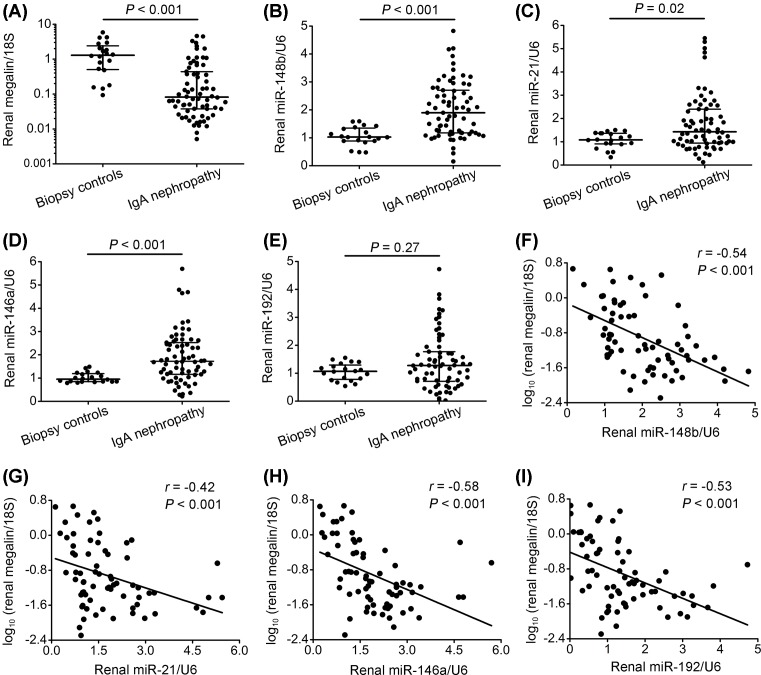
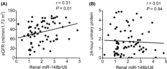
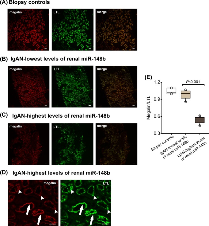
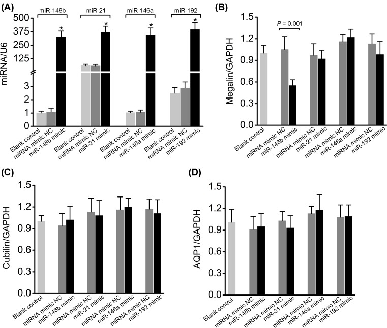
Megalin is essential for proximal tubule reabsorption of filtered proteins, hormones, and vitamins, and its dysfunction has been reported in IgA nephropathy (IgAN). miR-148b has been shown to regulate renal megalin expression in vitro and in animal models of kidney disease. We examined a potential role of miR-148b and other miRNAs in regulating megalin expression in IgAN by analyzing the association between megalin and miR-148b, miR-21, miR-146a, and miR-192 expression. Quantitative PCR (qPCR) analysis identified a marked increase in renal levels of several miRNAs, including miR-148b, miR-21, miR-146a, and a significant decrease in megalin mRNA levels in IgAN patients when compared with normal controls. By multiple linear regression analysis, however, only renal miR-148b was independently associated with megalin mRNA levels in IgAN. Proximal tubule megalin expression was further evaluated by immunofluorescence labeling of biopsies from the patients. The megalin expression was significantly lower in patients with highest levels of renal miR-148b compared with patients with lowest levels. To examine the direct effects of the miRNAs on megalin and other membrane proteins expression, proximal tubule LLC-PK1 cells were transfected with miR-148b, miR-21, miR-146a, or miR-192 mimics. Transfection with miR-148b mimic, but not the other three miRNA mimics inhibited endogenous megalin mRNA expression. No significant effect of any of the four miRNA mimics was observed on cubilin or aquaporin 1 (AQP1) mRNA expression. The findings suggest that miR-148b negatively regulates megalin expression in IgAN, which may affect renal uptake and metabolism of essential substances.
Keywords: IgA nephropathy; cell transfection; megalin; microRNA; renal proximal tubule.
© 2018 The Author(s).
Conflict of interest statement
The authors declare that there are no competing interests associated with the manuscript.
Figures
Similar articles
-
MicroRNA-148b regulates megalin expression and is associated with receptor downregulation in mice with unilateral ureteral obstruction.Am J Physiol Renal Physiol. 2017 Aug 1;313(2):F210-F217. doi: 10.1152/ajprenal.00585.2016. Epub 2017 Mar 22. Am J Physiol Renal Physiol. 2017. PMID: 28331063
-
Renal Megalin mRNA Downregulation Is Associated with CKD Progression in IgA Nephropathy.Am J Nephrol. 2022;53(6):481-489. doi: 10.1159/000524929. Epub 2022 Jun 3. Am J Nephrol. 2022. PMID: 35661648
-
Acute endotoxemia in mice induces downregulation of megalin and cubilin in the kidney.Kidney Int. 2012 Jul;82(1):53-9. doi: 10.1038/ki.2012.62. Epub 2012 Mar 21. Kidney Int. 2012. PMID: 22437417
-
Megalin and cubilin in proximal tubule protein reabsorption: from experimental models to human disease.Kidney Int. 2016 Jan;89(1):58-67. doi: 10.1016/j.kint.2015.11.007. Kidney Int. 2016. PMID: 26759048 Review.
-
Protein reabsorption in renal proximal tubule-function and dysfunction in kidney pathophysiology.Pediatr Nephrol. 2004 Jul;19(7):714-21. doi: 10.1007/s00467-004-1494-0. Epub 2004 May 14. Pediatr Nephrol. 2004. PMID: 15146321 Review.
Cited by
-
ICAM-1 related long noncoding RNA is associated with progression of IgA nephropathy and fibrotic changes in proximal tubular cells.Sci Rep. 2022 Jun 10;12(1):9645. doi: 10.1038/s41598-022-13521-6. Sci Rep. 2022. PMID: 35688937 Free PMC article.
-
The Non-Coding RNA Landscape in IgA Nephropathy-Where Are We in 2021?J Clin Med. 2021 May 28;10(11):2369. doi: 10.3390/jcm10112369. J Clin Med. 2021. PMID: 34071162 Free PMC article. Review.
-
Megalin: A Sidekick or Nemesis of the Kidney?J Am Soc Nephrol. 2025 Feb 1;36(2):293-300. doi: 10.1681/ASN.0000000572. Epub 2024 Nov 15. J Am Soc Nephrol. 2025. PMID: 39607686 Review.
-
MicroRNAs in IgA nephropathy.Ren Fail. 2021 Dec;43(1):1298-1310. doi: 10.1080/0886022X.2021.1977320. Ren Fail. 2021. PMID: 34547971 Free PMC article. Review.
-
Pathogenic Role of MicroRNA Dysregulation in Podocytopathies.Front Physiol. 2022 Jun 29;13:948094. doi: 10.3389/fphys.2022.948094. eCollection 2022. Front Physiol. 2022. PMID: 35845986 Free PMC article. Review.
References
Publication types
MeSH terms
Substances
LinkOut - more resources
Full Text Sources
Miscellaneous