Isolation and Characterization of the Novel Botulinum Neurotoxin A Subtype 6
- PMID: 30355669
- PMCID: PMC6200982
- DOI: 10.1128/mSphere.00466-18
Isolation and Characterization of the Novel Botulinum Neurotoxin A Subtype 6
Abstract
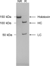
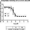
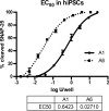
Botulinum neurotoxins (BoNTs), the most potent toxins known to humans and the causative agent of botulism, exert their effect by entering motor neurons and cleaving and inactivating SNARE proteins, which are essential for neurotransmitter release. BoNTs are proven, valuable pharmaceuticals used to treat more than 200 neuronal disorders. BoNTs comprise 7 serotypes and more than 40 isoforms (subtypes). BoNT/A1 is the only A-subtype used clinically due to its high potency and long duration of action. While other BoNT/A subtypes have been purified and described, only BoNT/A2 is being investigated as an alternative to BoNT/A1. Here we describe subtype BoNT/A6 with improved pharmacological properties compared to BoNT/A1. It was isolated from Clostridium botulinum CDC41370, which produces both BoNT/B2 and BoNT/A6. The gene encoding BoNT/B2 was genetically inactivated, and A6 was isolated to greater than 95% purity. A6 was highly potent in cultured primary rodent neuronal cultures and in human induced pluripotent stem cell-derived neurons, requiring 20-fold less toxin to cause 50% SNAP-25 cleavage than A1. Second, A6 entered hiPSCs faster and more efficiently than A1 and yet had a long duration of action similar to BoNT/A1. Third, BoNT/A6 had similar LD50 as BoNT/A1 after intraperitoneal injection in mice; however, local intramuscular injection resulted in less systemic toxicity than BoNT/A1 and a higher (i.m.) LD50, indicating its potential as a safer pharmaceutical. These data suggest novel characteristics of BoNT/A6 and its potential as an improved pharmaceutical due to more efficient neuronal cell entry, greater ability to remain localized at the injection site, and a long duration.IMPORTANCE Botulinum neurotoxins (BoNTs) have proved to be an effective treatment for a large number of neuropathic conditions. BoNTs comprise a large family of zinc metalloproteases, but BoNT/A1 is used nearly exclusively for pharmaceutical purposes. The genetic inactivation of a second BoNT gene in the native strain enabled expression and isolation of a single BoNT/A6 from cultures. Its characterization indicated that BoNT/A subtype A6 has a long duration of action comparable to A1, while it enters neurons faster and more efficiently and remains more localized after intramuscular injection. These characteristics of BoNT/A6 are of interest for potential use of BoNT/A6 as a novel BoNT-based therapeutic that is effective and has a fast onset, an improved safety profile, and a long duration of action. Use of BoNT/A6 as a pharmaceutical also has the potential to reveal novel treatment motifs compared to currently used treatments.
Keywords: BoNT; BoNT/A6; botulinum neurotoxin; cell entry; duration; potency; subtype.
Copyright © 2018 Moritz et al.
Figures


Similar articles
-
Comparative functional analysis of mice after local injection with botulinum neurotoxin A1, A2, A6, and B1 by catwalk analysis.Toxicon. 2019 Sep;167:20-28. doi: 10.1016/j.toxicon.2019.06.004. Epub 2019 Jun 7. Toxicon. 2019. PMID: 31181297 Free PMC article.
-
The Light Chain Defines the Duration of Action of Botulinum Toxin Serotype A Subtypes.mBio. 2018 Mar 27;9(2):e00089-18. doi: 10.1128/mBio.00089-18. mBio. 2018. PMID: 29588398 Free PMC article.
-
Purification and Characterization of Botulinum Neurotoxin FA from a Genetically Modified Clostridium botulinum Strain.mSphere. 2016 Feb 24;1(1):e00100-15. doi: 10.1128/mSphere.00100-15. eCollection 2016 Jan-Feb. mSphere. 2016. PMID: 27303710 Free PMC article.
-
A Comprehensive Structural Analysis of Clostridium botulinum Neurotoxin A Cell-Binding Domain from Different Subtypes.Toxins (Basel). 2023 Jan 18;15(2):92. doi: 10.3390/toxins15020092. Toxins (Basel). 2023. PMID: 36828407 Free PMC article. Review.
-
Progress in cell based assays for botulinum neurotoxin detection.Curr Top Microbiol Immunol. 2013;364:257-85. doi: 10.1007/978-3-642-33570-9_12. Curr Top Microbiol Immunol. 2013. PMID: 23239357 Free PMC article. Review.
Cited by
-
Effect of samarium oxide nanoparticles on virulence factors and motility of multi-drug resistant Pseudomonas aeruginosa.World J Microbiol Biotechnol. 2022 Aug 30;38(11):209. doi: 10.1007/s11274-022-03384-4. World J Microbiol Biotechnol. 2022. PMID: 36040540
-
Biological Toxins as the Potential Tools for Bioterrorism.Int J Mol Sci. 2019 Mar 8;20(5):1181. doi: 10.3390/ijms20051181. Int J Mol Sci. 2019. PMID: 30857127 Free PMC article. Review.
-
Botulinum Neurotoxins: History, Mechanism, and Applications. A Narrative Review.J Neurochem. 2025 Aug;169(8):e70187. doi: 10.1111/jnc.70187. J Neurochem. 2025. PMID: 40762356 Free PMC article. Review.
-
Catch and Anchor Approach To Combat Both Toxicity and Longevity of Botulinum Toxin A.J Med Chem. 2020 Oct 8;63(19):11100-11120. doi: 10.1021/acs.jmedchem.0c01006. Epub 2020 Sep 18. J Med Chem. 2020. PMID: 32886509 Free PMC article.
-
Critical Analysis of Neuronal Cell and the Mouse Bioassay for Detection of Botulinum Neurotoxins.Toxins (Basel). 2019 Dec 7;11(12):713. doi: 10.3390/toxins11120713. Toxins (Basel). 2019. PMID: 31817843 Free PMC article. Review.
References
Publication types
MeSH terms
Substances
Grants and funding
LinkOut - more resources
Full Text Sources
Medical
Research Materials

