Plasticity of Amino Acid Residue 145 Near the Receptor Binding Site of H3 Swine Influenza A Viruses and Its Impact on Receptor Binding and Antibody Recognition
- PMID: 30355680
- PMCID: PMC6321904
- DOI: 10.1128/JVI.01413-18
Plasticity of Amino Acid Residue 145 Near the Receptor Binding Site of H3 Swine Influenza A Viruses and Its Impact on Receptor Binding and Antibody Recognition
Abstract
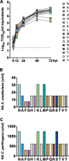
The hemagglutinin (HA), a glycoprotein on the surface of influenza A virus (IAV), initiates the virus life cycle by binding to terminal sialic acid (SA) residues on host cells. The HA gradually accumulates amino acid substitutions that allow IAV to escape immunity through a mechanism known as antigenic drift. We recently confirmed that a small set of amino acid residues are largely responsible for driving antigenic drift in swine-origin H3 IAV. All identified residues are located adjacent to the HA receptor binding site (RBS), suggesting that substitutions associated with antigenic drift may also influence receptor binding. Among those substitutions, residue 145 was shown to be a major determinant of antigenic evolution. To determine whether there are functional constraints to substitutions near the RBS and their impact on receptor binding and antigenic properties, we carried out site-directed mutagenesis experiments at the single-amino-acid level. We generated a panel of viruses carrying substitutions at residue 145 representing all 20 amino acids. Despite limited amino acid usage in nature, most substitutions at residue 145 were well tolerated without having a major impact on virus replication in vitro All substitution mutants retained receptor binding specificity, but the substitutions frequently led to decreased receptor binding. Glycan microarray analysis showed that substitutions at residue 145 modulate binding to a broad range of glycans. Furthermore, antigenic characterization identified specific substitutions at residue 145 that altered antibody recognition. This work provides a better understanding of the functional effects of amino acid substitutions near the RBS and the interplay between receptor binding and antigenic drift.IMPORTANCE The complex and continuous antigenic evolution of IAVs remains a major hurdle for vaccine selection and effective vaccination. On the hemagglutinin (HA) of the H3N2 IAVs, the amino acid substitution N 145 K causes significant antigenic changes. We show that amino acid 145 displays remarkable amino acid plasticity in vitro, tolerating multiple amino acid substitutions, many of which have not yet been observed in nature. Mutant viruses carrying substitutions at residue 145 showed no major impairment in virus replication in the presence of lower receptor binding avidity. However, their antigenic characterization confirmed the impact of the 145 K substitution in antibody immunodominance. We provide a better understanding of the functional effects of amino acid substitutions implicated in antigenic drift and its consequences for receptor binding and antigenicity. The mutation analyses presented in this report represent a significant data set to aid and test the ability of computational approaches to predict binding of glycans and in antigenic cartography analyses.
Keywords: H3 subtype; hemagglutinin; influenza vaccines; swine influenza; virus evolution.
This is a work of the U.S. Government and is not subject to copyright protection in the United States. Foreign copyrights may apply.
Figures


Similar articles
-
The Molecular Determinants of Antibody Recognition and Antigenic Drift in the H3 Hemagglutinin of Swine Influenza A Virus.J Virol. 2016 Aug 26;90(18):8266-80. doi: 10.1128/JVI.01002-16. Print 2016 Sep 15. J Virol. 2016. PMID: 27384658 Free PMC article.
-
Immune Escape Adaptive Mutations in Hemagglutinin Are Responsible for the Antigenic Drift of Eurasian Avian-Like H1N1 Swine Influenza Viruses.J Virol. 2022 Aug 24;96(16):e0097122. doi: 10.1128/jvi.00971-22. Epub 2022 Aug 2. J Virol. 2022. PMID: 35916512 Free PMC article.
-
Immune Escape Adaptive Mutations in the H7N9 Avian Influenza Hemagglutinin Protein Increase Virus Replication Fitness and Decrease Pandemic Potential.J Virol. 2020 Sep 15;94(19):e00216-20. doi: 10.1128/JVI.00216-20. Print 2020 Sep 15. J Virol. 2020. PMID: 32699084 Free PMC article.
-
Antibody Immunodominance: The Key to Understanding Influenza Virus Antigenic Drift.Viral Immunol. 2018 Mar;31(2):142-149. doi: 10.1089/vim.2017.0129. Epub 2018 Jan 22. Viral Immunol. 2018. PMID: 29356618 Free PMC article. Review.
-
Molecular Epidemiology and Evolutionary Dynamics of Human Influenza Type-A Viruses in Africa: A Systematic Review.Microorganisms. 2022 Apr 25;10(5):900. doi: 10.3390/microorganisms10050900. Microorganisms. 2022. PMID: 35630344 Free PMC article. Review.
Cited by
-
2018-2019 human seasonal H3N2 influenza A virus spillovers into swine with demonstrated virus transmission in pigs were not sustained in the pig population.J Virol. 2024 Dec 17;98(12):e0008724. doi: 10.1128/jvi.00087-24. Epub 2024 Nov 11. J Virol. 2024. PMID: 39526773 Free PMC article.
-
Plasticity of the Influenza Virus H5 HA Protein.mBio. 2021 Feb 9;12(1):e03324-20. doi: 10.1128/mBio.03324-20. mBio. 2021. PMID: 33563825 Free PMC article.
-
Amino acid 138 in the HA of a H3N2 subtype influenza A virus increases affinity for the lower respiratory tract and alveolar macrophages in pigs.PLoS Pathog. 2024 Feb 20;20(2):e1012026. doi: 10.1371/journal.ppat.1012026. eCollection 2024 Feb. PLoS Pathog. 2024. PMID: 38377132 Free PMC article.
-
Machine Learning Prediction and Experimental Validation of Antigenic Drift in H3 Influenza A Viruses in Swine.mSphere. 2021 Mar 17;6(2):e00920-20. doi: 10.1128/mSphere.00920-20. mSphere. 2021. PMID: 33731472 Free PMC article.
-
Genetic Characterization of Influenza A Viruses in Japanese Swine in 2015 to 2019.J Virol. 2020 Jul 1;94(14):e02169-19. doi: 10.1128/JVI.02169-19. Print 2020 Jul 1. J Virol. 2020. PMID: 32350072 Free PMC article.
References
-
- Matrosovich M, Tuzikov A, Bovin N, Gambaryan A, Klimov A, Castrucci MR, Donatelli I, Kawaoka Y. 2000. Early alterations of the receptor-binding properties of H1, H2, and H3 avian influenza virus hemagglutinins after their introduction into mammals. J Virol 74:8502–8512. doi:10.1128/JVI.74.18.8502-8512.2000. - DOI - PMC - PubMed
Publication types
MeSH terms
Substances
Grants and funding
LinkOut - more resources
Full Text Sources

