. 2019 Jan 4;93(2):e01849-18.
doi: 10.1128/JVI.01849-18. Print 2019 Jan 15.

Low-pH Endocytic Entry of the Porcine Alphaherpesvirus Pseudorabies Virus

Affiliations

Low-pH Endocytic Entry of the Porcine Alphaherpesvirus Pseudorabies Virus

Jonathan L Miller et al. J Virol. .

Abstract

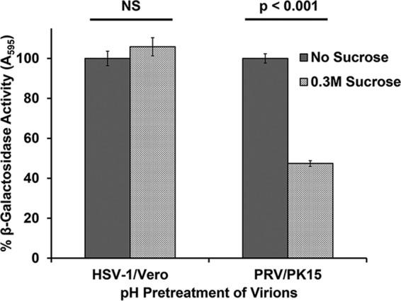
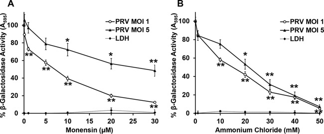
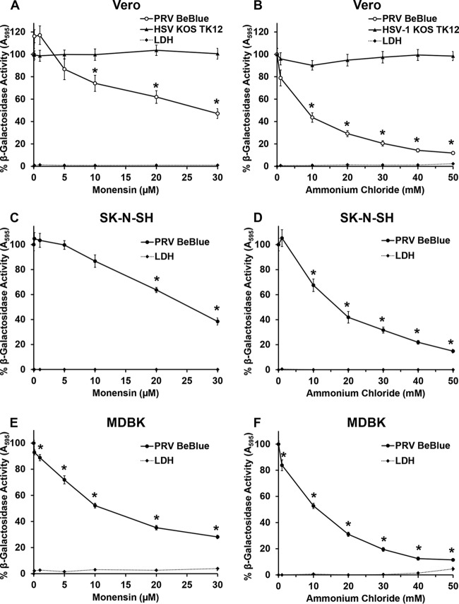
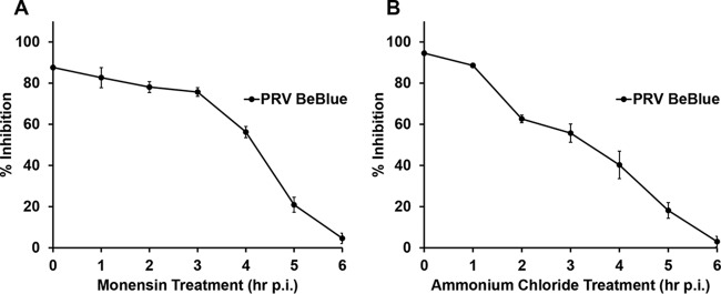
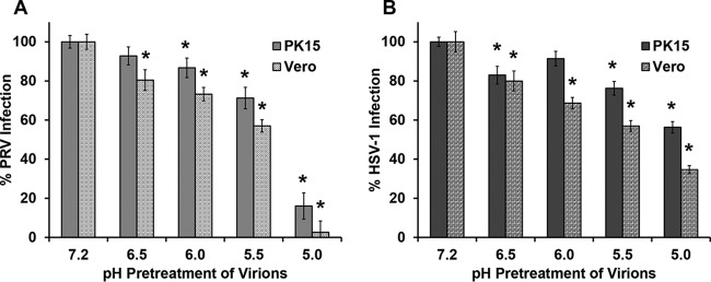
The alphaherpesvirus pseudorabies virus (PRV) is the causative agent of pseudorabies, a disease of great economic and welfare importance in swine. Other alphaherpesviruses, including herpes simplex virus (HSV), utilize low-pH-mediated endocytosis to enter a subset of cell types. We investigated whether PRV used this entry pathway in multiple laboratory model cell lines. Inhibition of receptor-mediated endocytosis by treatment with hypertonic medium prevented PRV entry. PRV entry into several cell lines, including porcine kidney (PK15) cells and African green monkey kidney (Vero) cells, was inhibited by noncytotoxic concentrations of the lysosomotropic agents ammonium chloride and monensin, which block the acidification of endosomes. Inactivation of virions by acid pretreatment is a hallmark of viruses that utilize a low-pH-mediated entry pathway. Exposure of PRV virions to pH 5.0 in the absence of host cell membranes reduced entry into PK15 and Vero cells by >80%. Together, these findings suggest that endocytosis followed by fusion with host membranes triggered by low endosomal pH is an important route of entry for PRV.IMPORTANCE PRV is a pathogen of great economic and animal welfare importance in many parts of the world. PRV causes neurological, respiratory, and reproductive disorders, often resulting in mortality of young and immunocompromised animals. Mortality, decreased production, and trade restrictions result in significant financial losses for the agricultural industry. Understanding the molecular mechanisms utilized by PRV to enter host cells is an important step in identifying novel strategies to prevent infection and spread. A thorough understanding of these mechanisms will contribute to a broader understanding of alphaherpesvirus entry. Here, we demonstrate PRV entry into multiple model cell lines via a low-pH endocytosis pathway. Together, these results provide a framework for elucidating the early events of the PRV replicative cycle.

Keywords: endocytosis; herpesviruses; low pH; pseudorabies virus; viral entry.


Figures

FIG 1
FIG 1
Inhibition of endocytic uptake of PRV from the cell surface. PRV BeBlue was prebound to cells for 1 h at 4°C. Cells were then treated with medium containing 0.3 M sucrose or complete DMEM and incubated at 37°C for 30 min. Cells were washed with PBS and treated with medium buffered to pH 4.7 to inactivate noninternalized virions. Infection proceeded for 6 h in the presence of normal culture medium. β-Galactosidase expression was calculated as a percentage of the activity in mock-treated cells. Values for β-galactosidase activity are the means of data from three independent experiments, each with quadruplicate samples, with standard errors. Treatment with 0.3 M sucrose was noncytotoxic under the experimental conditions, with lactate dehydrogenase (LDH) leakage values below 1% for both Vero and PK15 cells. The P values were determined using Student’s t test. NS, not significant (P > 0.05).
FIG 2
FIG 2
Effect of lysosomotropic agents on PRV entry into PK15 cells. PK15 cells were treated with monensin (A) or ammonium chloride (B) at the indicated concentrations for 20 min at 37°C. Cells were infected with PRV BeBlue at an MOI of 1 or 5 for 6 h in the continued presence of the agents. β-Galactosidase activity in the absence of the agent was set to 100%. LDH activity as a measure of cytotoxicity is reported as a percentage of the value for the detergent-lysed sample. Values for β-galactosidase activity are the means of data from three independent experiments, each with quadruplicate samples, with standard errors. Values for cytotoxicity are the means and standard errors of data from three independent experiments with triplicate samples. The P values relative to those with no drug treatment at the same MOI were determined with Student’s t test (*, P < 0.05; **, P < 0.01). For an MOI of 1, monensin treatment at 0.1 μM and ammonium chloride treatment at 1 mM were also significant (P < 0.01).
FIG 3
FIG 3
Effect of lysosomotropic agents on PRV entry into Vero, SK-N-SH, and MDBK cells. Vero (A and B), SK-N-SH (C and D), or MDBK (E and F) cells were treated with monensin or ammonium chloride at the indicated concentrations for 20 min at 37°C. Cells were infected with PRV BeBlue (MOI of 1) or HSV-1 KOS tk12 (MOI of 5) for 6 h in the continued presence of agents. β-Galactosidase activity of untreated, infected cells was set to 100%. LDH activity as a measure of cytotoxicity is reported as a percentage of the value for the detergent-lysed sample. Values for β-galactosidase activity are the means of data from three independent experiments, each with quadruplicate samples, with standard errors. Values for cytotoxicity are the means and standard errors of data from three independent experiments with triplicate samples. The P values relative to those with no drug treatment were determined using Student’s t test (*, P < 0.01).
FIG 4
FIG 4
Time course of lysosomotropic agent effects on PRV entry. PRV BeBlue was bound to confluent PK15 cells in 96-well plates at 4°C for 1 h. Noninternalized virus was inactivated with sodium citrate buffer, and 30 μM monensin (A) or 50 mM NH4Cl (B) was added at time points from 0 to 6 h p.i. Infection proceeded for a total of 6 h. Control wells for each time point were treated with sodium citrate buffer, and untreated medium was added to the cells. β-Galactosidase expression was calculated as a percentage of the activity in cells in control wells for each time point. Values for β-galactosidase activity are the means of data from three independent experiments, each with quadruplicate samples, with standard errors. Values for cytotoxicity are the means and standard errors of data from three independent experiments with triplicate samples.
FIG 5
FIG 5
Effect of mildly acidic pH on PRV infectivity. PRV BeBlue (A) or HSV-1 tk12 (B) was incubated at pHs ranging from 7.2 to 5.0 at 37°C for 30 min and then neutralized to pH 7.2. Treated virions were added to confluent PK15 or Vero cells in 96-well plates for 6 h. β-Galactosidase activity was measured to indicate virus entry, with maximum infectivity set to 100%. Values for β-galactosidase activity are the means of data from three independent experiments, each with quadruplicate samples, with standard errors. The P values relative to those with no drug treatment were determined using Student’s t test (*, P < 0.05).
FIG 6
FIG 6
Effect of MG132, a proteasome inhibitor, on PRV entry. PK15 cells were treated with MG132 for 20 min at 37°C. PRV BeBlue was added (MOI of 1) in the continued presence of MG132. At 6 h p.i., β-galactosidase activity of untreated, infected cells was set to 100%. Cytotoxicity is shown as percent LDH activity. Each experiment was performed with quadruplicate (entry) or triplicate (LDH) samples. Values are the means and standard errors of data from three independent experiments. The P values relative to those with no drug treatment were determined using Student’s t test (*, P < 0.05).

Similar articles

Cited by

References

    1. Muller T, Hahn EC, Tottewitz F, Kramer M, Klupp BG, Mettenleiter TC, Freuling C. 2011. Pseudorabies virus in wild swine: a global perspective. Arch Virol 156:1691–1705. doi:10.1007/s00705-011-1080-2. - DOI - PubMed
    1. Pomeranz LE, Reynolds AE, Hengartner CJ. 2005. Molecular biology of pseudorabies virus: impact on neurovirology and veterinary medicine. Microbiol Mol Biol Rev 69:462–500. doi:10.1128/MMBR.69.3.462-500.2005. - DOI - PMC - PubMed
    1. Enquist LW. 1999. Life beyond eradication: veterinary viruses in basic science. Arch Virol Suppl 15:87–109. - PubMed
    1. Lee JYS, Wilson MR. 1979. A review of pseudorabies (Aujeszky’s disease) in pigs. Can Vet J 20:65–69. - PMC - PubMed
    1. Mettenleiter TC. 1996. Immunobiology of pseudorabies (Aujeszky’s disease). Vet Immunol Immunopathol 54:221–229. doi:10.1016/S0165-2427(96)05695-4. - DOI - PubMed

Publication types

LinkOut - more resources