. 2019 Jan 4;93(2):e01835-18.
doi: 10.1128/JVI.01835-18. Print 2019 Jan 15.

The NS1 Protein of Influenza A Virus Participates in Necroptosis by Interacting with MLKL and Increasing Its Oligomerization and Membrane Translocation

Affiliations

The NS1 Protein of Influenza A Virus Participates in Necroptosis by Interacting with MLKL and Increasing Its Oligomerization and Membrane Translocation

Amit Gaba et al. J Virol. .

Abstract

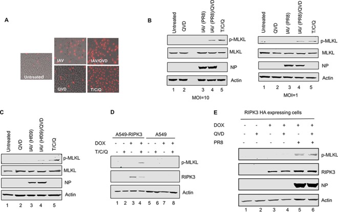
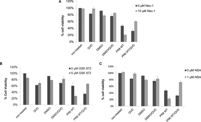
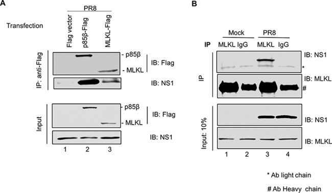
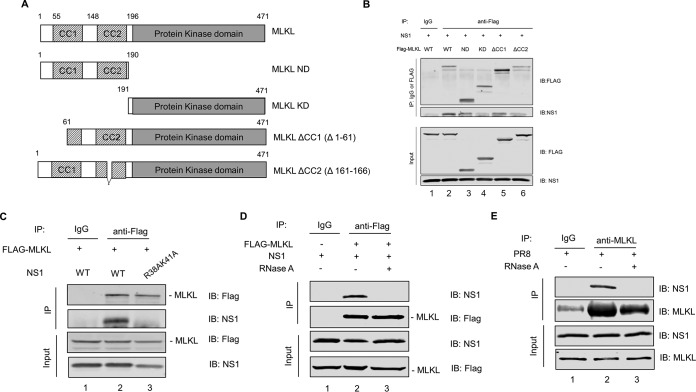
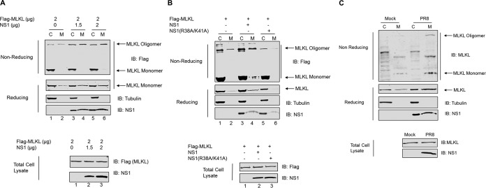
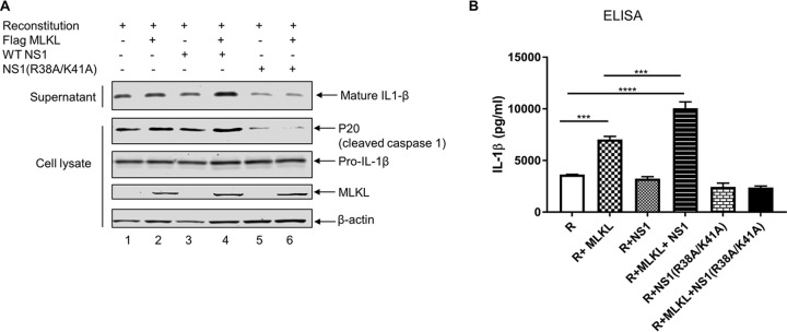
Elimination of infected cells by programmed cell death is a well-recognized host defense mechanism to control the spread of infection. In addition to apoptosis, necroptosis is also one of the mechanisms of cell death that can be activated by viral infection. Activation of necroptosis leads to the phosphorylation of mixed-lineage kinase domain-like protein (MLKL) by receptor-interacting protein kinase 3 (RIPK3) and results in MLKL oligomerization and membrane translocation, leading to membrane disruption and a loss of cellular ion homeostasis. It has recently been reported that influenza A virus (IAV) infection induces necroptosis. However, the underlying mechanism of the IAV-mediated necroptosis process, particularly the roles of IAV proteins in necroptosis, remains unexplored. Here, we report that IAV infection induces necroptosis in macrophages and epithelial cells. We demonstrate that the NS1 protein of IAV interacts with MLKL. Coiled-coil domain 2 of MLKL has a predominant role in mediating the MLKL interaction with NS1. The interaction of NS1 with MLKL increases MLKL oligomerization and membrane translocation. Moreover, the MLKL-NS1 interaction enhances MLKL-mediated NLRP3 inflammasome activation, leading to increased interleukin-1β (IL-1β) processing and secretion.IMPORTANCE Necroptosis is a programmed cell death that is inflammatory in nature owing to the release of danger-associated molecular patterns from the ruptured cell membrane. However, necroptosis also constitutes an important arm of host immune responses. Thus, a balanced inflammatory response determines the disease outcome. We report that the NS1 protein of IAV participates in necroptosis by interacting with MLKL, resulting in increased MLKL oligomerization and membrane translocation. These results reveal a novel function of the NS1 protein and the mechanism by which IAV induces necroptosis. Moreover, we show that this interaction enhances NLRP3 inflammasome activation and IL-1β processing and secretion. This information may contribute to a better understanding of the role of necroptosis in IAV-induced inflammation.

Keywords: MLKL; NS1 protein; inflammasome; influenza A virus; necroptosis.


Figures

FIG 1
FIG 1
IAV infection induces necroptosis in THP1 cells and the A549 cell line inducibly expressing human RIPK3. (A) Representative images of differentiated THP1 cells that were either left untreated, infected with IAV (PR8) at an MOI of 1, treated with QVD, treated with QVD for 1 h and then infected with IAV, or treated with the combination of T/C/Q. Cell death was assessed by PI staining at 16 h p.i. (B) THP1 cells were infected with IAV (PR8) at the indicated MOIs or treated as described above for panel A, and at 8 h posttreatment, cells were harvested and examined for phosphorylated MLKL by Western blotting. (C) THP1 cells were infected with IAV (Hf09) at an MOI of 10 or treated as described above for panel A. At 8 h posttreatment, cells were harvested and examined for phosphorylated MLKL by Western blotting. (D) A549 cells and an A549 cell line inducibly expressing HA-tagged human RIPK3 were cultured in the absence or presence of 0.5 μg/ml of doxycycline (DOX) for 12 h and either left untreated or treated with the combination of T/C/Q (TNF-α, cycloheximide, and QVD). At 8 h posttreatment, cells were harvested and examined for RIPK3 and phosphorylated MLKL by Western blotting. (E) The A549 cell line inducibly expressing human RIPK3 was cultured in the absence or presence of 0.5 μg/ml of doxycycline for 12 h, and cells were either left untreated or treated with QVD for 1 h and then either mock infected or infected with IAV (PR8) at an MOI of 10. At 8 h p.i., cells were harvested and examined for phosphorylated MLKL by Western blotting.
FIG 2
FIG 2
IAV-induced cell death involves both apoptosis and necroptosis. Differentiated THP1 cells were either left untreated or treated for 1 h with the caspase inhibitor QVD (20 μM) and one of the following pharmacological inhibitors: Nec-1 (10 μM) for RIPK1 (A), GSK872 (5 μM) for RIPK3 (B), and NSA (1 μM) for MLKL (C). Following chemical treatment, the cells were either mock infected or infected with PR8 virus at an MOI of 5. At 18 to 20 h p.i., cell death was assessed by measuring the ATP concentration in cells using a Cell Titer-Glo luminescent viability assay.
FIG 3
FIG 3
NS1 protein of IAV interacts with MLKL. (A) HEK293T cells were transfected with a Flag vector or the p85β-Flag or MLKL-Flag plasmid. At 24 h posttransfection, cells were infected with PR8 virus at an MOI of 10. Cell lysates were prepared at 6 h p.i. and subjected to IP with anti-Flag antibody. Precipitated proteins were subjected to Western blotting using antibodies against Flag and NS1 proteins. (B) Differentiated THP1 cells were either mock infected or infected with PR8 virus at an MOI of 10. Cell lysates were prepared at 8 h p.i. and subjected to IP with either normal rabbit IgG or anti-MLKL antibody (Ab). Precipitated proteins were subjected to Western blotting using antibodies against MLKL and NS1 proteins. IB, immunoblotting.
FIG 4
FIG 4
CC2 of MLKL has a predominant role in mediating MLKL interaction with NS1. (A) Flag-tagged plasmids expressing different truncated MLKL proteins were constructed to identify the domains critical for the interaction of MLKL with the NS1 protein. A schematic representation of plasmids expressing mutant MLKL proteins is shown. (B) HEK293T cells were cotransfected with an NS1-expressing plasmid and either the Flag-tagged MLKL-expressing plasmid or one of the truncated MLKL-expressing plasmids. Cell lysates were prepared at 36 h posttransfection and subjected to IP with either normal rabbit IgG or anti-Flag antibody. Precipitated proteins were subjected to Western blotting using antibodies against the Flag tag or the NS1 protein. (C) HEK293T cells were cotransfected with the Flag-tagged MLKL-expressing plasmid and either the WT NS1- or NS1(R38A/K41A)-expressing plasmid. Cell lysates were prepared at 36 h posttransfection and subjected to IP with either normal rabbit IgG or anti-Flag antibody. Precipitated proteins were subjected to Western blotting using antibodies against the Flag tag and NS1 protein. (D) HEK293T cells were cotransfected with the Flag-tagged MLKL-expressing plasmid and NS1-expressing plasmid. At 36 h posttransfection, cell lysates were prepared, treated with RNase A at 10 μg/ml for 30 min at 4°C or left untreated, and subjected to IP with either normal rabbit IgG or anti-Flag antibody. Precipitated proteins were subjected to Western blotting using antibodies against the Flag tag and NS1 protein. (E) Differentiated THP1 cells were infected with PR8 virus at an MOI of 10. At 8 h p.i., cell lysates were prepared, treated with RNase A or left untreated, and subjected to IP with either normal rabbit IgG or anti-MLKL antibody. Precipitated proteins were subjected to Western blotting using antibodies against MLKL and NS1 proteins.
FIG 5
FIG 5
NS1 enhances MLKL oligomerization and membrane translocation. (A) HEK293T cells were cotransfected with the Flag-tagged MLKL-expressing plasmid and increasing amounts of the NS1-expressing plasmid. At 36 h posttransfection, cell lysates were separated into the total, cytoplasmic (C), or membrane (M) fraction. The cell lysates were resolved on SDS-PAGE gels with (reducing) or without (nonreducing) β-mercaptoethanol and analyzed by Western blotting using the indicated antibodies. (B) HEK293T cells were cotransfected with the Flag-tagged MLKL-expressing plasmid and either the WT NS1- or NS1(R38A/K41A)-expressing plasmid. At 36 h posttransfection, cell lysates were harvested and subjected to assays as described above for panel A. (C) Differentiated THP1 cells were either mock infected or infected with PR8 virus at an MOI of 10. At 8 h p.i., cell lysates were harvested and subjected to assays as described above for panel A.
FIG 6
FIG 6
Interaction of NS1 with MLKL enhances MLKL-mediated NLRP3 inflammasome activation and IL-1β processing and secretion. (A) HEK293T cells were transfected with plasmids expressing NLRP3 inflammasome components and pro-IL-1β along with the indicated plasmids expressing null (empty vector), MLKL, NS1, or NS1(R38A/K41A). The supernatant and cell lysates were collected at 20 h posttransfection and analyzed by Western blotting with the indicated antibodies. (B) HEK293T cells were transfected as described above for panel A, and IL-1β levels in the supernatants of the above-described samples were measured by an ELISA. R, reconstitution (pcDNA-NLRP3 plus pcDNA-ASC plus pCMV-Flag-pro-caspase 1 plus pcDNA-pro-IL-1β). ***, P < 0.001; ****, P < 0.0001. Results are representative of data from three independent experiments performed in duplicates.

Similar articles

Cited by

References

    1. Sridharan H, Upton JW. 2014. Programmed necrosis in microbial pathogenesis. Trends Microbiol 22:199–207. doi:10.1016/j.tim.2014.01.005. - DOI - PubMed
    1. Galluzzi L, Vanden Berghe T, Vanlangenakker N, Buettner S, Eisenberg T, Vandenabeele P, Madeo F, Kroemer G. 2011. Programmed necrosis from molecules to health and disease. Int Rev Cell Mol Biol 289:1–35. doi:10.1016/B978-0-12-386039-2.00001-8. - DOI - PubMed
    1. Li J, McQuade T, Siemer AB, Napetschnig J, Moriwaki K, Hsiao YS, Damko E, Moquin D, Walz T, McDermott A, Chan FK, Wu H. 2012. The RIP1/RIP3 necrosome forms a functional amyloid signaling complex required for programmed necrosis. Cell 150:339–350. doi:10.1016/j.cell.2012.06.019. - DOI - PMC - PubMed
    1. Cai Z, Jitkaew S, Zhao J, Chiang HC, Choksi S, Liu J, Ward Y, Wu LG, Liu ZG. 2014. Plasma membrane translocation of trimerized MLKL protein is required for TNF-induced necroptosis. Nat Cell Biol 16:55–65. doi:10.1038/ncb2883. - DOI - PMC - PubMed
    1. Wang H, Sun L, Su L, Rizo J, Liu L, Wang LF, Wang FS, Wang X. 2014. Mixed lineage kinase domain-like protein MLKL causes necrotic membrane disruption upon phosphorylation by RIP3. Mol Cell 54:133–146. doi:10.1016/j.molcel.2014.03.003. - DOI - PubMed

Publication types

MeSH terms

Substances

LinkOut - more resources