. 2019 Jan 4;93(2):e01265-18.
doi: 10.1128/JVI.01265-18. Print 2019 Jan 15.

A MicroRNA Derived from Adenovirus Virus-Associated RNAII Promotes Virus Infection via Posttranscriptional Gene Silencing

Affiliations

A MicroRNA Derived from Adenovirus Virus-Associated RNAII Promotes Virus Infection via Posttranscriptional Gene Silencing

K Wakabayashi et al. J Virol. .

Abstract

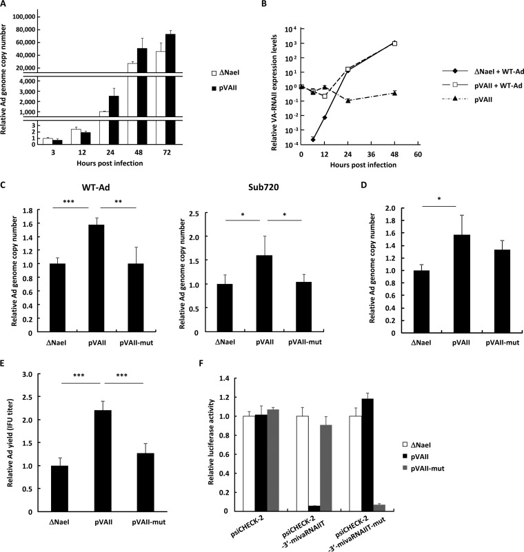
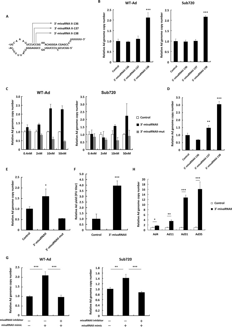
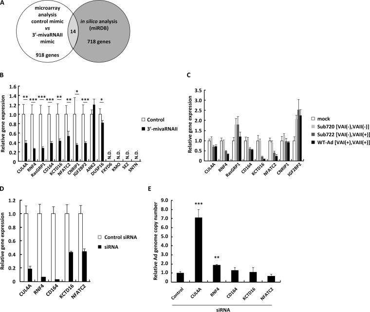
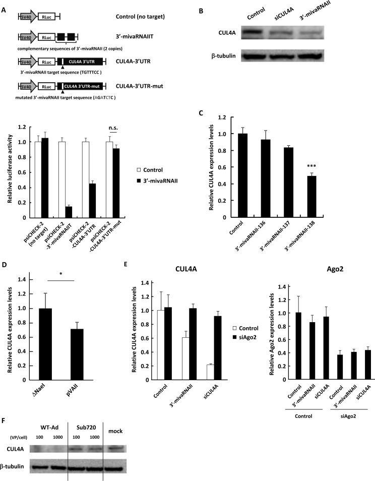
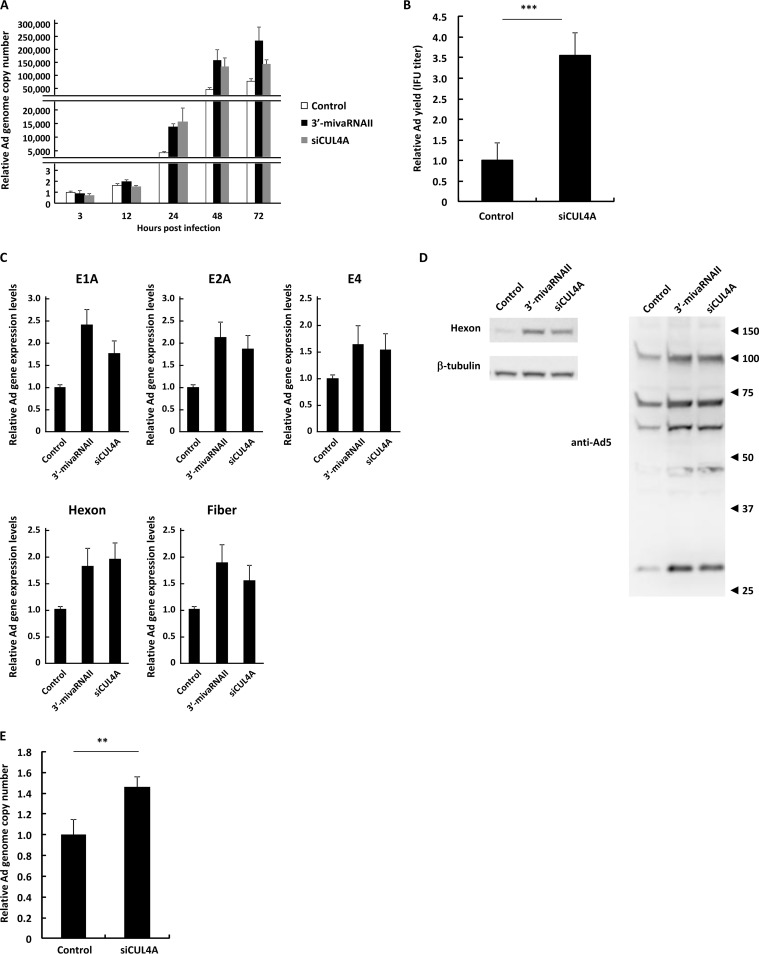
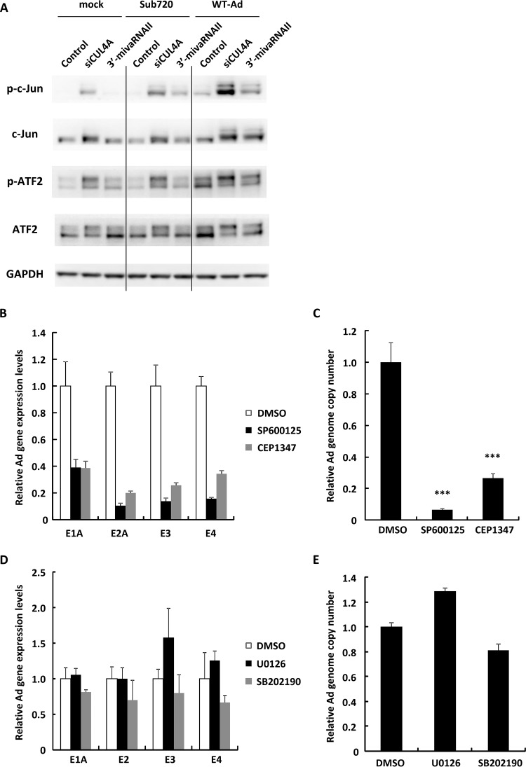
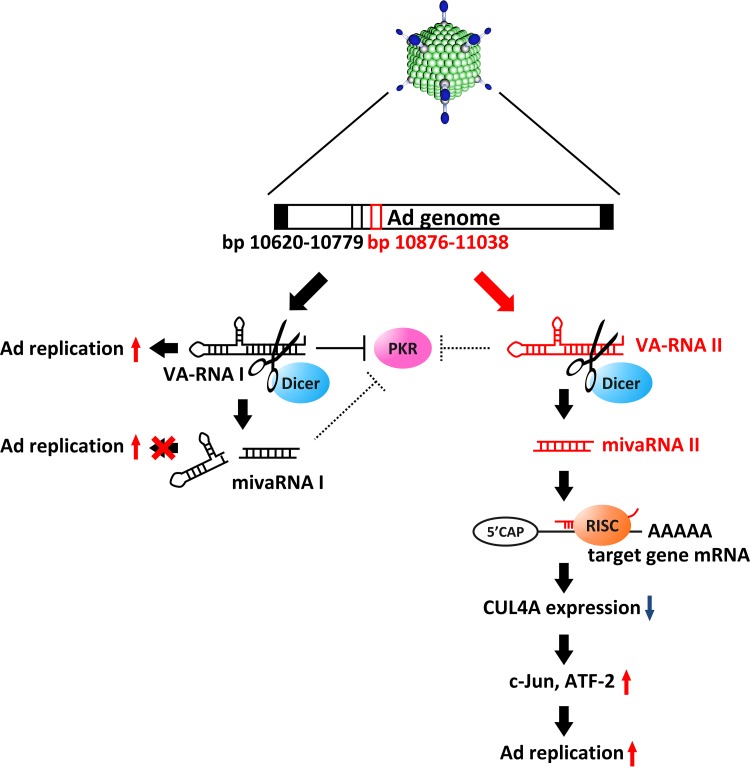
The adenovirus (Ad) serotype 5 genome encodes two noncoding small RNAs (virus-associated RNAs I and II [VA-RNAI and -II]), which are approximately 160-nucleotide (nt) RNAs transcribed by RNA polymerase III. It is well known that VA-RNAI supports Ad infection via the inhibition of double-stranded RNA-dependent protein kinase (PKR), which recognizes double-stranded RNA and acts as an antiviral system. Recent studies revealed that VA-RNAs are processed into VA-RNA-derived microRNAs (miRNAs) (mivaRNAI and -II); however, we and another group recently demonstrated that mivaRNAI does not promote Ad replication. On the other hand, the roles of VA-RNAII and mivaRNAII in Ad replication have remained to be clarified. In this study, we demonstrated mivaRNAII-mediated promotion of Ad replication. Transfection with chemically synthesized 3'-mivaRNAII-138, one of the most abundant forms of mivaRNAII, significantly enhanced Ad replication, while the other species of mivaRNAII did not. We identified 8 putative target genes of 3'-mivaRNAII-138 by microarray analysis and in silico analysis. Among the 8 candidates, knockdown of the cullin 4A (CUL4A) gene, which encodes a component of the ubiquitin ligase complex, most significantly enhanced Ad replication. CUL4A expression was significantly suppressed by 3'-mivaRNAII-138 via posttranscriptional gene silencing, indicating that CUL4A is a target gene of 3'-mivaRNAII-138 and mivaRNAII functions as a viral miRNA promoting Ad infection. It has been reported that CUL4A is involved in degradation of c-Jun, which acts as a transcription factor in the Jun-N-terminal kinase (JNK) signaling cascade. Treatment with JNK inhibitors dramatically suppressed Ad replication, suggesting that mivaRNAII-mediated downregulation of CUL4A enhanced JNK signaling and thereby promoted Ad infection.IMPORTANCE Several types of viruses encode viral miRNAs which regulate host and/or viral gene expression via posttranscriptional gene silencing, leading to efficient viral infection. Adenovirus (Ad) expresses miRNAs derived from VA-RNAs (mivaRNAI and -II); however, recent studies have revealed that processing of VA-RNAI into mivaRNAI inhibits Ad replication. Conversely, we demonstrate here that mivaRNAII significantly promotes Ad replication and that mivaRNAII-mediated suppression of CUL4A expression via posttranscriptional gene silencing induces accumulation of c-Jun, leading to promotion of Ad infection. These results exhibited the significance of VA-RNAII for supporting Ad infection through a mechanism complementary to that of VA-RNAI. These observations could provide important clues toward a new perspective on host-virus interaction. Moreover, Ad is widely used as a basic framework for viral vectors and oncolytic viruses. Our findings will help to regulate Ad infection and will promote the development of novel Ad vectors and oncolytic Ad.

Keywords: JNK signaling; adenoviruses; cullin 4A; microRNA; posttranscriptional gene silencing.


Figures

FIG 1
FIG 1
Ad replication is upregulated by overexpression of VA-RNAII. (A) HeLa cells were transfected with a control plasmid (pAdVAntage-ΔNaeI; ΔNaeI) or a VA-RNAII-expressing plasmid (pVAII) for 48 h, followed by infection with WT-Ad at 100 VPs/cell. After 3, 12, 24, 48, and 72 h of incubation, the Ad genome copy numbers were determined by qPCR analysis and expressed as relative values (ΔNaeI, 3 h postinfection = 1). (B) HeLa cells were transfected with ΔNaeI or pVAII for 48 h, followed by infection with WT-Ad at 100 VPs/cell. After 0, 6, 12, 24, and 48 h of incubation following Ad infection, the copy numbers of VA-RNAII were determined by qRT-PCR analysis and expressed as relative values (pVAII, 0 h postinfection = 1). (C) HeLa cells were transfected with ΔNaeI, pVAII, or pVAII-mut for 48 h, followed by infection with Ad (WT-Ad or Ad lacking VA-RNAI, II; Sub720) at 100 VPs/cell. After 24 h of incubation, the Ad genome copy numbers were determined and expressed as relative values (ΔNaeI = 1). (D) A549 cells were transfected with VA-RNAII-expressing plasmids (ΔNaeI, pVAII, and pVAII-mut) for 48 h, followed by infection with WT-Ad at 100 VPs/cell. Ad genome copy numbers were determined 24 h after infection and expressed as relative values (ΔNaeI = 1). (E) HeLa cells were transfected with VA-RNAII-expressing plasmids (ΔNaeI, pVAII, and pVAII-mut) for 48 h, followed by infection with WT-Ad at 100 VPs/cell. After 24 h of incubation, infectious unit (IFU) titers of progeny WT-Ad in the cells were determined and expressed as relative values (ΔNaeI = 1). (F) HeLa cells were cotransfected with VA-RNAII-expressing plasmids (ΔNaeI, pVAII, and pVAII-mut) and reporter plasmids (psiCHECK-2, siCHECK-2-3′-mivaRNAIIT, and psiCHECK-2-3′-mivaRNAIIT-mut). After 48 h of incubation, luciferase activities were determined. RLuc activities were normalized to Fluc activities. These data are expressed as means ± SD (n = 4). *, P < 0.05; **, P < 0.01; ***, P < 0.001 (Student’s t test).
FIG 2
FIG 2
3′-mivaRNAII promotes Ad replication. (A) A schematic diagram of the processing of VA-RNA II by dicer, producing 3′-mivaRNAII-136, -137, and -138. (B) HeLa cells were transfected with 3′-mivaRNAII-136, -137, or -138 mimics at 50 nM for 48 h, followed by infection with Ad (WT-Ad and Sub720) at 100 VPs/cell. Ad genome copy numbers were determined 24 h after infection and expressed as relative values (control = 1). (C) HeLa cells were transfected with 3′-mivaRNAII (3′-mivaRNAII-138) and 3′-mivaRNAII-mut mimics at the indicated concentrations for 48 h, followed by infection with Ad (WT-Ad and Sub720) at 100 VPs/cell. Ad genome copy numbers were determined 24 h after infection and expressed as relative values (control = 1 at the respective concentration). (D) A549 cells were transfected with 3′-mivaRNAII-136, -137, or -138 mimics at 50 nM for 48 h, followed by infection with WT-Ad at 100 VPs/cell. Ad genome copy numbers were determined 24 h after infection and expressed as relative values (control = 1). (E) A549 cells were transfected with 3′-mivaRNAII-138 and -mut mimics at 50 nM for 48 h, followed by infection with WT-Ad at 100 VPs/cell. Ad genome copy numbers were determined 24 h after infection and expressed as relative values (control = 1). (F) HeLa cells were transfected with 3′-mivaRNAII (3′-mivaRNAII-138) mimic at 50 nM and incubated for 48 h, followed by infection with WT-Ad at 100 VPs/cell. After 24 h of incubation, IFU titers of progeny WT-Ad in the cells were determined and expressed as relative values (control = 1). (G) HeLa cells were cotransfected with 3′-mivaRNAII (3′-mivaRNAII-138) mimic and negative control inhibitor or 3′-mivaRNAII inhibitor at 30 nM each for 48 h, followed by infection with WT-Ad at 100 VPs/cell. Ad genome copy numbers were determined 24 h after infection and expressed as relative values (control = 1). (H) HeLa cells were transfected with 3′-mivaRNAII (3′-mivaRNAII-138) mimic at 50 nM for 48 h, followed by infection with Ad4, Ad11, Ad31, or Ad35 at 100 VPs/cell. Ad genome copy numbers were determined 24 h after infection and expressed as relative values (control = 1). These data are expressed as means ± SD (n = 4).
FIG 3
FIG 3
Identification of 3′-mivaRNA II target genes. (A) A schematic diagram of analysis for identification of mivaRNAII target genes. (B) HeLa cells were transfected with 3′-mivaRNAII (3′-mivaRNAII-138) mimic at 50 nM. After 48 h of incubation, mRNA levels of the indicated genes were determined by qRT-PCR analysis. (C) HeLa cells were infected with Ad (WT-Ad, Sub722, and Sub720) at 1,000 VPs/cell. After 48 h of incubation, mRNA levels of the indicated genes were determined by qRT-PCR analysis. (D and E) HeLa cells were transfected with siRNAs targeting the indicated genes at 50 nM. (D) After 48 h of incubation, mRNA levels of the indicated genes were determined by qRT-PCR analysis. (E) After 48 h of incubation, cells were infected with WT-Ad at a multiplicity of infection (MOI) of 5. Ad genome copy numbers in the cells were determined 24 h after infection and expressed as relative values (control = 1). These data are expressed as means ± SD (A to C, F, and G, n = 4; D and E, n = 3).
FIG 4
FIG 4
3′-mivaRNAII suppresses CUL4A expression via posttranscriptional gene silencing. (A) HeLa cells were cotransfected with 3′-mivaRNAII (3′-mivaRNAII-138) mimic and the indicated reporter plasmids, described in the upper portion. Luciferase activities were determined 48 h after transfection. RLuc activities were normalized to Fluc activities. The mutated nucleotides in the sequences complementary to the seed sequences of 3′-mivaRNAII are shown in gray. SV40, SV40 promoter; n.s., not significant. (B) HeLa cells were transfected with 3′-mivaRNAII (3′-mivaRNAII-138) mimic, or siCUL4A at 50 nM. After 48 h of incubation, protein levels of CUL4A were evaluated by Western blotting analysis. (C) HeLa cells were transfected with 3′-mivaRNAII-136, -137, or -138 mimics at 50 nM. After 48 h of incubation, mRNA levels of CUL4A were determined by qRT-PCR analysis. (D) HeLa cells were transfected with ΔNaeI or pVAII. After 48 h of incubation, the mRNA levels of CUL4A were determined by qRT-PCR analysis. (E) HeLa cells were cotransfected with a 3′-mivaRNAII mimic (3′-mivaRNAII-138), siCUL4A, and siAgo2 at 50 nM. After 48 h of incubation, the mRNA levels of CUL4A and Ago2 were determined by qRT-PCR analysis. (F) HeLa cells were infected with Ad (WT-Ad and Sub720) at 100 or 1,000 VPs/cell. After 48 h of incubation, protein levels of CUL4A were evaluated by Western blotting analysis. These data are expressed as means ± SD (A and C, n = 4).
FIG 5
FIG 5
Suppression of CUL4A gene expression promotes Ad infection. (A) HeLa cells were transfected with a 3′-mivaRNAII mimic (3′-mivaRNAII-138) or siCUL4A at 50 nM for 48 h, followed by infection with WT-Ad at 100 VPs/cell. After 3, 12, 24, 48, or 72 h of incubation, the Ad genome copy numbers were determined and expressed as relative values (control, 3 h postinfection = 1). (B) HeLa cells were transfected with siCUL4A at 50 nM for 48 h, followed by infection with WT-Ad at 100 VPs/cell. After 24 h of incubation, IFU titers of progeny WT-Ad were determined and expressed as relative values (control = 1). (C) HeLa cells were transfected with a 3′-mivaRNAII mimic (3′-mivaRNAII-138) or siCUL4A at 50 nM for 48 h, followed by infection with WT-Ad at 100 VPs/cell. mRNA levels of the Ad genes were determined by qRT-PCR analysis at 12 h (E1A, E2A, and E4) or 24 h (hexon and fiber) after infection. (D) HeLa cells were transfected with a 3′-mivaRNAII mimic (3′-mivaRNAII-138) or siCUL4A at 50 nM for 48 h, followed by infection with WT-Ad at 100 VPs/cell. After 24 h of incubation, the protein levels of the Ad genes were evaluated by Western blotting analysis. Numbers at the right of the membranes indicate the protein sizes in kilodaltons. Note that the sizes of the Ad major capsid proteins, hexon, penton base, and fiber, are 108 kDa, 63 kDa, and 61 kDa, respectively. (E) A549 cells were transfected with siCUL4A at 50 nM for 48h, followed by infection with WT-Ad at 100 VPs/cell. After 24 h of incubation, Ad genome copy numbers were determined and expressed as relative values (control = 1). These data are expressed as means ± SD (n = 4).
FIG 6
FIG 6
JNK signaling is involved in mivaRNAII-mediated promotion of Ad infection. (A) HeLa cells were transfected with 3′-mivaRNAII (3′-mivaRNAII-138) mimic or siCUL4A at 50 nM for 48 h, followed by infection with Ad (WT-Ad, Sub720) at 100 VPs/cell. After 24 h of incubation, protein levels of c-Jun and ATF2 were evaluated by Western blotting analysis. (B and C) HeLa cells were treated with JNK inhibitors (10 mM SP600125 or 20 mM CEP1347), followed by infection with WT-Ad at 100 VPs/cell. (B) After 12 h of incubation, mRNA levels of the Ad genes were determined by qRT-PCR analysis. (C) After 24 h of incubation, Ad genome copy numbers were determined and expressed as relative values (dimethyl sulfoxide [DMSO] = 1). (D and E) HeLa cells were treated with 10 mM U0126 (an ERK inhibitor) or 20 mM SB202190 (a p38 inhibitor) and infected with WT-Ad at 100 VPs/cell. (D) After 12 h of incubation, mRNA levels of the Ad genes were determined by qRT-PCR analysis. (E) After 24 h of incubation, Ad genome copy numbers were determined. These data are expressed as means ± SD (B and C, n = 4; D and E, n = 3).
FIG 7
FIG 7
Model of VA-RNAII-mediated promotion of Ad replication via posttranscriptional gene silencing of CUL4A. After Ad infection, VA-RNAI and -II are rapidly transcribed and promote Ad infection in different ways. VA-RNAI inhibits the activation of PKR, which plays a key role in antiviral responses. VA-RNAII functions as a precursor of mivaRNAII, which promotes Ad infection, while mivaRNAI does not support Ad infection. Suppression of CUL4A, the highest-potential target of mivaRNAII, amplifies JNK signaling via stabilization of c-Jun and ATF2, leading to promotion of Ad replication.

Similar articles

Cited by

References

    1. Ambros V. 2004. The functions of animal microRNAs. Nature 431:350–355. doi:10.1038/nature02871. - DOI - PubMed
    1. Macfarlane L-AA, Murphy PR. 2010. MicroRNA: biogenesis, function and role in cancer. Curr Genomics 11:537–561. doi:10.2174/138920210793175895. - DOI - PMC - PubMed
    1. Kincaid RP, Sullivan CS. 2012. Virus-encoded microRNAs: an overview and a look to the future. PLoS Pathog 8:e1003018. doi:10.1371/journal.ppat.1003018. - DOI - PMC - PubMed
    1. Grundhoff A, Sullivan CS. 2011. Virus-encoded microRNAs. Virology 411:325–343. doi:10.1016/j.virol.2011.01.002. - DOI - PMC - PubMed
    1. Gupta A, Gartner JJ, Sethupathy P, Hatzigeorgiou AG, Fraser NW. 2006. Anti-apoptotic function of a microRNA encoded by the HSV-1 latency-associated transcript. Nature 442:82–85. doi:10.1038/nature04836. - DOI - PubMed

Publication types