. 2019 Jan 4;93(2):e01520-18.
doi: 10.1128/JVI.01520-18. Print 2019 Jan 15.

Encapsidation of Viral RNA in Picornavirales: Studies on Cowpea Mosaic Virus Demonstrate Dependence on Viral Replication

Affiliations

Encapsidation of Viral RNA in Picornavirales: Studies on Cowpea Mosaic Virus Demonstrate Dependence on Viral Replication

Inga Kruse et al. J Virol. .

Abstract

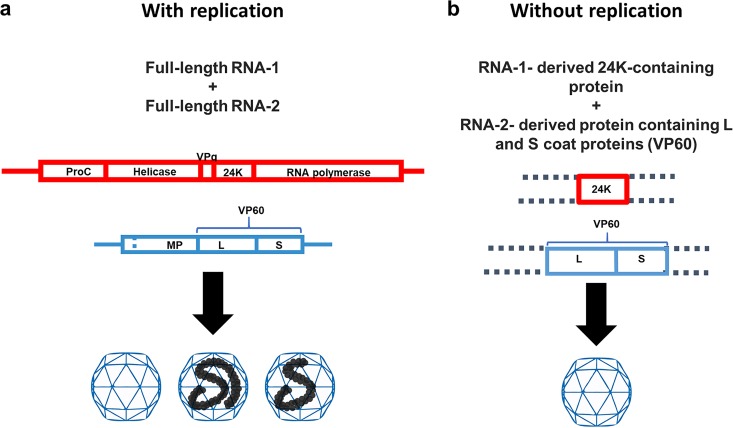
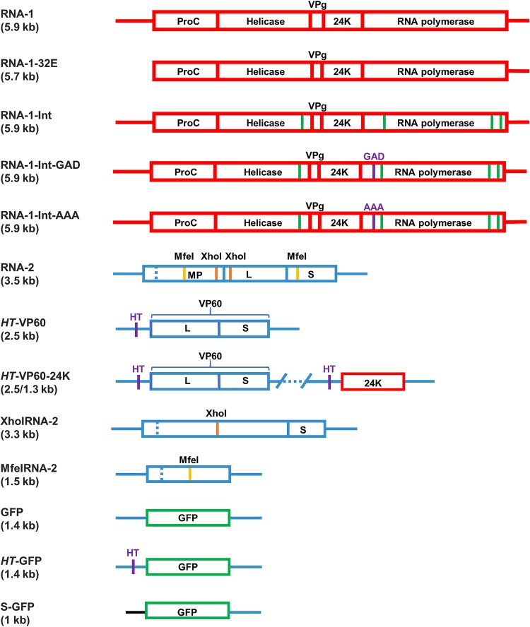
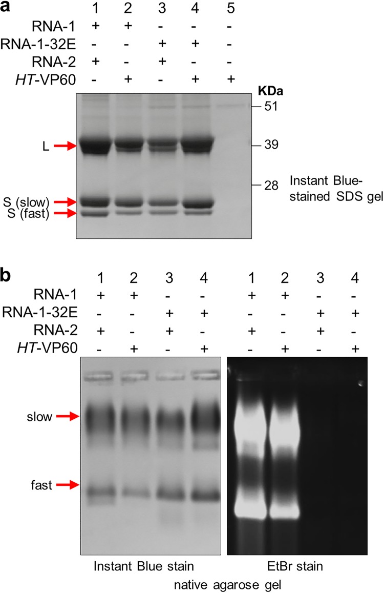
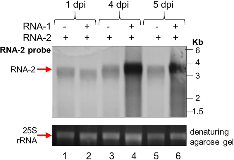
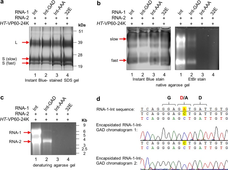
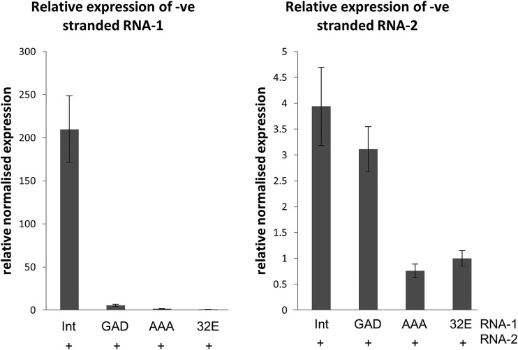
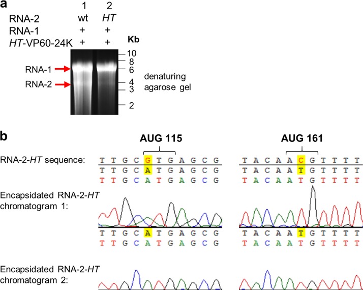
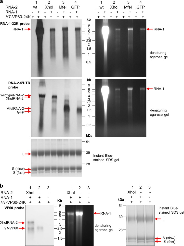
To elucidate the linkage between replication and encapsidation in Picornavirales, we have taken advantage of the bipartite nature of a plant-infecting member of this order, cowpea mosaic virus (CPMV), to decouple the two processes. RNA-free virus-like particles (empty virus-like particles [eVLPs]) can be generated by transiently coexpressing the RNA-2-encoded coat protein precursor (VP60) with the RNA-1-encoded 24,000-molecular-weight (24K) protease, in the absence of the replication machinery (K. Saunders, F. Sainsbury, and G. P. Lomonossoff, Virology 393:329-337, 2009, https://doi.org/10.1016/j.virol.2009.08.023). We have made use of the ability to produce assembled capsids of CPMV in the absence of replication to examine the putative linkage between RNA replication and packaging in the Picornavirales We have created a series of mutant RNA-1 and RNA-2 molecules and have assessed the effects of the mutations on both the replication and packaging of the viral RNAs. We demonstrate that mutations that affect replication have a concomitant impact on encapsidation and that RNA-1-mediated replication is required for encapsidation of both RNA-1 and RNA-2. This close coupling between replication and encapsidation provides a means for the specific packaging of viral RNAs. Moreover, we demonstrate that this feature of CPMV can be used to specifically encapsidate custom RNA by placing a sequence of choice between the RNA-2 sequences required for replication.IMPORTANCE The mechanism whereby members of the order Picornavirales specifically package their genomic RNAs is poorly understood. Research with monopartite members of the order, such as poliovirus, indicated that packaging is linked to replication, although the presence of "packaging signals" along the length of the viral RNA has also been suggested. Thanks to the bipartite nature of the CPMV genome, which allows the manipulation of RNA-1 without modifying RNA-2, we show here that this specificity is due to a functional link between the two processes of viral replication and encapsidation. This has important implications for our understanding of the fundamental molecular biology of Picornavirales and opens the door to novel research and therapeutic applications in the field of custom RNA packaging and delivery technologies.

Keywords: Picornavirales; RNA packaging; RNA virus; cowpea mosaic virus; plant viruses; viral encapsidation; viral replication.


Figures

FIG 1
FIG 1
The bipartite CPMV replication and packaging system. (a) When full-length, replication-competent RNA-1 and RNA-2 are coexpressed, three species of particles are formed: empty (top component), RNA-2-containing (middle component), and RNA-1-containing (bottom component) species. (b) The coexpression of any 24K protease-containing sequence with any VP60-containing sequence in the absence of viral replication will yield empty particles indistinguishable from the top component (4, 23).
FIG 2
FIG 2
Diagrams of constructs used in this study. Boxes represent open reading frames, and lines represent UTRs. (Top) RNA-1-based constructs (red). Green lines represent introns introduced into the RNA-1 sequence to alleviate toxicity of the DNA constructs in bacteria. Point mutations of the replicase GDD motif are shown in purple, and the amino acid sequence that replaces GDD is indicated. (Bottom) RNA-2-based constructs (blue). The HT mutations shown in the 5′ UTR of the HT-VP60 construct are a pair of point mutations that greatly increase the translational efficiency of the downstream open reading frame (49). XhoI-RNA-2 and MfeI-RNA-2 are truncated by restriction digestion with the respective enzymes and subsequent religation. The GFP substitution is shown in green, and the synthetic 5′ UTR substituted for the 5′ UTR of RNA-2 in the S-GFP construct is shown in black. All constructs expressed from a pEAQ plasmid also contain a P19 silencing suppressor cassette on the same T-DNA (not shown). The constructs expressed from pBIN plasmids (RNA-1 from pBinPS1NT, RNA-1-32E from pBinPS32E, and RNA-2 from pBinPS2NT) were coinfiltrated with pBIN61-P19 to supply P19. All constructs are expressed in plants transiently via Agrobacterium-mediated expression.
FIG 3
FIG 3
A replication-competent version of RNA-1 is necessary for RNA encapsidation. CPMV particles were purified from N. benthamiana leaves agroinfiltrated with pBinPS1NT and pBinPS2NT (lanes 1 in each gel), pBinPS1NT and pEAQ-HT-VP60 (lanes 2), pBinP32E and pBinPS2NT (lanes 3), and pBinP32E and pEAQ-HT-VP60 (lanes 4) or with pEAQ-HT-VP60 alone (lanes 5). In each case, the CPMV RNAs expressed within the leaves are indicated. The purified particles were examined by either denaturing SDS-PAGE followed by staining with Instant Blue (a) or electrophoresis on a nondenaturing agarose gel (b) followed by staining with either Instant Blue (left) to visualize protein or ethidium bromide (EtBr) (right) to visualize nucleic acid. The positions of the large (L) coat protein and two forms of the small [S (slow) and S (fast)] coat protein are indicated to the left of the gel in panel a. Note that in panel b, CPMV particles separate into distinct electrophoretic populations based on the presence or absence of the labile 24 amino acids at the C terminus of the small coat protein (2) as seen in panel a.
FIG 4
FIG 4
RNA-2 is abundant in the cell even in the absence of RNA-1. (Top) Northern blot of total RNA extracted from agroinfiltrated leaf material probed with an RNA probe specific for RNA-2 positive strands. Leaves were either agroinfiltrated with pEAQ-RNA-2 alone or coinfiltrated with both pBinPS1NT and pEAQ-RNA-2 and were harvested at 1, 4, and 5 days postinfiltration (dpi). (Bottom) Ethidium bromide-stained denaturing agarose gel prior to transfer of the RNAs to the membrane, showing the levels of 25S rRNA present in each sample as a loading control.
FIG 5
FIG 5
GDD mutants of RNA-1 abolish viral symptoms. Agroinfiltration was used to coexpress pEAQ-RNA-2 and an RNA-1-based construct, as indicated. Photographs were taken from above at 30 days postinfiltration to show any symptoms on the upper, systemic leaves. RNA-1-Int (expressing a wild-type replicase) causes systemic symptoms in upper leaves of N. benthamiana when coexpressed with RNA-2, as seen by mottled yellowing and curling of the upper leaves (top left-hand panel). In contrast, replicase mutants RNA-1-Int-GAD and RNA-1-Int-AAA, just like RNA-1-32E, do not cause symptoms when coexpressed with RNA-2.
FIG 6
FIG 6
GDD mutants of RNA-1 are encapsidation deficient. Particles were purified from leaves agroinfiltrated with pEAQ-RNA-2 and an RNA-1-based construct, as indicated. The same preparation of purified particles was used for the gels shown here. (a) Protein content of particles visualized by SDS-PAGE and Instant Blue staining. The correctly processed large (L) coat protein and two electrophoretic forms of the small (S) coat protein present in all samples are indicated. (b) Purified particles from the same preparations as in panel a were analyzed in duplicate on a native agarose gel. (Left) Half of the gel was stained with Instant Blue protein stain to reveal equal loading of particles; (right) the other half of the gel was stained with ethidium bromide to visualize encapsidated nucleic acid. The positions of the L coat protein and two forms of the S coat protein are indicated to the left of the gel in panel a. Note that in panel b, CPMV particles separate into distinct electrophoretic populations based on the presence or absence of the labile 24 amino acids at the C terminus of the small coat protein (2) as seen in panel a. (c) RNA extracted from equal amounts of purified particles analyzed on an ethidium bromide-stained denaturing agarose RNA gel to reveal encapsidated RNA-1 and RNA-2. (d) Nucleic acid sequencing was carried out on the encapsidated RNA-1-Int-GAD seen in lane 2 in panel c, and this revealed that the encapsidated RNA has preserved the GAD mutation and has not reverted to wild-type GDD. Duplicate sequencing chromatograms are aligned to the wild-type sequence encoding GDD to highlight the point mutation, with the relevant amino acids indicated above. Sequencing was carried out by Eurofins Scientific, and sequence alignment was carried out using Vector NTI Advance 11.5.3.
FIG 7
FIG 7
The 87K GAD mutation reduces replication efficiency, while the AAA mutation abolishes it. Total RNA was extracted from leaves agroinfiltrated with pEAQ-RNA-2 and an RNA-1-based construct, as indicated. Gene-specific qRT-PCR was carried out to quantify negative (-ve)-stranded RNA-1 and RNA-2 (replication intermediates) in the different samples. Data from three replicate experiments were analyzed using Bio-Rad CFX software to show normalized expression of negative-stranded RNA-1 (left) and RNA-2 (right) relative to RNA-1-32E. Error bars represent standard errors of the means.
FIG 8
FIG 8
RNA-2-HT is encapsidated and reverts to the wild type. (a) RNA from purified particles electrophoresed on an ethidium bromide-stained denaturing agarose gel. Particles were purified from leaves after agroinfiltration with pBinPS1NT, together with pEAQ-HT-VP60-24K and an RNA-2 construct, as indicated. (b) Sequence analysis of the RNA-2 5′ UTR at the positions of the HT mutations. Sequencing was performed on RNA-2 extracted from particles from leaves coinfiltrated with pBinPS1NT, pEAQ-HT-VP60-24K, and pEAQ-RNA-2-HT and shows that the HT mutations have reverted to the wild type prior to encapsidation.
FIG 9
FIG 9
Truncated RNA-2 constructs can be encapsidated in the presence of RNA-1. (a) Northern blots of RNA packaged in particles produced with different versions of RNA-2 with and without RNA-1. RNA was extracted from particles purified from leaves agroinfiltrated with HT-VP60-24K and an RNA-2 construct, as indicated, in the presence or absence of pBinPS1NT. In each case, the RNA was extracted from 3 mg purified particles, and the resulting RNA was split equally on two duplicate denaturing agarose gels for subsequent Northern blotting. (Left) Detection of RNA-1 (top) or RNA-2 (middle) with probes annealing to the 32K ProC sequence of RNA-1 or the RNA-2 5′ UTR, respectively; (right) denaturing agarose gels before transfer to nylon membranes; (bottom) purified particle preparations visualized on an Instant Blue-stained denaturing SDS-PAGE gel serving as a control for processing of VP60 and the use of equal amounts of particles for each RNA extraction. (b) HT-VP60 is encapsidated in the presence of RNA-1. The presence of encapsidated RNA was analyzed in purified particles extracted from plants transiently expressing pEAQ-HT-VP60-24K with or without pEAQ-XhoI-RNA-2 and with or without pBinPS1NT. RNA was extracted from 2 mg purified particles, and the resulting RNA was loaded onto a denaturing agarose gel for subsequent Northern blotting. (Left) Immunoblot detection of VP60 with the probe annealing to a sequence within the VP60 coding region that is partially removed in the construct resulting from XhoI digestion; (middle) denaturing agarose gel before transfer to a nylon membrane; (right) Instant Blue-stained protein on a denaturing SDS-PAGE gel serving as a control for processing of the VP60 protein precursor and the use of equal amounts of particles in each RNA extraction.
FIG 10
FIG 10
Heterologous RNA can be encapsidated when bordered by RNA-2 UTRs. (a) Expression of GFP following agroinfiltration of N. benthamiana leaves with the constructs indicated. GFP fluorescence was visualized under UV light at 7 dpi, with the empty pEAQ vector (e.v.) agroinfiltrated as a negative control. (b) RNA extracted from particles purified from plants agroinfiltrated with pEAQ-HT-VP60-24K together with GFP constructs flanked by different 5′ UTRs, as indicated, in the presence or absence of pBinPS1NT. In each case, RNA was extracted from 1.5 mg purified particles, and the resulting RNA was analyzed by denaturing agarose gel electrophoresis and subsequent Northern blotting. (Top left) Northern blotting using a probe specific for the coding region of GFP; (top right) agarose gel before transfer to nylon membranes stained with ethidium bromide; (bottom) Instant Blue-stained SDS-PAGE gel used to reveal processing of VP60 and protein content of the particle preparations. (c) Sequence analysis of the RNA-2 5′ UTR at the positions of the HT mutations. Sequencing was performed on HT-GFP RNA shown in lane 2 in panel b, from particles from leaves coinfiltrated with pBinPS1NT, pEAQ-HT-VP60-24K, and pEAQ-HT-GFP. The duplicate chromatograms show that the HT mutations have not reverted to the wild type prior to encapsidation and are still HT.

Similar articles

Cited by

References

    1. Natarajan P, Lander GC, Shepherd CM, Reddy VS, Brooks CL III, Johnson JE. 2005. Exploring icosahedral virus structures with VIPER. Nat Rev Microbiol 3:809–817. doi:10.1038/nrmicro1283. - DOI - PubMed
    1. Hesketh EL, Meshcheriakova Y, Dent KC, Saxena P, Thompson RF, Cockburn JJ, Lomonossoff GP, Ranson NA. 2015. Mechanisms of assembly and genome packaging in an RNA virus revealed by high-resolution cryo-EM. Nat Commun 6:10113. doi:10.1038/ncomms10113. - DOI - PMC - PubMed
    1. Shakeel S, Westerhuis BM, Domanska A, Koning RI, Matadeen R, Koster AJ, Bakker AQ, Beaumont T, Wolthers KC, Butcher SJ. 2016. Multiple capsid-stabilizing interactions revealed in a high-resolution structure of an emerging picornavirus causing neonatal sepsis. Nat Commun 7:11387. doi:10.1038/ncomms11387. - DOI - PMC - PubMed
    1. Hesketh EL, Meshcheriakova Y, Thompson RF, Lomonossoff GP, Ranson NA. 2017. The structures of a naturally empty cowpea mosaic virus particle and its genome-containing counterpart by cryo-electron microscopy. Sci Rep 7:539. doi:10.1038/s41598-017-00533-w. - DOI - PMC - PubMed
    1. Jiang P, Liu Y, Ma HC, Paul AV, Wimmer E. 2014. Picornavirus morphogenesis. Microbiol Mol Biol Rev 78:418–437. doi:10.1128/MMBR.00012-14. - DOI - PMC - PubMed

Publication types

LinkOut - more resources