. 2018 Dec 28;293(52):19942-19956.
doi: 10.1074/jbc.RA118.005627. Epub 2018 Oct 24.

A combined computational and experimental approach reveals the structure of a C/EBPβ-Spi1 interaction required for IL1B gene transcription

Affiliations

A combined computational and experimental approach reveals the structure of a C/EBPβ-Spi1 interaction required for IL1B gene transcription

Sree H Pulugulla et al. J Biol Chem. .

Abstract

We previously reported that transcription of the human IL1B gene, encoding the proinflammatory cytokine interleukin 1β, depends on long-distance chromatin looping that is stabilized by a mutual interaction between the DNA-binding domains (DBDs) of two transcription factors: Spi1 proto-oncogene at the promoter and CCAAT enhancer-binding protein (C/EBPβ) at a far-upstream enhancer. We have also reported that the C-terminal tail sequence beyond the C/EBPβ leucine zipper is critical for its association with Spi1 via an exposed residue (Arg-232) located within a pocket at one end of the Spi1 DNA-recognition helix. Here, combining in vitro interaction studies with computational docking and molecular dynamics of existing X-ray structures for the Spi1 and C/EBPβ DBDs, along with the C/EBPβ C-terminal tail sequence, we found that the tail sequence is intimately associated with Arg-232 of Spi1. The Arg-232 pocket was computationally screened for small-molecule binding aimed at IL1B transcription inhibition, yielding l-arginine, a known anti-inflammatory amino acid, revealing a potential for disrupting the C/EBPβ-Spi1 interaction. As evaluated by ChIP, cultured lipopolysaccharide (LPS)-activated THP-1 cells incubated with l-arginine had significantly decreased IL1B transcription and reduced C/EBPβ's association with Spi1 on the IL1B promoter. No significant change was observed in direct binding of either Spi1 or C/EBPβ to cognate DNA and in transcription of the C/EBPβ-dependent IL6 gene in the same cells. These results support the notion that disordered sequences extending from a leucine zipper can mediate protein-protein interactions and can serve as druggable targets for regulating gene promoter activity.

Keywords: CCAAT-enhancer–binding protein (C/EBP); ETS transcription factor family; cytokine; gene transcription; inflammation; interleukin 1 (IL-1); molecular docking; molecular dynamics; protein–protein interaction.


Conflict of interest statement

The authors declare that they have no conflicts of interest with the contents of this article

Figures

Figure 1.
Figure 1.
Computational docking using the ab initio ZDOCK server. Shown are six different views of a composite backbone ribbon structure presenting the general orientation of the top eight C/EBPβ leucine zipper coiled-coil dimers (long blue coils representing aa 296–336) with an invariant Spi1 DBD, represented as three α-helices (short blue coils) and four β-strands (red bands). The Spi1 α3 DNA-recognition helix in two views is labeled with a red arrow pointing to the location of Arg-232 (R232). A green arrow locates the position of the amino end of the Spi1 DBD (residue 171). The first 170 N-terminal residues of the full-length Spi1 protein TD is not contained within the solved X-ray structure. The four structures in the top row are related by incremental 90° rotations, whereas the lower row presents top and bottom views. Because of its flexible behavior in the absence of DNA, the aa 266–295 DNA-binding basic region of the bZIP is not shown in the figure.
Figure 2.
Figure 2.
Summary of GST interaction studies between the C/EBPβ leucine zipper (aa 259–345) and the Spi1 DBD. The location of Arg-232 in the α3 helix is indicated by a white band. A, schematic representation and relative binding of GST–Spi1 ETS domain DBD fragments to [35S]methionine-labeled C/EBPβ leucine zipper. B, SDS-polyacrylamide gels presenting the degree of interaction by autoradiography. The relative amount of input protein by staining with Coomassie Brilliant Blue is shown in Fig. S8. C, X-ray crystal structure (PDB code 1PUE) of Spi1 bound to DNA (gray space-filled) presenting specific GST protein–protein interaction results shown as blue and green structural elements, as indicated in A, with their general location on backbone ribbon (top panel) and space-filling (bottom panel). The Arg-232 side chain is shaded red, and the location of the N-terminal lysine of the Spi1 DBD is labeled K171.
Figure 3.
Figure 3.
Computational docking using the data-driven HADDOCK server. Shown are two views of the top four docking solutions for the C/EBPβ leucine zipper coiled-coil dimer (red coils) to Spi1 (pink surface) bound to DNA (gray surface). The diagram to the right presents the interactions between Spi1 DBD and each C/EBPβ (aa 266–336) chain.
Figure 4.
Figure 4.
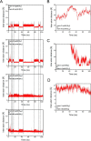
NAMD simulation for the solvated completely docked Spi1–C/EBPβ with C-terminal tails that generated the final structure shown in Fig. 5A. A, the interatom distances for different conformations of the salt bridge between the ζ carbon of Arg-232 on Spi1 (chain C) and C-terminal carboxylate carbon of Cys-345 on C/EBPβ chain B during the NAMD run. B–D, the interatom distance between the ζ 1 nitrogen of Lys-224 on Spi1 (chain C) and backbone carboxylate carbon of Cys-345 on C/EBPβ chain A (B); the ζ nitrogen of Lys-198 on Spi1 (chain C) and backbone carboxylate carbon of Cys-345 on C/EBPβ chain A (C); and the ζ 1 nitrogen of Lys-248 on Spi1 (chain C) and side-chain carboxylate carbon of Cys-336 on C/EBPβ chain A (D).
Figure 5.
Figure 5.
Details of the interaction between Spi1 and C/EBPβ resulting from NAMD simulation. A shows three views of the DNA–Spi1DBD–C/EBPβbZIP+Tails–DNA structure at the end of the 110-ns NAMD simulation. The white dotted box in the bottom-side view locates the region where the major Arg-232–Cys-345 and Arg-235–His-344 interactions occur between Spi1 and C/EBPβ. The C/EBPβ A and B chains are colored red and yellow, respectively. Spi1 is colored blue. B, detailed structure of the interaction between Arg-232 of the Spi1 with Cys-345 at the C terminus of C/EBPβ chain B and Arg-235 of Spi1 with His-344 of C/EBPβ chain B at the end of the NAMD run. Backbone colors are as described for A. C, specific atomic distances tracked during the NAMD simulation. The first plot (left panel) tracks interatom distances between the ζ carbon of Arg-232 on Spi1 and C-terminal carboxylate carbon of Cys-345 on the C/EBPβ chain B. The second plot (right panel) tracks interatom distances between the Eta1 nitrogen of Arg-235 on Spi1 (chain C) and the backbone carboxylate oxygen of His-344 on C/EBPβ chain B. D, a modified figure derived from our previously published report (5) demonstrating the relative importance of Arg-232 and Arg-235 as revealed by in vitro protein–protein interaction studies.
Figure 6.
Figure 6.
The Spi1–C/EBPβ interaction surface. A, structural interaction diagram shows the key interacting residues in C/EBPβ chain A (blue), C/EBPβ chain B (green), and Spi1 (red). The overall contact surface is marked by a yellow swath. H-bonds are indicated by black dots, and electrostatic interactions are indicated by blue dashes. B, summary of all intermolecular interactions between Spi1 and C/EBPβ.
Figure 7.
Figure 7.
A potential binding mode for l-arginine docked into the Spi1–C/EBPβ chain B interaction pocket. A, two-dimensional interaction schematic. B, three perspective views. Locations of relevant amino acids, DNA backbone phosphates, and l-arginine are labeled. C, pharmacophore model demonstrating the favorable accessibility and chemical interaction potential for C/EBPβ chain B over chain A.
Figure 8.
Figure 8.
l-Arginine inhibits recruitment of C/EBPβ to Spi1 and transcription of the IL1B gene in living cells. A, relative mRNA expression levels for the IL1B gene in THP-1 cells treated with 1 μg/ml LPS for 2.5 h following 12 h of pretreatment with indicated concentrations of l-arginine. B, relative mRNA expression levels for the IL1B and IL6 genes in THP-1 cells titrated with LPS, as indicated, for 2.5 h following the 12-h pretreatment with 10 mm l-arginine. The mRNA data presented are relative to unstimulated THP-1 cells (vehicle) and were normalized to cells treated with 1 μg/ml LPS for 2.5 h following 12 h of pretreatment with 0 mm l-arginine. C and D, ChIP studies on the IL1B gene for C/EBPβ (C) and Spi1 (D) in THP-1 cells that are treated with 1 μg/ml LPS for 2.5 h following 12 h of pretreatment with 10 mm l-arginine. The amplicon −19 on IL1B gene is the Spi1-binding site, and +4858 (IL1B) is downstream background control. The amplicon −77 on IL6 gene is direct C/EBPβ-binding site. The ChIP data were normalized to amplicon −19 on IL1B gene in unstimulated THP-1 cells. The primer sequences are indicated in Table S1. The standard error for all significant samples is representative of at least three biological replicates with p value indicated as follows: *, p < 0.05; and **, p < 0.01.
Figure 9.
Figure 9.
Four rotational views of the final DNA–C/EBPβ–Spi1–DNA composite model. Shown are the locations of the two bZIP chains and the Spi1 wHTH DBD, along with the likely long-range enhancer–promoter DNA interaction, for four 90° rotational views in association with DNA segments representing the long-range enhancer and promoter sites of the IL1B gene.

References

    1. Donner A. J., Ebmeier C. C., Taatjes D. J., and Espinosa J. M. (2010) CDK8 is a positive regulator of transcriptional elongation within the serum response network. Nat. Struct. Mol. Biol. 17, 194–201 10.1038/nsmb.1752 - DOI - PMC - PubMed
    1. Adamik J., Wang K. Z., Unlu S., Su A. J., Tannahill G. M., Galson D. L., O'Neill L. A., and Auron P. E. (2013) Distinct mechanisms for induction and tolerance regulate the immediate early genes encoding interleukin 1β and tumor necrosis factor α. PLoS One 8, e70622 10.1371/journal.pone.0070622 - DOI - PMC - PubMed
    1. Pugh B. F., and Venters B. J. (2016) Genomic organization of human transcription initiation complexes. PLoS One 11, e0149339 10.1371/journal.pone.0149339 - DOI - PMC - PubMed
    1. Yang Z., Wara-Aswapati N., Chen C., Tsukada J., and Auron P. E. (2000) NF-IL6 (C/EBPβ) vigorously activates il1b gene expression via a Spi-1 (PU.1) protein–protein tether. J. Biol. Chem. 275, 21272–21277 10.1074/jbc.M000145200 - DOI - PubMed
    1. Listman J. A., Wara-aswapati N., Race J. E., Blystone L. W., Walker-Kopp N., Yang Z., and Auron P. E. (2005) Conserved ETS domain arginines mediate DNA binding, nuclear localization, and a novel mode of bZIP interaction. J. Biol. Chem. 280, 41421–41428 10.1074/jbc.M509143200 - DOI - PubMed

Publication types

MeSH terms