. 2018 Oct 18;11(10):dmm035469.
doi: 10.1242/dmm.035469.

Effective CRISPR/Cas9-based nucleotide editing in zebrafish to model human genetic cardiovascular disorders

Affiliations

Effective CRISPR/Cas9-based nucleotide editing in zebrafish to model human genetic cardiovascular disorders

Federico Tessadori et al. Dis Model Mech. .

Abstract

The zebrafish (Danio rerio) has become a popular vertebrate model organism to study organ formation and function due to its optical clarity and rapid embryonic development. The use of genetically modified zebrafish has also allowed identification of new putative therapeutic drugs. So far, most studies have relied on broad overexpression of transgenes harboring patient-derived mutations or loss-of-function mutants, which incompletely model the human disease allele in terms of expression levels or cell-type specificity of the endogenous gene of interest. Most human genetically inherited conditions are caused by alleles carrying single nucleotide changes resulting in altered gene function. Introduction of such point mutations in the zebrafish genome would be a prerequisite to recapitulate human disease but remains challenging to this day. We present an effective approach to introduce small nucleotide changes in the zebrafish genome. We generated four different knock-in lines carrying distinct human cardiovascular-disorder-causing missense mutations in their zebrafish orthologous genes by combining CRISPR/Cas9 with a short template oligonucleotide. Three of these lines carry gain-of-function mutations in genes encoding the pore-forming (Kir6.1, KCNJ8) and regulatory (SUR2, ABCC9) subunits of an ATP-sensitive potassium channel (KATP) linked to Cantú syndrome (CS). Our heterozygous zebrafish knock-in lines display significantly enlarged ventricles with enhanced cardiac output and contractile function, and distinct cerebral vasodilation, demonstrating the causality of the introduced mutations for CS. These results demonstrate that introducing patient alleles in their zebrafish orthologs promises a broad application for modeling human genetic diseases, paving the way for new therapeutic strategies using this model organism.

Keywords: ABCC9; CRISPR/Cas9; Cantú syndrome; Genome editing; KCNJ8; Point mutation; Zebrafish.


Conflict of interest statement

Competing interestsThe authors declare no competing or financial interests.

Figures

Fig. 1.
Fig. 1.
Generation of patient-specific KI lines in zebrafish. (A) Stepwise procedure followed to establish the KI lines described in this study with corresponding minimal timeline for each step. (B) Schematics showing the targeted genomic sequence for the introduction of the c.A193G substitution resulting in the p.V65M in the kcnj8 genomic sequence. The PAM sequence is highlighted in gray, the specific section of the sgRNA is highlighted in cyan and the modified codon is underlined. Red text indicates substituted nucleotide. Note that in this case the substitution is located on the PAM sequence. (C) Sequencing traces for wild-type and heterozygous kcnj8+/V65M zebrafish. Asterisk denotes the substituted nucleotide in the heterozygous kcnj8+/V65M sequencing trace.
Fig. 2.
Fig. 2.
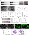
Heterozygous kcnj8+/V65M mutation induces CS-related cardiac anomalies and cerebral vasodilation in zebrafish. (A) Representative images illustrating the morphology of 5 dpf wild-type and kcnj8+/V65M mutants as seen from a left lateral (top) and dorsal (bottom) view. Boxes designate imaged areas that were used to assess cardiac function: the cardinal vein (1) and the heart (2). The ventricular area of the heart is highlighted, with the long axis and short axis of the ventricle indicated by dashed lines. a, atrium; ba, bulbous ateriosus; cv, cardinal vein; da, dorsal aorta; v, ventricle. (B) Quantification of cardiac function using individual characteristic confocal sections from a time series of the embryonic cardiac cycle at 5 dpf. Pericardial edema was quantified by measuring pericardial area using striking morphological landmarks, indicated by white boxes. Ventricular area was subtracted. Arrows show accumulation of fluid in kcnj8+/V65M mutants. Dotted red lines indicate ventricle (v) and bulbous arteriosus (ba). (C) Tracking of individual red blood cells (RBCs) measuring blood flow velocity in the cardinal vein. RBCs were tracked for ten frames using ImageJ (NIH) and the plugin MTrackJ (Meijering et al., 2012). One representative image of each genotype is shown. Black arrow indicates the direction of RBC movement. (D) Quantification of vascular dilations in a Tg(kdrl:GFP) background. Representative confocal images of the circular structure comprising the BCA and PCS in wild-type and heterozygous 5 dpf fish are outlined in red. The arrowheads indicate distinct regions of vasodilation. 3D reconstruction of vascular structure in Imaris was used to calculate vessel volume. (E) Ventricular area in heterozygous kcnj8+/V65M mutants. Representative heart histology of adult kcnj8+/V65M mutants and respective wild-type siblings after H&E staining. Exemplary depiction of one WT and one kcnj8+/V65M heart. For assessment of ventricular chamber size, tissue sections showing the largest ventricular area were selected and area was quantified using ImageJ (NIH). For all graphs, significance was determined by two-tailed unpaired Student's t-test or Mann–Whitney two-tailed U-test: *P≤0.05; **P≤0.01; ***P≤0.001; ****P≤0.0001. The black horizontal bar indicates the mean value for each condition. Sample sizes: (B) kcnj8+/+, n=21; kcnj8+/V65M, n=14; (C) kcnj8+/+, n=10; kcnj8+/V65M, n=7; (D) kcnj8+/+, n=12; kcnj8+/V65M, n=20; (E) kcnj8+/+, n=6; kcnj8+/V65M, n=6. Scale bars: (A) 1 mm (top and middle) and 50 µm (bottom); (B) 50 µm; (C) 10 μm; (D) 50 µm; (E) 500 µm. All embryos analyzed originated from group matings of adult zebrafish.

Similar articles

Cited by

References

    1. Armstrong G. A. B., Liao M., You Z., Lissouba A., Chen B. E. and Drapeau P. (2016). Homology directed knockin of point mutations in the zebrafish tardbp and fus genes in ALS using the CRISPR/Cas9 system. PLoS ONE 11, e0150188 10.1371/journal.pone.0150188 - DOI - PMC - PubMed
    1. Ashcroft F. M. (2010). New uses for old drugs: neonatal diabetes and sulphonylureas. Cell Metab. 11, 179-181. 10.1016/j.cmet.2010.02.004 - DOI - PubMed
    1. Bedell V. M., Wang Y., Campbell J. M., Poshusta T. L., Starker C. G., Krug R. G. II, Tan W., Penheiter S. G., Ma A. C., Leung A. Y. et al. (2012). In vivo genome editing using a high-efficiency TALEN system. Nature 491, 114-118. 10.1038/nature11537 - DOI - PMC - PubMed
    1. Brownstein C. A., Towne M. C., Luquette L. J., Harris D. J., Marinakis N. S., Meinecke P., Kutsche K., Campeau P. M., Yu T. W., Margulies D. M. et al. (2013). Mutation of KCNJ8 in a patient with Cantu syndrome with unique vascular abnormalities-support for the role of K(ATP) channels in this condition. Eur. J. Med. Genet. 56, 678-682. 10.1016/j.ejmg.2013.09.009 - DOI - PMC - PubMed
    1. Cahill T. J., Ashrafian H. and Watkins H. (2013). Genetic cardiomyopathies causing heart failure. Circ. Res. 113, 660-675. 10.1161/CIRCRESAHA.113.300282 - DOI - PubMed

Publication types