. 2018 Oct 24;10(464):eaam7019.
doi: 10.1126/scitranslmed.aam7019.

Clostridioides difficile uses amino acids associated with gut microbial dysbiosis in a subset of patients with diarrhea

Affiliations

Clostridioides difficile uses amino acids associated with gut microbial dysbiosis in a subset of patients with diarrhea

Eric J Battaglioli et al. Sci Transl Med. .

Abstract

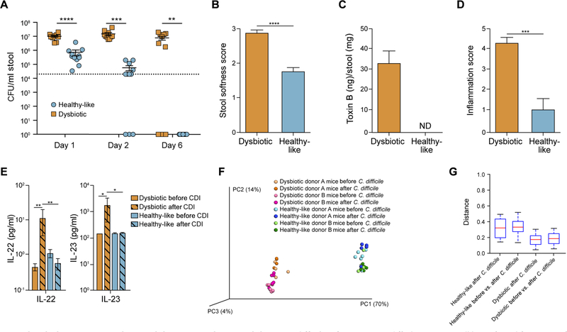
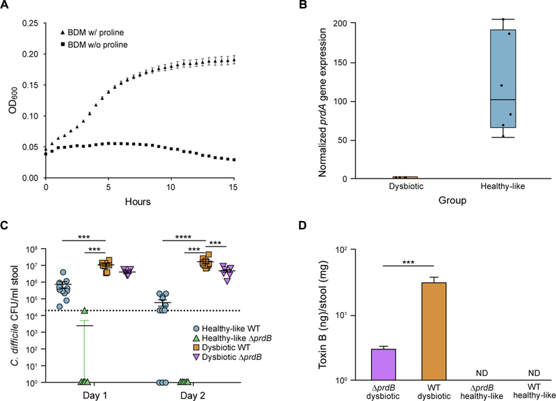
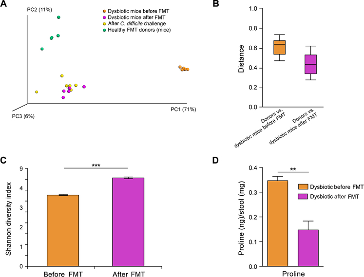
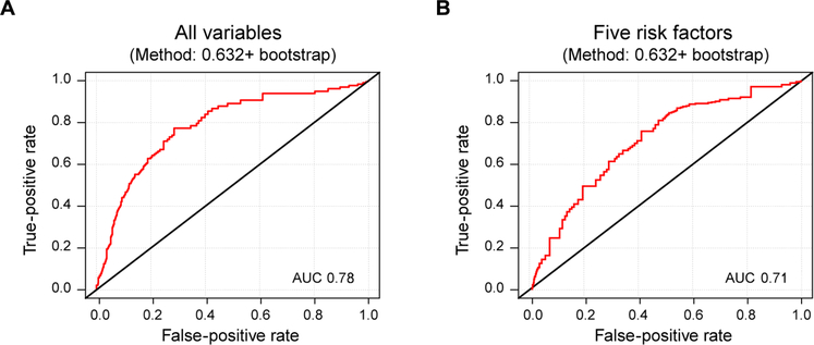
The gut microbiota plays a critical role in pathogen defense. Studies using antibiotic-treated mice reveal mechanisms that increase susceptibility to Clostridioides difficile infection (CDI), but risk factors associated with CDI in humans extend beyond antibiotic use. Here, we studied the dysbiotic gut microbiota of a subset of patients with diarrhea and modeled the gut microbiota of these patients by fecal transplantation into germ-free mice. When challenged with C. difficile, the germ-free mice transplanted with fecal samples from patients with dysbiotic microbial communities showed increased gut amino acid concentrations and greater susceptibility to CDI. A C. difficile mutant that was unable to use proline as an energy source was unable to robustly infect germ-free mice transplanted with a dysbiotic or healthy human gut microbiota. Prophylactic dietary intervention using a low-proline or low-protein diet in germ-free mice colonized by a dysbiotic human gut microbiota resulted in decreased expansion of wild-type C. difficile after challenge, suggesting that amino acid availability might be important for CDI. Furthermore, a prophylactic fecal microbiota transplant in mice with dysbiosis reduced proline availability and protected the mice from CDI. Last, we identified clinical risk factors that could potentially predict gut microbial dysbiosis and thus greater susceptibility to CDI in a retrospective cohort of patients with diarrhea. Identifying at-risk individuals and reducing their susceptibility to CDI through gut microbiota-targeted therapies could be a new approach to preventing C. difficile infection in susceptible patients.


Conflict of interest statement

Competing interests: R.P. has received funding from BioFire, Check-Points, Curetis, 3M, Merck, Hutchison Biofilm Medical Solutions, Accelerate Diagnostics, Allergan, and The Medicines Company and also serves as a consultant to Curetis, Roche, Qvella, and Diaxonhit. R.P. is a co-inventor on a patent on Bordetella pertussis/parapertussis PCR with royalties paid by MOLBIOL GmbH, has a patent on a device/method for sonication with royalties paid by Samsung to Mayo Clinic, and has a patent on an anti-biofilm substance. In addition, R.P. serves on an Actelion data monitoring board, receives travel reimbursement and an editor’s stipend from the American Society for Microbiology and Infectious Diseases Society of America, and honoraria from the USMLE, Up-To-Date and the Infectious Diseases Board Review Course. S.K. has received funding from Merck and serves as a consultant to Rebiotix and Summit Pharmaceuticals. D.S.P. consults for Seres Therapeutics, Salix Pharmaceuticals, Cubist Pharmaceuticals, Merck, Janssen, and Otsuka Pharmaceutical. P.C.K., E.J.B., and V.L.H. are co-inventors on a patent no. WO2017053544 A1 (filed 22 September 2016, published 30 March 2017) entitled “Methods and materials for using biomarkers which predict susceptibility to Clostridium difficile infection.”

Figures

Fig. 1.
Fig. 1.. A subset of patients with diarrhea have a dysbiotic gut microbiota.
(A) β-Diversity (unweighted UniFrac) of the gut microbiota of healthy control individuals (n = 118) compared to patients with diarrhea clustered on the basis of partitioning around medoids (PAM) [cluster H (healthy-like), n = 78 and cluster D (dysbiotic), n = 37]. (B) Unweighted UniFrac distances between healthy-like and a dysbiotic gut microbiota from patients with diarrhea compared to a healthy control gut microbiota. The plotted median with interquartile range (IQR) and SD (Bonferroni-corrected P < 0.0001, t test) is shown. (C) α-Diversity as indicated by the Shannon diversity index is shown for dysbiotic and healthy-like gut microbiotas from patients with diarrhea. Plotted averages with SEM (***P < 0.0005, t test). (D) Heatmap showing significantly different microbial taxa between healthy-like and dysbiotic gut microbial communities. The operational taxonomic unit (OTU) number is featured after the genus (all Bonferroni-corrected P < 0.02, Wilcoxon rank-sum test). (E and F) β-Diversity (unweighted UniFrac) of the gut microbiota from (E) healthy control individuals (n = 118), dysbiotic patients with diarrhea (n = 37), and patients with C. difficile infection (n = 95); (F) unweighted UniFrac distance between the dysbiotic gut microbiota of patients with diarrhea versus the gut microbiota of healthy controls or those with CDI. Plotted median with IQR and SD (Bonferroni-corrected P < 0.0001, t test).
Fig. 2.
Fig. 2.. Mice with a dysbiotic gut microbiota exhibit increased susceptibility to C. difficile infection.
(A) C. difficile CFUs per milliliter of stool from germ-free mice colonized with either a healthy-like (n = 11) or dysbiotic (n = 10) gut microbiota from patients with diarrhea are shown. Data points represent individual animals with lines indicating average and SEM. Assay limit of detection (LOD) indicated by horizontal dotted line at 2 × 10⁴ CFU/ml stool (**P < 0.005, ***P < 0.0005, ****P < 0.00005, Holm-Šídák test). (B) Stool consistency for germ-free mice transplanted with a healthy-like (n = 11) or dysbiotic (n = 10) gut microbiota, 2 days after C. difficile challenge. Plotted means and SEM (****P < 0.0001, Mann-Whitney test). (C) C. difficile toxin B concentrations measured by quantitative ELISA in stool from germ-free mice transplanted with a healthy-like or dysbiotic human gut microbiota, 6 days after C. difficile challenge. Plotted means and SEM; ND, not detected. (D) Average proximal colon inflammation score in germ-free mice transplanted with a healthy-like (n = 11) or dysbiotic (n = 10) human gut microbiota, after C. difficile challenge. Plotted means and SEM (***P < 0.0005, Mann-Whitney test). (E) IL-22 and IL-23 concentrations measured from full thickness tissue collected from the proximal colon of germ-free mice transplanted with a dysbiotic (n = 5) or healthy-like (n = 5) human gut microbiota before and 7 days after C. difficile challenge. Plotted means and SEM (*P < 0.05, **P < 0.005, Mann-Whitney test). (F) β-Diversity and (G) weighted UniFrac distance comparisons for dysbiotic and healthy-like human gut microbial communities after transplant into germ-free mice before and 2 days after C. difficile challenge [Bonferroni-corrected P = 1 (dysbiotic), Bonferroni-corrected P = 1 (healthy-like), Student’s t test].
Fig. 3.
Fig. 3.. C. difficile exploits increased availability of amino acids in the dysbiotic gut microbiota.
(A) A subset of pathway gene expression based on whole gut microbial community gene expression (RNA-seq) normalized using shallow metagenomic sequencing of stool from germ-free mice transplanted with healthy-like (n = 6) or dysbiotic (n = 6) human gut microbiota before C. difficile challenge. (B) Amino acid concentrations in stool from mice transplanted with a healthy-like (n = 5) or dysbiotic (n = 8) human gut microbiota. Plotted averages and SEM (*P < 0.05, **P < 0.005, Mann-Whitney test); ns, not significant; ND, not detected. (C) C. difficile growth kinetics in basal defined medium (BDM) containing 0, 0.01, or 0.1% deoxycholic acid (DCA) and amino acid concentrations at 100, 50, or 25% those of standard media concentrations. Plotted averages and SEM.
Fig. 4.
Fig. 4.. Proline affects C. difficile colonization in germ-free mice transplanted with a dysbiotic or healthy-like human gut microbiota.
(A) C. difficile growth kinetics indicated optical density (OD) at 600 nm in the presence or absence of proline in basal defined medium without glucose. Plotted means and SEM. (B) D-proline reductase A ( prdA) expression normalized to metagenomic read counts in the healthy-like and dysbiotic gut microbiota of transplanted mice before C. difficile challenge. (C) ΔprdB mutant C. difficile CFU/ml stool from germ-free mice transplanted with a dysbiotic (n = 7) or healthy-like (n = 8) human gut microbiota after C. difficile challenge is shown. Colonization of the transplanted mice with wild-type (WT) C. difficile from Fig. 2A is also shown. Data points represent individual animals with lines indicating average and SEM. Assay limit of detection (LOD) indicated by a horizontal dotted line at 2 × 10⁴ CFU/ml stool (***P < 0.005, ****P < 0.0005, two-way ANOVA). (D) C. difficile toxin B concentrations were measured by quantitative ELISA in the stool of mice transplanted with a dysbiotic (n = 7) or a healthy-like (n = 8) human gut microbiota 6 days after challenge with ΔprdB mutant C. difficile or wild-type C. difficile (data from Fig. 2C). Plotted means and SEM (***P < 0.0005, Mann-Whitney test).
Fig. 5.
Fig. 5.. Fecal microbiota transplant from healthy individuals reduces free proline and susceptibility of transplanted mice to C. difficile infection.
(A) β-Diversity (weighted UniFrac) of mice transplanted with a dysbiotic human gut microbiota, before and after a fecal microbiota transplant (FMT) from healthy individuals (n = 6). (B) Distances (weighted UniFrac) between FMT healthy donors and mice transplanted with a dysbiotic gut microbiota were significantly decreased after FMT (Bonferroni-corrected P < 0.0001, Student’s t test). (C) α-Diversity of the dysbiotic gut microbiota in transplanted mice before and after FMT (n = 6). Plotted averages and SEM (***P < 0.0005, Student’s t test). (D) Proline concentrations in stool from mice transplanted with a dysbiotic gut microbiota, before and after FMT (n = 6). Plotted averages and SEM (**P < 0.005, Mann-Whitney test).
Fig. 6.
Fig. 6.. Five clinical risk factors may predict gut microbial dysbiosis and susceptibility to C. difficile infection in patients with diarrhea.
(A) Receiver operating characteristic (ROC) curve based on five clinical risk factors that may be predictive of gut microbiota dysbiosis. Recent antibiotics (OR, 5.21; 95% CI, 2.14 to 12.71; P < 0.001), immunosuppression (OR, 2.87; 95% CI, 1.27 to 6.48; P = 0.012), current hospitalization (OR, 6.17; 95% CI, 2.22 to 17.15; P < 0.001), recent hospitalization (OR, 4.87; 95% CI, 1.72 to 13.74; P = 0.003), and prior C. difficile infection (OR, 9.26; 95% CI, 2.37 to 36.20; P = 0.001). Area under the curve (AUC), 0.78 (see table S4). (B) ROC curve based on five clinical risk factors that may be predictive of C. difficile infection. Recent antibiotics (OR, 3.35; 95% CI, 2.78 to 4.03; P = 6.21 × 10−37), immunosuppression (OR, 2.47; 95% CI, 2.06 to 2.96; P = 8.42 × 10⁻²³), current hospitalization (OR, 2.94; 95% CI, 2.40 to 3.61; P = 8.45 × 10⁻²⁵), recent hospitalization (OR, 3.32; 95% CI, 2.72 to 4.06; P = 1.85 × 10⁻³¹), and prior C. difficile infection (OR, 5.84; 95% CI, 4.42 to 7.72; P = 1.66 × 10⁻³⁵). AUC, 0.71.

Similar articles

Cited by

References

    1. Schulz MD, Atay C, Heringer J, Romrig FK, Schwitalla S, Aydin B, Ziegler PK, Varga J, Reindl W, Pommerenke C, Salinas-Riester G, Böck A, Alpert C, Blaut M, Polson SC, Brandl L, Kirchner T, Greten FR, Polson SW, Arkan MC, High-fat-diet-mediated dysbiosis promotes intestinal carcinogenesis independently of obesity. Nature 514, 508–512 (2014). - PMC - PubMed
    1. Britton RA, Young VB, Role of the intestinal microbiota in resistance to colonization by Clostridium difficile. Gastroenterology 146, 1547–1553 (2014). - PMC - PubMed
    1. Maeda Y, Kurakawa T, Umemoto E, Motooka D, Ito Y, Gotoh K, Hirota K, Matsushita M, Furuta Y, Narazaki M, Sakaguchi N, Kayama H, Nakamura S, Iida T, Saeki Y, Kumanogoh A, Sakaguchi S, Takeda K, Dysbiosis contributes to arthritis development via activation of autoreactive T cells in the intestine. Arthritis Rheumatol. 68, 2646–2661 (2016). - PubMed
    1. Rajilić-Stojanović M, Jonkers DM, Salonen A, Hanevik K, Raes J, Jalanka J, de Vos WM, Manichanh C, Golic N, Enck P, Philippou E, Iraqi FA, Clarke G, Spiller RC, Penders J, Intestinal microbiota and diet in IBS: Causes, consequences, or epiphenomena? Am. J. Gastroenterol. 110, 278–287 (2015). - PMC - PubMed
    1. Keeney KM, Yurist-Doutsch S, Arrieta MC, Finlay BB, Effects of antibiotics on human microbiota and subsequent disease. Annu. Rev. Microbiol 68, 217–235 (2014). - PubMed

Publication types

MeSH terms