. 2018 Oct 24;10(11):398.
doi: 10.3390/cancers10110398.

The Intrinsic and Extrinsic Implications of RANKL/RANK Signaling in Osteosarcoma: From Tumor Initiation to Lung Metastases

Affiliations

The Intrinsic and Extrinsic Implications of RANKL/RANK Signaling in Osteosarcoma: From Tumor Initiation to Lung Metastases

Benjamin Navet et al. Cancers (Basel). .

Abstract

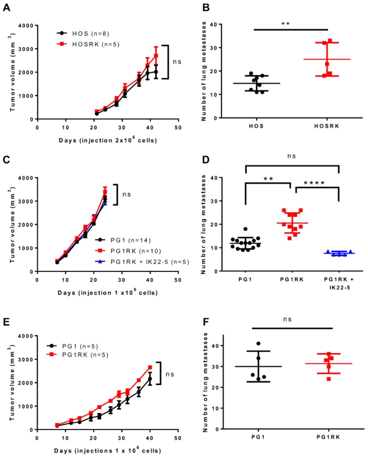
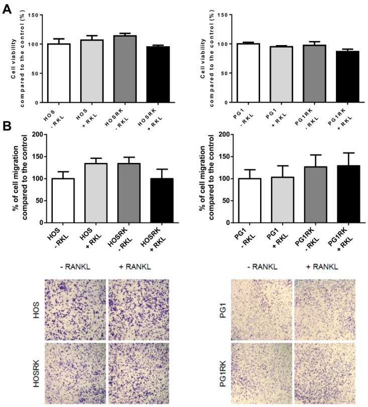
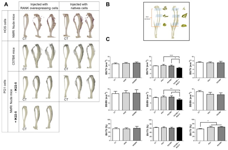
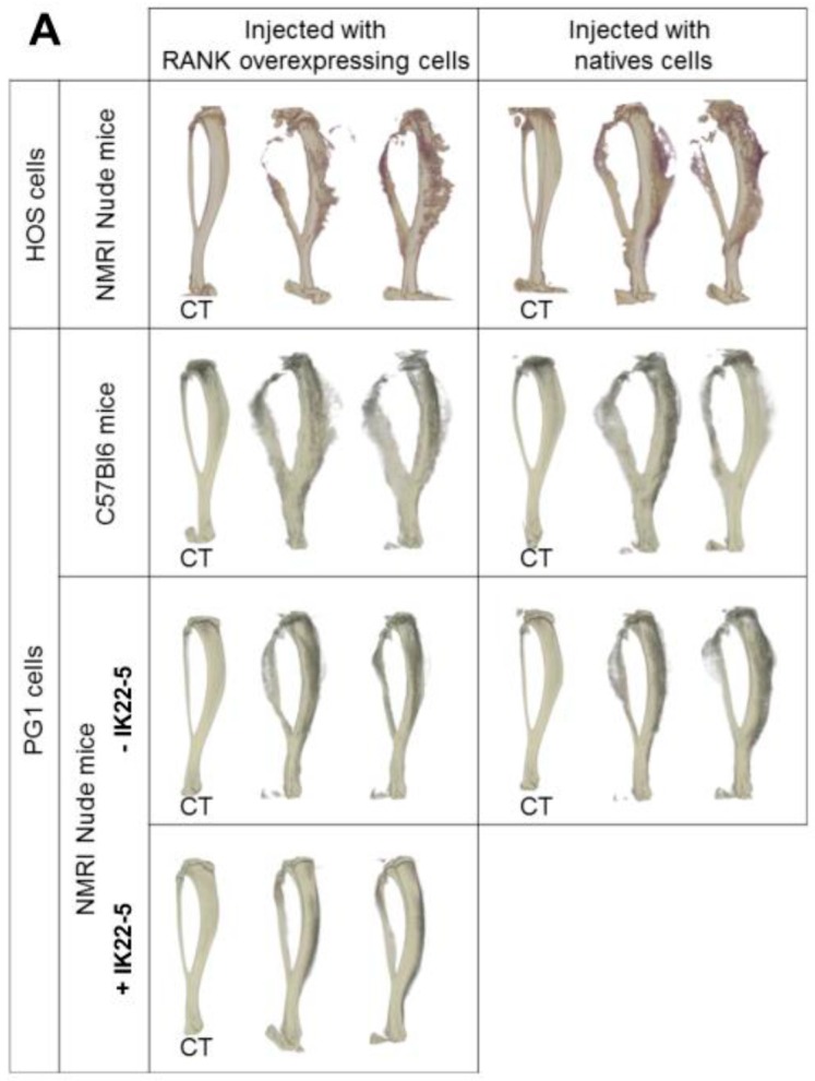
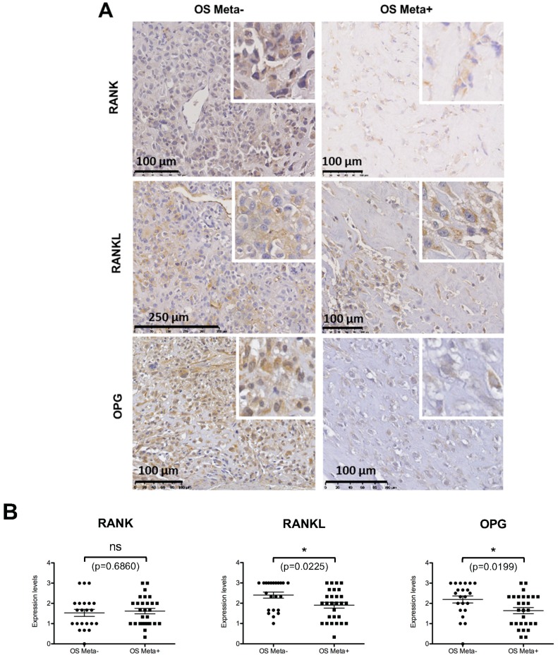
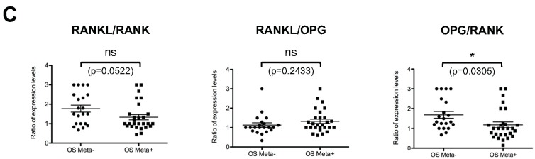
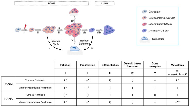
Background: Osteosarcoma is the most frequent form of malignant pediatric bone tumor. Despite the current therapeutic arsenal, patient life-expectancy remains low if metastases are detected at the time of diagnosis, justifying research into better knowledge at all stages of osteosarcoma ontogenesis and identification of new therapeutic targets. Receptor Activator of Nuclear factor κB (RANK)expression has been reported in osteosarcoma cells, raising the question of Receptor Activator of Nuclear factor κB Ligand (RANKL)/RANK signaling implications in these tumor cells (intrinsic), in addition to previously reported implications through osteoclast activation in the tumor microenvironment (extrinsic). Methods: Based on in vitro and in vivo experimentations using human and mouse osteosarcoma cell lines, the consequences on the main cellular processes of RANK expression in osteosarcoma cells were analyzed. Results: The results revealed that RANK expression had no impact on cell proliferation and tumor growth, but stimulated cellular differentiation and, in an immune-compromised environment, increased the number of lung metastases. The analysis of RANKL, RANK and osteoprotegerin (OPG) expressions in biopsies of a cohort of patients revealed that while RANK expression in osteosarcoma cells was not significantly different between patients with or without metastases at the time of diagnosis, the OPG/RANK ratio decreased significantly. Conclusion: Altogether, these results are in favor of RANKL-RANK signaling inhibition as an adjuvant for the treatment of osteosarcoma.

Keywords: RANKL/RANK; T-lymphocyte; bone; metastases; osteosarcoma.


Conflict of interest statement

The authors declare no conflict of interest.

Figures

Figure 1
Figure 1
Impact of Receptor Activator of Nuclear factor κB (RANK) over-expression in osteosarcoma cells on tumor growth and the number of lung metastases. No significant difference was observed concerning tumor growth regardless of the cell-line considered (K-HOS (A), MOS-J PG1 (C,E)) or the immune status of the host mouse strain (Nude (A,C) or C57BL/6 (E)). However, regarding the number of lung metastases, a significant increase was observed regardless of the RANK over-expressing cell-line considered, in immune-deficient Nude mice (B,D) but not in immune-competent C57BL/6 mice (F). Moreover, injections of a Receptor Activator of Nuclear factor κB Ligand (RANKL)-blocking antibody (IK22.5) in Nude mice made it possible to reduce the number of lung metastases obtained with RANK expressing PG1 (D). n: number of mice in each group. Growth curves (A,C,E) are shown as the mean ± SEM. All data analysis was performed with the Kruskal Wallis test. ns: not significant; **: p < 0.01; ****: p < 0.0001.
Figure 2
Figure 2
Consequences of RANK over-expression in osteosarcoma cells on cell viability (A) and migration (B). A moderate decrease (tendency) in the cell viability in response to the addition of RANKL to the culture medium was observed for both RANK over-expressing HOS cells and PG1 cells (A). For cell migration, which was evaluated with Boyden chambers, no significant impact of RANK over-expression and RANKL stimulation was observed, regardless of the cell-line considered (B).
Figure 3
Figure 3
Impact of RANK over-expression in osteosarcoma cells on bone structure (A) and bone parameters (B,C). Whatever tumor cell-line or host mouse strain was considered, representative three dimension images did not make it possible to observe any difference concerning bone resorption and osteoid tissue formation in relationship with RANK over-expression (A). As expected, IK22-5 RANKL blocking antibody injections made it possible to protect the bone from resorption (A). The bone parameters BS/TV, BS/BV and BV/TV were measured on 450 sections whose positions are presented in (B). The results showed no difference between the cells over-expressing or not RANK, whatever parameter was considered (C). The IK22-5 RANKL blocking antibody made it possible to significantly reduce the BS/TV and BS/BV parameters, with no impact on the BV/TV parameter. A significant difference was observed concerning the BV/TV parameter only in C57BL/6 mice when comparing contra-lateral control tibias (CT) and tibias with tumors independently of RANK over-expression by tumor cells. The data in (C) are shown as the mean ± SD. Data analyses were performed using the Kruskal Wallis test. ns: no significant. *: p < 0.05. **: p < 0.01. ***: p < 0.001.
Figure 4
Figure 4
Consequences of T-cell specific invalidation of Rankl in recipient mice on RANK over-expressing PG1 tumor growth, number of metastases and bone parameters. Rankl depletion in mouse T-cells (LCK-CRE) was validated by PCR on gDNA extracted from tails and T cells (A). PCR data confirmed the effective recombination, specifically in T-cells. After injection of one million PG1 cells over-expressing RANK, tumor growth (B) and the number of lung metastases formed (C) were compared between mice invalidated for Rank in T-cells (n = 4) and control mice (n = 5) showing no difference. Representative three-dimensional images of tibias with tumors (and their controls, C) did not make it possible to observe any differences regarding bone resorption or tumor osteoid tissue formation (D). Micro-CT analysis of the BS/TV (mm-1), BS/BV (mm-1) and BV/TV (%) parameters of the tibias revealed no differences. However, an increase in BV/TV was observed comparatively to contralateral safe tibias, independently of the mouse genotype (E). The data in (E) are shown as the mean ± SD. Data analyses were performed using the Kruskal Wallis test. ns: no significant. *: p < 0.05.
Figure 4
Figure 4
Consequences of T-cell specific invalidation of Rankl in recipient mice on RANK over-expressing PG1 tumor growth, number of metastases and bone parameters. Rankl depletion in mouse T-cells (LCK-CRE) was validated by PCR on gDNA extracted from tails and T cells (A). PCR data confirmed the effective recombination, specifically in T-cells. After injection of one million PG1 cells over-expressing RANK, tumor growth (B) and the number of lung metastases formed (C) were compared between mice invalidated for Rank in T-cells (n = 4) and control mice (n = 5) showing no difference. Representative three-dimensional images of tibias with tumors (and their controls, C) did not make it possible to observe any differences regarding bone resorption or tumor osteoid tissue formation (D). Micro-CT analysis of the BS/TV (mm-1), BS/BV (mm-1) and BV/TV (%) parameters of the tibias revealed no differences. However, an increase in BV/TV was observed comparatively to contralateral safe tibias, independently of the mouse genotype (E). The data in (E) are shown as the mean ± SD. Data analyses were performed using the Kruskal Wallis test. ns: no significant. *: p < 0.05.
Figure 5
Figure 5
Tissue array analysis of RANK, RANKL and OPG expressions in a cohort of 50 biopsies of patients with (n = 28) and without (n = 22) metastases at time of diagnosis. Representative views of the different immuno-stainings are presented (A) with enlargement inset to clearly show the stained osteosarcoma cells. Statistical analyses (Student test) of the staining based on the percentage of stained osteosarcoma cells revealed a significantly lower number of positive cells for RANKL and OPG in the metastatic patient group, while no difference was observed between the two groups concerning the number of RANK-expressing cells (B). Statistical analysis of the three ratios between these factors, established individually for each patient, showed that only the OPG/RANK ratio was significantly different between the two groups with lower values in the metastatic patient group (C). ns: not significant. *: p < 0.05. p values are given for each test.
Figure 5
Figure 5
Tissue array analysis of RANK, RANKL and OPG expressions in a cohort of 50 biopsies of patients with (n = 28) and without (n = 22) metastases at time of diagnosis. Representative views of the different immuno-stainings are presented (A) with enlargement inset to clearly show the stained osteosarcoma cells. Statistical analyses (Student test) of the staining based on the percentage of stained osteosarcoma cells revealed a significantly lower number of positive cells for RANKL and OPG in the metastatic patient group, while no difference was observed between the two groups concerning the number of RANK-expressing cells (B). Statistical analysis of the three ratios between these factors, established individually for each patient, showed that only the OPG/RANK ratio was significantly different between the two groups with lower values in the metastatic patient group (C). ns: not significant. *: p < 0.05. p values are given for each test.
Figure 6
Figure 6
Schematic representation of the different stages of osteosarcoma from initiation to lung metastasis, and the implications of intrinsic and extrinsic RANKL and RANK in these different stages. +: implication revealed; 0: No implication; *: part of the vicious cycle; **: part of the escape bolstering; ∞: according to Chen et al., 2016.

Similar articles

Cited by

References

    1. Heymann D. Bone Cancer. Academic Press; San Diego, CA, USA: 2010. Preface; p. ix.
    1. Bielack S.S., Hecker-Nolting S., Blattmann C., Kager L. Advances in the management of osteosarcoma. F1000Research. 2016;5:2767. doi: 10.12688/f1000research.9465.1. - DOI - PMC - PubMed
    1. Lindsey B.A., Markel J.E., Kleinerman E.S. Osteosarcoma Overview. Rheumatol. Ther. 2017;4:25–43. doi: 10.1007/s40744-016-0050-2. - DOI - PMC - PubMed
    1. Nataraj V., Rastogi S., Khan S.A., Sharma M.C., Agarwala S., Vishnubhatla S., Bakhshi S. Prognosticating metastatic osteosarcoma treated with uniform chemotherapy protocol without high dose methotrexate and delayed metastasectomy: A single center experience of 102 patients. Clin. Transl. Oncol. 2016;18:937–944. doi: 10.1007/s12094-015-1467-8. - DOI - PubMed
    1. Allison D.C., Carney S.C., Ahlmann E.R., Hendifar A., Chawla S., Fedenko A., Angeles C., Menendez L.R. A Meta-Analysis of Osteosarcoma Outcomes in the Modern Medical Era. Sarcoma. 2012;2012:704872. doi: 10.1155/2012/704872. - DOI - PMC - PubMed

LinkOut - more resources