Review
. 2018 Oct 24;23(11):2748.
doi: 10.3390/molecules23112748.

Thermodynamic Model for B-Z Transition of DNA Induced by Z-DNA Binding Proteins

Affiliations
Review

Thermodynamic Model for B-Z Transition of DNA Induced by Z-DNA Binding Proteins

Ae-Ree Lee et al. Molecules. .

Abstract

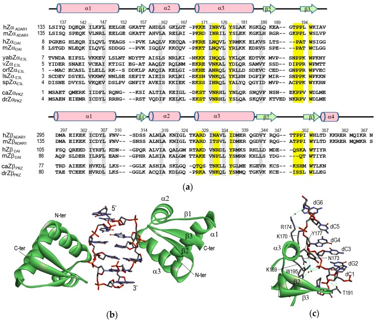
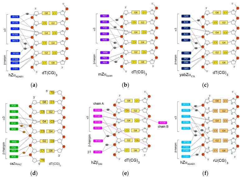
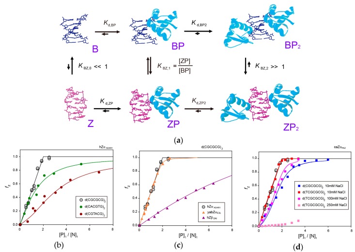
Z-DNA is stabilized by various Z-DNA binding proteins (ZBPs) that play important roles in RNA editing, innate immune response, and viral infection. In this review, the structural and dynamics of various ZBPs complexed with Z-DNA are summarized to better understand the mechanisms by which ZBPs selectively recognize d(CG)-repeat DNA sequences in genomic DNA and efficiently convert them to left-handed Z-DNA to achieve their biological function. The intermolecular interaction of ZBPs with Z-DNA strands is mediated through a single continuous recognition surface which consists of an α3 helix and a β-hairpin. In the ZBP-Z-DNA complexes, three identical, conserved residues (N173, Y177, and W195 in the Zα domain of human ADAR1) play central roles in the interaction with Z-DNA. ZBPs convert a 6-base DNA pair to a Z-form helix via the B-Z transition mechanism in which the ZBP first binds to B-DNA and then shifts the equilibrium from B-DNA to Z-DNA, a conformation that is then selectively stabilized by the additional binding of a second ZBP molecule. During B-Z transition, ZBPs selectively recognize the alternating d(CG)n sequence and convert it to a Z-form helix in long genomic DNA through multiple sequence discrimination steps. In addition, the intermediate complex formed by ZBPs and B-DNA, which is modulated by varying conditions, determines the degree of B-Z transition.

Keywords: B-Z transition; DNA-protein interaction; Z-DNA; Z-DNA binding protein.


Conflict of interest statement

The authors declare no conflicts of interest.

Figures

Figure 1
Figure 1
(a) Multiple sequence alignment of ZBPs: hZαADAR1, hZβADAR1, human ADAR1; mZαADAR1, mZβADAR1, murine ADAR1; hZαDAI, hZβDAI, human DAI; mZαDAI, mZβDAI, murine DAI; yabZαE3L, Yaba-like disease virus E3L; vZαE3L, vaccinia virus E3L; orfZαE3L, orf virus E3L; lsZαE3L, lumpy skin disease virus E3L; spZαE3L, swinepox virus E3L; caZαPKZ, caZβPKZ, goldfish PKZ; drZαPKZ, drZβPKZ, zebrafish PKZ. Numbering and secondary structural elements for hZαADAR1 and hZβADAR1 are shown above the sequence. Yellow and gray bars indicate residues important for Z-DNA recognition and protein folding, respectively. (b) Overview of the hZαADAR1 domain bound to left-handed Z-DNA (PDB id: 1QBJ) [7]. (c) View of the DNA recognition surface of hZαADAR1 (PDB id: 1QBJ) [7]. The green lines indicate the H-bonding interactions. In (b,c), the backbone structure of hZαADAR1 domain and Z-DNA duplex, d(TCGCGCG)2, are represented by the green ribbon and element-based stick presentation, respectively.
Figure 2
Figure 2
Protein residues involved Z-DNA/Z-RNA interactions in (a) hZαADAR1–dT(CG)3 [7], (b) mZαDLM1–dT(CG)3 [29], (c) yabZαE3L–dT(CG)3 [30], (d) caZαPKZ–dT(CG)3 [31], (e) hZβDAI–dT(CG)3 [32], and (f) hZαADAR1–rU(CG)3 complexes [33]. Intermolecular H-bonds and van der Waals contacts are indicated by solid lines and open circles, respectively. The water molecules in key positions within the protein–DNA interface are indicated by ovals.
Figure 3
Figure 3
(a) Mechanism for the B-Z conformational transition of a 6-bpDNA by two ZBPs [41]. (b) Relative Z-DNA populations (fZ) of d(CGCGCG)2 (grey circle) [41], d(CACGTG)2 (dark green circle) [42], and d(CGTACG)2 (brown circle) [42] induced by hZαADAR1 in NMR buffer (pH = 8.0) containing 100 mM NaCl as a function of [P]t/[N]t ratio. (c) fZ of d(CGCGCG)2 induced by hZαADAR1 (grey circle) [41], yabZαE3L (orange triangle) [43], and hZβDAI (purple triangle) [44] in NMR buffer (pH = 8.0) containing 100 mM NaCl as a function of [P]t/[N]t ratio. (d) fZ of d(CGCGCG)2 at 10 mM NaCl (blue square) and d(TCGCGCG)2 at 10 mM (red square), 100 mM (pink square), and 250 mM NaCl (light pink square) induced by caZαPKZ [45].

Similar articles

Cited by

References

    1. Pohl F.M., Jovin T.M. Salt-induced co-operative conformational change of a synthetic DNA: Equilibrium and kinetic studies with poly(dG-dC) J. Mol. Biol. 1972;67:375–396. doi: 10.1016/0022-2836(72)90457-3. - DOI - PubMed
    1. Wang A.H., Quigley G.J., Kolpak F.J., Crawford J.L., van Boom J.H., van der Marel G., Rich A. Molecular structure of a left-handed double helical DNA fragment at atomic resolution. Nature. 1979;282:680–686. doi: 10.1038/282680a0. - DOI - PubMed
    1. Herbert A., Rich A. The biology of left-handed Z-DNA. J. Biol. Chem. 1996;271:11595–11598. doi: 10.1074/jbc.271.20.11595. - DOI - PubMed
    1. Herbert A., Rich A. Left-handed Z-DNA: Structure and function. Genetica. 1999;106:37–47. doi: 10.1023/A:1003768526018. - DOI - PubMed
    1. Liu L.F., Wang J.C. Supercoiling of the DNA template during transcription. Proc. Natl. Acad. Sci. USA. 1987;84:7024–7027. doi: 10.1073/pnas.84.20.7024. - DOI - PMC - PubMed

LinkOut - more resources