

Additive Manufacturing for Guided Bone Regeneration: A Perspective for Alveolar Ridge Augmentation
- PMID: 30355988
- PMCID: PMC6274711
- DOI: 10.3390/ijms19113308
Additive Manufacturing for Guided Bone Regeneration: A Perspective for Alveolar Ridge Augmentation
Abstract
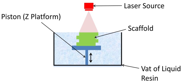
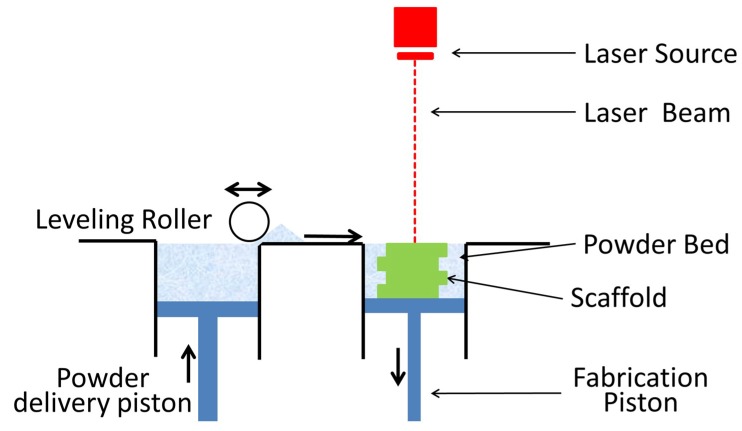
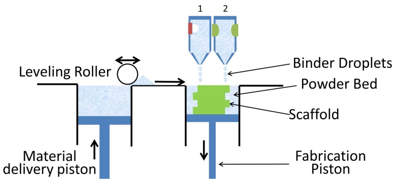
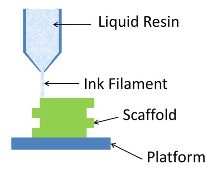
Three-dimensional (3D) printing has become an important tool in the field of tissue engineering and its further development will lead to completely new clinical possibilities. The ability to create tissue scaffolds with controllable characteristics, such as internal architecture, porosity, and interconnectivity make it highly desirable in comparison to conventional techniques, which lack a defined structure and repeatability between scaffolds. Furthermore, 3D printing allows for the production of scaffolds with patient-specific dimensions using computer-aided design. The availability of commercially available 3D printed permanent implants is on the rise; however, there are yet to be any commercially available biodegradable/bioresorbable devices. This review will compare the main 3D printing techniques of: stereolithography; selective laser sintering; powder bed inkjet printing and extrusion printing; for the fabrication of biodegradable/bioresorbable bone tissue scaffolds; and, discuss their potential for dental applications, specifically augmentation of the alveolar ridge.
Keywords: 3D printing; additive manufacturing; bone augmentation; bone regeneration; bone scaffold; dentistry.
Conflict of interest statement
The authors declare no conflict of interest.
Figures
Similar articles
-
Alveolar bone regeneration using a 3D-printed patient-specific resorbable scaffold for dental implant placement: A case report.Clin Oral Implants Res. 2024 Dec;35(12):1655-1668. doi: 10.1111/clr.14340. Epub 2024 Aug 7. Clin Oral Implants Res. 2024. PMID: 39109582 Free PMC article.
-
Three-dimensional (3D) printed scaffold and material selection for bone repair.Acta Biomater. 2019 Jan 15;84:16-33. doi: 10.1016/j.actbio.2018.11.039. Epub 2018 Nov 24. Acta Biomater. 2019. PMID: 30481607 Review.
-
3D-printed Bioresorbable Scaffold for Periodontal Repair.J Dent Res. 2015 Sep;94(9 Suppl):153S-7S. doi: 10.1177/0022034515588303. Epub 2015 Jun 29. J Dent Res. 2015. PMID: 26124215 No abstract available.
-
Marginal and internal fit of 3D printed resin graft substitutes mimicking alveolar ridge augmentation: An in vitro pilot study.PLoS One. 2019 Apr 15;14(4):e0215092. doi: 10.1371/journal.pone.0215092. eCollection 2019. PLoS One. 2019. PMID: 30986268 Free PMC article.
-
Regenerative Medicine for Periodontal and Peri-implant Diseases.J Dent Res. 2016 Mar;95(3):255-66. doi: 10.1177/0022034515618887. Epub 2015 Nov 25. J Dent Res. 2016. PMID: 26608580 Free PMC article. Review.
Cited by
-
The Granule Size Mediates the In Vivo Foreign Body Response and the Integration Behavior of Bone Substitutes.Materials (Basel). 2021 Dec 1;14(23):7372. doi: 10.3390/ma14237372. Materials (Basel). 2021. PMID: 34885527 Free PMC article.
-
Comparison of the Validity of Enzymatic and Immunohistochemical Detection of Tartrate-resistant Acid Phosphatase (TRAP) in the Context of Biocompatibility Analyses of Bone Substitutes.In Vivo. 2022 Sep-Oct;36(5):2042-2051. doi: 10.21873/invivo.12930. In Vivo. 2022. PMID: 36099106 Free PMC article.
-
Nano-Hydroxyapatite Composite Scaffolds Loaded with Bioactive Factors and Drugs for Bone Tissue Engineering.Int J Mol Sci. 2023 Jan 9;24(2):1291. doi: 10.3390/ijms24021291. Int J Mol Sci. 2023. PMID: 36674810 Free PMC article. Review.
-
In Vivo Biocompatibility Investigation of an Injectable Calcium Carbonate (Vaterite) as a Bone Substitute including Compositional Analysis via SEM-EDX Technology.Int J Mol Sci. 2022 Jan 21;23(3):1196. doi: 10.3390/ijms23031196. Int J Mol Sci. 2022. PMID: 35163120 Free PMC article.
-
Hyaluronic Acid/Bone Substitute Complex Implanted on Chick Embryo Chorioallantoic Membrane Induces Osteoblastic Differentiation and Angiogenesis, but not Inflammation.Int J Mol Sci. 2018 Dec 19;19(12):4119. doi: 10.3390/ijms19124119. Int J Mol Sci. 2018. PMID: 30572565 Free PMC article.
References
Publication types
MeSH terms
LinkOut - more resources
Full Text Sources
Other Literature Sources
