

Pneumomediastinum, Tracheal Diverticulum, and Probable Asthma: Coincidence or Possible Association? A Case Report
- PMID: 30356031
- PMCID: PMC6213822
- DOI: 10.12659/AJCR.911413
Pneumomediastinum, Tracheal Diverticulum, and Probable Asthma: Coincidence or Possible Association? A Case Report
Abstract
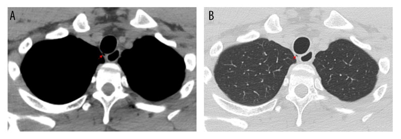
BACKGROUND Many conditions and triggers have been identified and associated with spontaneous pneumomediastinum (SPM), including asthma, strenuous exercise, chronic obstructive pulmonary disease, diabetic ketoacidosis, inhalational drugs, and other activities associated with the Valsalva maneuver. Among rare findings reported in patients with SPM is tracheal diverticulum. We present a case of SPM that on further evaluation was noted to have a tracheal diverticulum, together with a possible diagnosis of asthma. CASE REPORT A 25-year-old male was admitted to the hospital for dyspnea and chest pain. Based on initial assessment, laboratory findings, and imaging, he was diagnosed with SPM. Recovery was successful, and the patient was discharged 3 days later. Follow-up at 2 weeks revealed an abnormality on imaging and abnormal pulmonary function tests. A computed tomography scan revealed a tracheal diverticulum located on the right posterolateral region of the trachea at T1 level. Pulmonary function tests abnormalities included: high fractional exhaled nitric oxide (FeNO), high lung clearance index (LCI), and elevated diffusing capacity of the lungs for carbon monoxide (DLCO). CONCLUSIONS Although the patient presented with a normal spirometry, the FeNO, LCI, and DLCO findings proved valuable and suggested a possible diagnosis of asthma. The anatomic weakness associated with the tracheal diverticulum could have been the breaking point of sustained increased pressure in the airways, due to a possible asthma exacerbation. In retrospective, we hypothesized this to be a series of events that ultimately ended as a pneumomediastinum.
Conflict of interest statement
None.
Figures
Similar articles
-
Pneumomediastinum related to distal tracheal diverticulum.Anaesthesia. 2013 Apr;68(4):432-3. doi: 10.1111/anae.12193. Anaesthesia. 2013. PMID: 23488851 No abstract available.
-
Tracheal diverticulum masquerading as pneumomediastinum in a trauma victim.Am J Emerg Med. 2015 Feb;33(2):310.e1-3. doi: 10.1016/j.ajem.2014.08.014. Epub 2014 Aug 8. Am J Emerg Med. 2015. PMID: 25239696
-
The accidental discovery of a tracheal diverticulum.Intensive Care Med. 2019 Feb;45(2):259-260. doi: 10.1007/s00134-018-5316-4. Epub 2018 Jul 24. Intensive Care Med. 2019. PMID: 30043274 No abstract available.
-
Tracheal diverticulum: a case report and literature review.Am J Otolaryngol. 2014 Jul-Aug;35(4):542-5. doi: 10.1016/j.amjoto.2014.03.015. Epub 2014 Mar 26. Am J Otolaryngol. 2014. PMID: 24767473 Review.
-
Tracheal diverticulum: a report of 4 cases.Ear Nose Throat J. 2009 Jan;88(1):E11. Ear Nose Throat J. 2009. PMID: 19172558 Review.
Cited by
-
Valsalva Maneuver during Computed Tomography for the Diagnosis of Tracheal Diverticulum: A Case Report.Cureus. 2024 Oct 30;16(10):e72726. doi: 10.7759/cureus.72726. eCollection 2024 Oct. Cureus. 2024. PMID: 39618673 Free PMC article.
-
Tracheal Diverticulum in SARS-CoV-2 Patients on Non-Invasive Ventilation A not so "Spontaneous" Cause of Pneumomediastinum? An imaging Pictorial Presentation of Two Cases with Review of Literature.Acta Med Litu. 2021;28(2):302-307. doi: 10.15388/Amed.2021.28.2.19. Epub 2021 Dec 22. Acta Med Litu. 2021. PMID: 35474930 Free PMC article.
-
Chronic Cough Revealing a Tracheal Diverticulum: A Case Report.Cureus. 2022 Jul 18;14(7):e26958. doi: 10.7759/cureus.26958. eCollection 2022 Jul. Cureus. 2022. PMID: 35989740 Free PMC article.
-
Tracheal Diverticulum Mimicking Pneumomediastinum: A Case Report Emphasizing the Importance of Differential Diagnosis in Chest Imaging Evaluation.Cureus. 2023 May 21;15(5):e39297. doi: 10.7759/cureus.39297. eCollection 2023 May. Cureus. 2023. PMID: 37346209 Free PMC article.
References
-
- Caceres M, Ali SZ, Braud R, Weiman D, Garrett HE., Jr Spontaneous pneumomediastinum: A comparative study and review of the literature. Ann Thorac Surg. 2008;86:962–66. - PubMed
-
- Dajer-Fadel WL, Arguero-Sanchez R, Ibarra-Perez C, Navarro-Reynoso FP. Systematic review of spontaneous pneumomediastinum: A survey of 22 years’ data. Asian Cardiovasc Thorac Ann. 2014;22:997–1002. - PubMed
