. 2018 Oct 24;9(1):4421.
doi: 10.1038/s41467-018-06652-w.

SOX17 regulates uterine epithelial-stromal cross-talk acting via a distal enhancer upstream of Ihh

Affiliations

SOX17 regulates uterine epithelial-stromal cross-talk acting via a distal enhancer upstream of Ihh

Xiaoqiu Wang et al. Nat Commun. .

Abstract

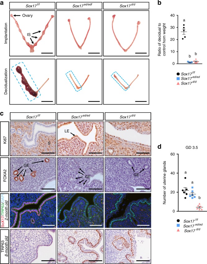
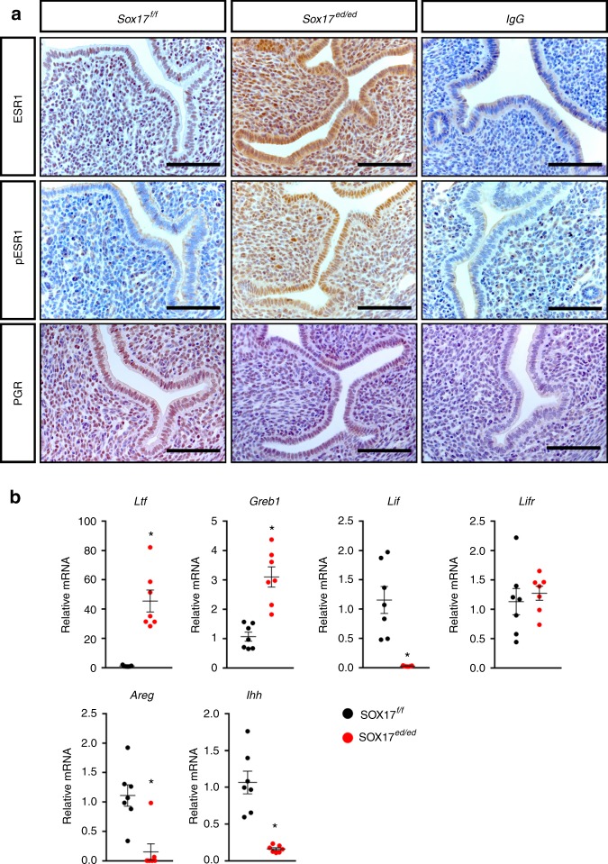
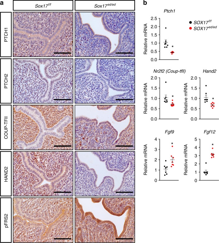
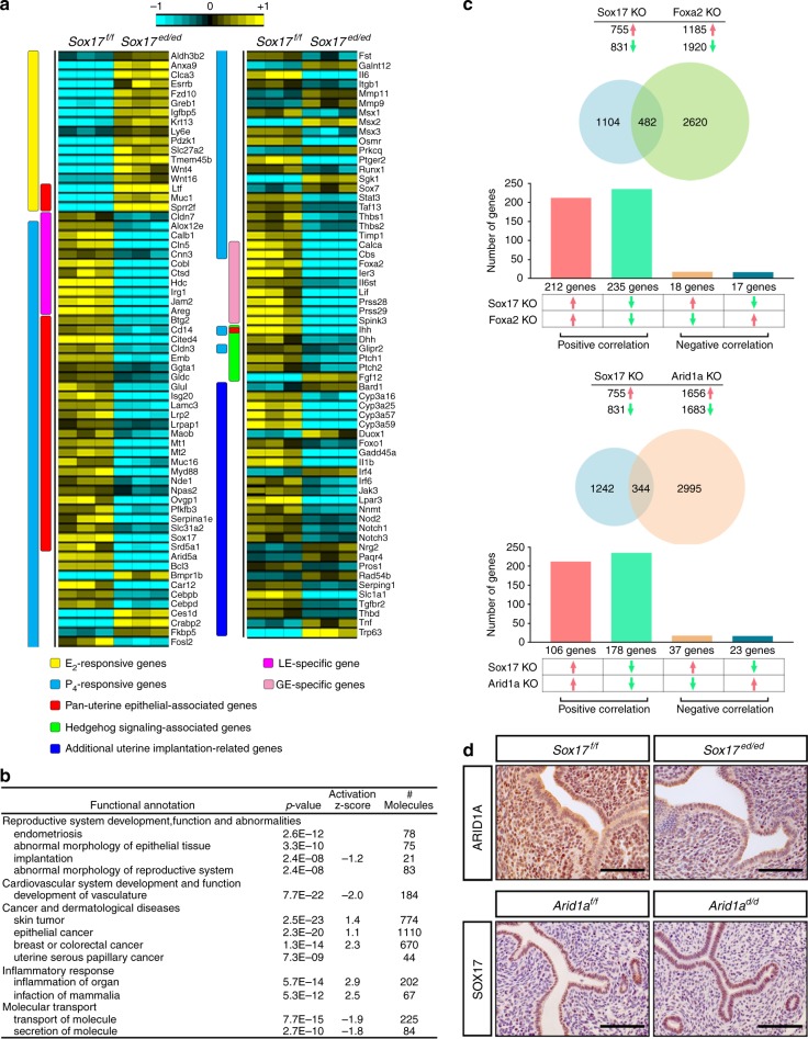
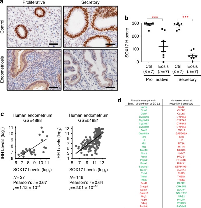
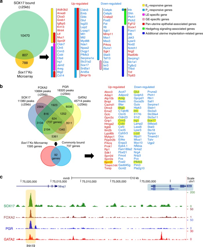
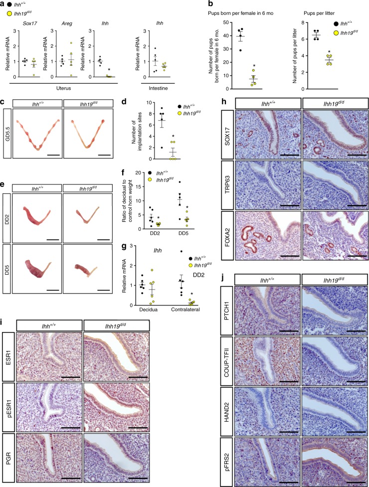
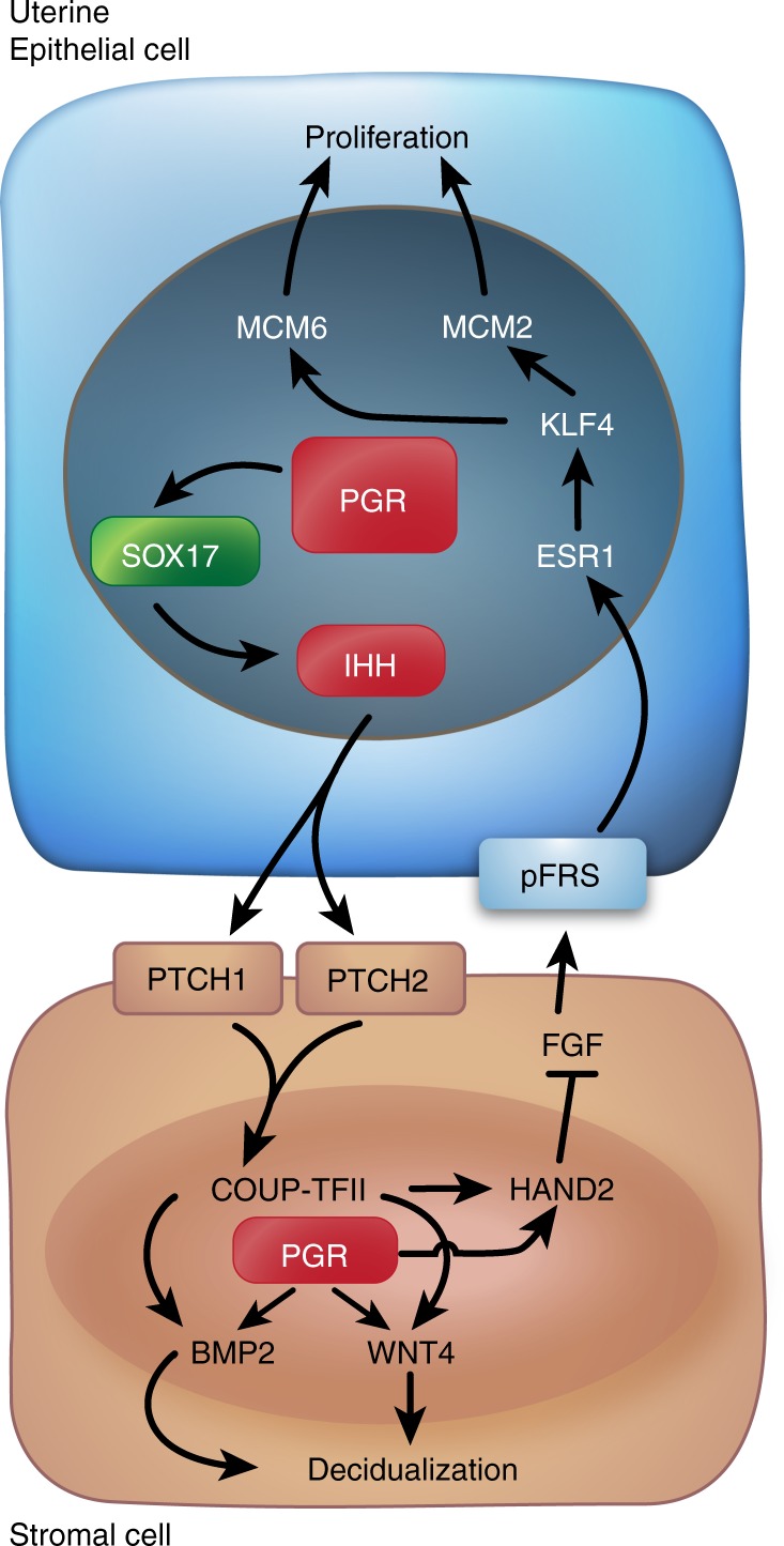
Mammalian pregnancy depends on the ability of the uterus to support embryo implantation. Previous studies reveal the Sox17 gene as a downstream target of the Pgr-Gata2-dependent transcription network that directs genomic actions in the uterine endometrium receptive for embryo implantation. Here, we report that ablating Sox17 in the uterine epithelium impairs leukemia inhibitory factor (LIF) and Indian hedgehog homolog (IHH) signaling, leading to failure of embryo implantation. In vivo deletion of the SOX17-binding region 19 kb upstream of the Ihh locus by CRISPR-Cas technology reduces Ihh expression specifically in the uterus and alters proper endometrial epithelial-stromal interactions, thereby impairing pregnancy. This SOX17-binding interval is also bound by GATA2, FOXA2, and PGR. This cluster of transcription factor binding is common in 737 uterine genes and may represent a key regulatory element essential for uterine epithelial gene expression.


Conflict of interest statement

The authors declare no competing interests.

Figures

Fig. 1
Fig. 1
Phenotype of conditional deletion of Sox17 in the adult mouse uterus. a Embryo implantation sites (top panel) and artificial deciduoma formation (bottom panel) in Sox17f/f (control), Sox17ed/ed (LtfiCreSox17f/f), and Sox17d/d (PgrCreSox17f/f) female mice (n = 5). Scale bars: 10 mm. IS, implantation site. b Ratio of decidual to control horn weight in Sox17f/f (black dots), Sox17ed/ed (blue squares), and Sox17d/d (pink triangles) mice (n = 5). c Immunohistochemical staining of Ki67 (row 1), FOXA2 (row 2), TRP63 (row 3) in the 2-month old uteri of Sox17f/f, Sox17ed/ed, and Sox17d/d female mice at GD 3.5 (n = 3). Row 4 is TRP63 staining in the 8-month old uteri (n = 3). Scale bars: 100 µm. S, stroma; LE, luminal epithelium; GE, glandular epithelium. d Number of endometrial glands in the 2-month old uteri from Sox17f/f (black dots), Sox17ed/ed (blue squares), and Sox17d/d (pink triangles) mice at GD 3.5 (n = 3 mice × 2 sections per mice = 6). Different superscript letters denote significant (P < 0.05, ANOVA with Tukey’s post hoc test) differences. Data are presented as means ± S.E.M
Fig. 2
Fig. 2
Dysregulated estrogen and progesterone signaling in Sox17-deficient uterus during the window of receptivity. a Immunohistochemical staining of ESR1, phosphorylated ESR1 and PGR in GD 3.5 uteri from Sox17f/f and Sox17ed/ed mice (n = 3). IgG staining as the negative control. Scale bars: 100 µm. b Quantification of ESR target genes (Ltf, Greb1, Lif, Lifr), as well as PGR target genes (Areg, Ihh) in GD 3.5 uteri from Sox17f/f (black dots) and Sox17ed/ed (red dots) mice (n = 7). *P < 0.05 (Student’s t test). Data are presented as means ± S.E.M
Fig. 3
Fig. 3
Alteration of Indian Hedgehog (IHH) signaling pathways during the window of receptivity. a Immunohistochemical staining of IHH receptors PTCH1 and PTCH2, as well as downstream mediator COUP-TFII, HAND2, and phosphorylated FRS in GD 3.5 uteri from Sox17f/f and Sox17ed/ed mice (n = 3). Scale bars: 100 µm. b Quantification of IHH signaling-associated genes in GD 3.5 uteri from Sox17f/f (black dots) and Sox17ed/ed (red dots) mice (n = 7). *P < 0.05 (Student’s t test). Data are presented as means ± S.E.M
Fig. 4
Fig. 4
SOX17 regulation of uterine transcriptome. a Heatmap of uterine function-associated genes differentially regulated between Sox17f/f and Sox17ed/ed mouse uteri at GD 3.5 generated from microarray analysis. b Enrichment of functional annotation in differentially expressed genes between Sox17f/f and Sox17ed/ed mouse uteri at GD 3.5 by IPA analysis. c Overlaps and correlations of Sox17 KO with Foxa2 KO or Arid1a KO transcriptomes generated by NextBio gene expression profile comparison. d Immunohistochemical staining of ARID1A in GD 3.5 uteri from Sox17f/f and Sox17ed/ed mice (n = 3; row 1), as well as SOX17 in GD 3.5 uteri from Arid1af/f and Arid1ad/d (PgrCreArid1af/f) mice (n = 3; row 2) Scale bars: 100 µm
Fig. 5
Fig. 5
A conserved SOX17 pathway for endometrial receptivity in humans. Immunohistochemical staining a and H-score quantification b of SOX17 in human endometrial section from women with and without endometriosis at proliferative and secretory phase of the menstrual cycle. Scale bars: 50 µm. *P < 0.001 (ANOVA with Tukey’s post hoc test). Data are presented as means ± S.E.M. c Correlation of levels between IHH and SOX17 in human endometrium (GSE4888 and GSE51981). d Comparison of genes altered in the Sox17-ablated uteri at GD 3.5 with human endometrial receptivity biomarkers. Green font denotes gene downregulation; red font denotes gene upregulation
Fig. 6
Fig. 6
Binding and regulation of Ihh by analysis of SOX17 uterine cistrome. a SOX17 ChIP-seq using Sox17f/f mice identified to 11277 genes with binding intervals within 25 kb of the gene boundary. Of these genes, 807 were differentially regulated in the Sox17 KO microarray at GD 3.5, and 78 genes were directly associated with uterine receptivity and implantation. b Analyses of SOX17, FOXA2, PGR, and GATA2 ChIP-seq data sets identified 624 common peaks, which assigned to 737 genes. Of these genes, 92 genes were identified as SOX17 target genes with shared binding sites by FOXA2, PGR, and GATA2. c Genome browser tracks of SOX17, FOXA2, PGR, and GATA2 binding at putative enhancers for Ihh gene in P4-treated or GD 3.5 uteri. UCSC Genome Browser views showing the mapped read coverage of SOX17, FOXA2, PGR, and GATA2 ChIP-seq data. The Ihh gene locus and the regions targeted by gRNAs for CRISPR/Cas9 deletion in vivo are highlighted in blue and yellow, respectively. Ihh19, putative enhancer 19 kb upstream of Ihh gene locus
Fig. 7
Fig. 7
The SOX17-binding region 19 kb upstream of Ihh gene governs the window of uterine receptivity. a Quantification of Sox17, Areg, Ihh gene expression in uteri and/or intestine from Ihh+/+ and Ihh19d/d female mice at GD2.5 (n = 5). b Pups born from Ihh+/+ and Ihh19d/d female mice during 6-month breeding trial (n = 4). c, d Embryo implantation sites (c) and quantification (d) in uteri from Ihh+/+ and Ihh19d/d female mice at GD5.5 (n = 5). Scale bars: 10 mm. e, f Artificial deciduoma formation (e) and ratio of decidual to control horn weight (f) in Ihh+/+ and Ihh19d/d female mice at DD2 (n = 6) and DD5 (n = 5). Scale bars: 10 mm. g Quantification of Ihh gene in decidual and contralateral horn from Ihh+/+ and Ihh19d/d female mice at DD2 (n = 6). h-j Immunohistochemical staining of SOX17, TRP63, FOXA2, ESR1, pESR1, PGR, PTCH1, COUP-TFII, HAND2, and pFRS2 in GD 3.5 uteri from Ihh+/+ and Ihh19d/d mice (n = 3). Scale bars: 100 µm. Black dots denote Ihh+/+ and yellow dots denote Ihh19d/d. *P < 0.05 (Student’s t test for a, b, d, and ANOVA with Tukey’s post hoc test for f, g). Data are presented as means ± S.E.M
Fig. 8
Fig. 8
SOX17 regulates IHH signaling pathway to govern uterine epithelial–stromal interactions during the window of receptivity

Similar articles

Cited by

References

    1. Lydon JP, et al. Mice lacking progesterone receptor exhibit pleiotropic reproductive abnormalities. Genes Dev. 1995;9:2266–2278. doi: 10.1101/gad.9.18.2266. - DOI - PubMed
    1. Rubel CA, et al. A Gata2-dependent transcription network regulates uterine progesterone responsiveness and endometrial function. Cell Rep. 2016;17:1414–1425. doi: 10.1016/j.celrep.2016.09.093. - DOI - PMC - PubMed
    1. Lee K, et al. Indian hedgehog is a major mediator of progesterone signaling in the mouse uterus. Nat. Genet. 2006;38:1204–1209. doi: 10.1038/ng1874. - DOI - PubMed
    1. Rubel CA, Franco HL, Jeong JW, Lydon JP, DeMayo FJ. GATA2 is expressed at critical times in the mouse uterus during pregnancy. Gene. Expr. Patterns. 2012;12:196–203. doi: 10.1016/j.gep.2012.03.004. - DOI - PubMed
    1. Rubel CA, et al. Research resource: Genome-wide profiling of progesterone receptor binding in the mouse uterus. Mol. Endocrinol. 2012;26:1428–1442. doi: 10.1210/me.2011-1355. - DOI - PMC - PubMed

Publication types

MeSH terms