. 2018 Oct 24;8(1):15731.
doi: 10.1038/s41598-018-34004-7.

Amplification of the Melanocortin-1 Receptor in Nephrotic Syndrome Identifies a Target for Podocyte Cytoskeleton Stabilization

Affiliations

Amplification of the Melanocortin-1 Receptor in Nephrotic Syndrome Identifies a Target for Podocyte Cytoskeleton Stabilization

Lovisa Bergwall et al. Sci Rep. .

Abstract

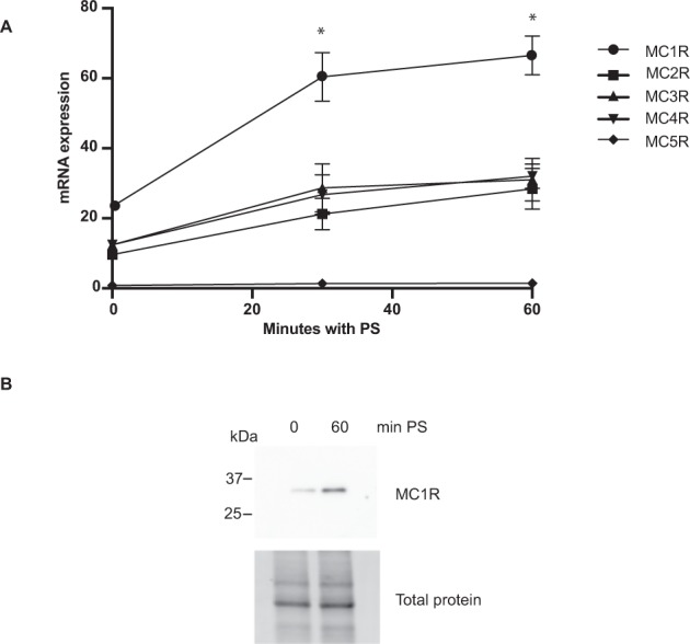
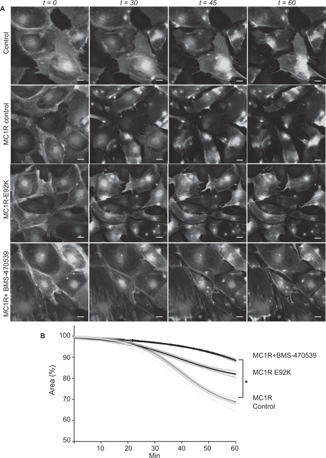
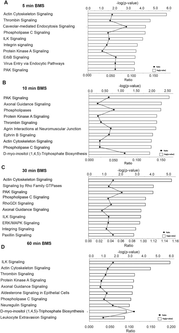
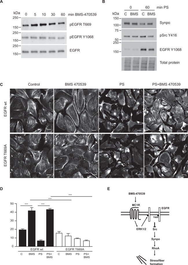
The melanocortin-1 receptor (MC1R) in podocytes has been suggested as the mediator of the ACTH renoprotective effect in patients with nephrotic syndrome with the mechanism of action beeing stabilization of the podocyte actin cytoskeleton. To understand how melanocortin receptors are regulated in nephrotic syndrome and how they are involved in restoration of filtration barrier function, melanocortin receptor expression was evaluated in patients and a rat model of nephrotic syndrome in combination with cell culture analysis. Phosphoproteomics was applied and identified MC1R pathways confirmed using biochemical analysis. We found that glomerular MC1R expression was increased in nephrotic syndrome, both in humans and in a rat model. A MC1R agonist protected podocytes from protamine sulfate induced stress fiber loss with the top ranked phoshoproteomic MC1R activated pathway beeing actin cytoskeleton signaling. Actin stabilization through the MC1R consisted of ERK1/2 dependent phosphorylation and inactivation of EGFR signaling with stabilization of synaptopodin and stressfibers in podocytes. These results further explain how patients with nephrotic syndrome show responsiveness to MC1R receptor activation by decreasing EGFR signaling and as a consequence restore filtration barrier function by stabilizing the podocyte actin cytoskeleton.


Conflict of interest statement

The authors declare no competing interests.

Figures

Figure 1
Figure 1
Glomerular damage increases MC1R expression. (A) Microarray expression data of the melanocortin receptors 1–5 (MCR) from glomeruli of FSGS and MN patients compared to healthy control-living donors. Significant Analysis of Microarray (SAM) with horizontal line representing mean value, *P < 0.5, **P < 0.01, ***P < 0.001). (B) Insitu hybridized MC1R in control and PAN treated rats (upper panel) showing positive cells identified by the Visiopharm quantitative image analysis software, highlighted in orange. (C) Positive glomerular MC1R in situ hybridized cells/ glomerulus in control (n = 3) and PAN treated rats (n = 8), (unpaired two-tailed t-test, **p < 0.01.). Scale bar 50 µm.
Figure 2
Figure 2
Protamine sulfate induced podocyte injury increases MC1R expression. (A) mRNA expression of melanocortinreceptor 1–5 (MCR) following protamine sulfate (PS) exposure. Increased MC1R mRNA expression is observed after 30 and 60 min of 600 ug/ml PS when compared to non-treated podocytes (n = 3, Students T-test, *P < 0.05), neither of the other MCRs (2–5) are significantly regulated by PS. (B) Protein expression of MC1R was upregulated at 60 min PS showing total protein as loading control (n = 3).
Figure 3
Figure 3
MC1R protects podocytes from actin disassembly induced by protamine sulfate. Experiments to analyze actin cytoskeleton formation were performed using LifeAct®, which is a 17-amino acid peptide sequence fused to GFP which binds selectively to F-actin. The experiments were performed on wild type (wt) podocytes, podocytes overexpressing wt MC1R or a constitutively active MC1R mutant (MC1R-E92K). To confirm the functionality of the overexpressed MC1Rs, cAMP assays were performed following BMS-470539 treatment (Supplement Fig. 1). (A) Protamine sulfate (PS) induced actin cytoskeletal reorganization in cultured podocytes presented as time-lapse micrographs during treatment with PS. Representative images ofpodocytes are shown at baseline and during treatment with PS 600 µg/ml for 15, 30, 45 and 60 min. Podocytes overexpression mCherry (control), overexpressing MC1R treated with vehicle (MC1R control), MC1R mutant E92K overexpressing podocytes (E92K) and podocytes overexpressing MC1R treated with 10 nM BMS-470539 for 1 h prior to addition of PS (MC1R + BMS-470539 1 h). (B) Quantification of the relative podocyte area during exposure to PS using the visiopharm software (n = 3). The analyzed areas are presented over time. Scale bar 20 µm.
Figure 4
Figure 4
MC1R phosphorylated signaling pathways. The ten highest significantly regulated phosphorylated signaling pathways identified by Ingenuity Pathway Analysis (IPA) in MC1R overexpressing podocytes stimulated with 10 nM BMS-470539 for (A) 5 min, (B) 10 min, (C) 30 min and (D) 60 min. The bars express the −log (p-value) for the pathway. The dots shown on the lower axis represent identified proteins/total proteins in the pathway. The signaling pathways were generated through the use of IPA (QIAGEN Inc., https://www.qiagenbioinformatics.com/products/ingenuity-pathway-analysis) by using the algorithms previously described by Kramer et al..
Figure 5
Figure 5
MC1R top ranked phosphorylated network at 5 min BMS-470539. The top ranked phosphorylated networks identified by IPA analysis after 5 min of BMS-470539 treatment of MC1R overexpressing podocytes. Green labeled proteins indicate that at least one peptide from that protein has been identified to be significantly phosphorylated when compared to vehicle treated controls. *More than two significantly phosphorylated peptides. The networks analyses were generated through the use of IPA (QIAGEN Inc., https://www.qiagenbioinformatics.com/products/ingenuity-pathway-analysis) by using the algorithms previously described by Kramer et al..
Figure 6
Figure 6
MC1R protection of actin stressfibers are promoted through ERK 1/2. The importance of ERK 1/2 in stress fiber stabilization downstream of the MC1R in podocytes was demonstrated using an inhibitor of ERK 1/2 (PD98059) in PS exposed podocytes treated with BMS-470539. (A) Western blots showing phosphorylated ERK 1/2 and total ERK1/2 in MC1R overexpressing podocytes treated with 0.5, 10, 30 and 60 min of BMS-470539. (B) Actin stressfibers visualized using phalloidin. (C) Actin stressfibers counted in podocytes exposed for 1 h to vehicle (control), ERK 1/2 inhibitor (PD98059), 1 h Protamine sulfate 600 ug/ml (PS), 1 h Protamine sulfate and MC1R agonist (PS + BMS-470539) and 1 h Protamine sulfate, MC1R agonist and ERK 1/2 inhibitor (PS + BMS-470539 + PD98059). The ERK 1/2 inhibitor alone did not affect stressfibers when compared to control cells. (n = 3, ***p < 0.001, ANOVA.) Scale bar 20 µm.
Figure 7
Figure 7
MC1R promote inhibition of EGFR dependent loss of actin stress fiber. (A) Western blots showing phosphorylation of EGFR on Threonine 699 (pEGFR T669) and Tyrosine 1068 (pEGFR Y1068) together with total levels of EGFR (EGFR) in podocytes treated with 0,5, 10, 30 and 60 min of BMS-470539. (B) Western blots on protamine sulfate treated podocytes (PS) in combination with BMS-470539 or vehicle (C) probed for synaptopodin (Synpo), phosphorylated Src on tyrosine 416 (pSrc Y416) and EGFR Y1068. Total protein is used as loading control. (C) Visualised actin stressfibers and (D) Actin stressfibers calculated in podoctes either overexpressing the EGFR wildtype (EGFR wt) or the point mutated T669A EGFR (EGFR T669A) treated with vehicle (C), protamine sulfate (PS), MC1R agonist BMS-470539 (BMS) or protamine sulfate and BMS-470539 (PS + BMS). (n = 3, ***p < 0.001, ANOVA.) Scale bar 20 µm. (E) MC1R stabilizes stressfibers by decreasing EGFR activation: a model. BMS-470539 activates the MC1R receptor promoting ERK 1/2 phosphorylation which phosphorylates EGFR T669 and inhibits its activation of Src. This results in stabilization of synaptopodin and RhoA induced stress fiber formation.

Similar articles

Cited by

References

    1. Berg AL, Nilsson-Ehle P, Arnadottir M. Beneficial effects of ACTH on the serum lipoprotein profile and glomerular function in patients with membranous nephropathy. Kidney Int. 1999;56:1534–1543. doi: 10.1046/j.1523-1755.1999.00675.x. - DOI - PubMed
    1. Madan A, et al. Acthar gel in the treatment of nephrotic syndrome: a multicenter retrospective case series. BMC Nephrol. 2016;17:37. doi: 10.1186/s12882-016-0241-7. - DOI - PMC - PubMed
    1. Bomback AS, et al. Treatment of nephrotic syndrome with adrenocorticotropic hormone (ACTH) gel. Drug Des Devel Ther. 2011;5:147–153. doi: 10.2147/DDDT.S17521. - DOI - PMC - PubMed
    1. Lorusso P, et al. Low-dose synthetic adrenocorticotropic hormone-analog therapy for nephrotic patients: results from a single-center pilot study. Int J Nephrol Renovasc Dis. 2015;8:7–12. doi: 10.2147/IJNRD.S74349. - DOI - PMC - PubMed
    1. Lindskog A, et al. Melanocortin 1 receptor agonists reduce proteinuria. J Am Soc Nephrol. 2010;21:1290–1298. doi: 10.1681/ASN.2009101025. - DOI - PMC - PubMed

Publication types