. 2018 Oct 24;9(11):1090.
doi: 10.1038/s41419-018-1124-6.

Reprogramming mechanisms influence the maturation of hematopoietic progenitors from human pluripotent stem cells

Affiliations

Reprogramming mechanisms influence the maturation of hematopoietic progenitors from human pluripotent stem cells

Hye-Ryeon Heo et al. Cell Death Dis. .

Abstract

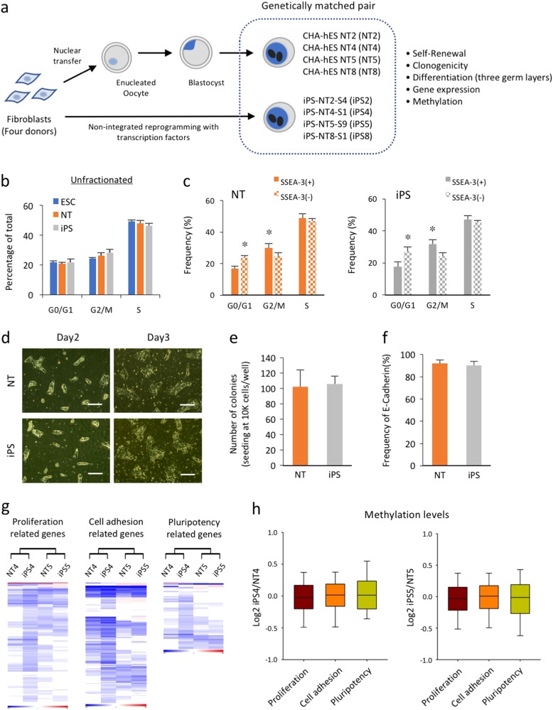
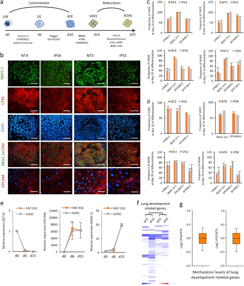
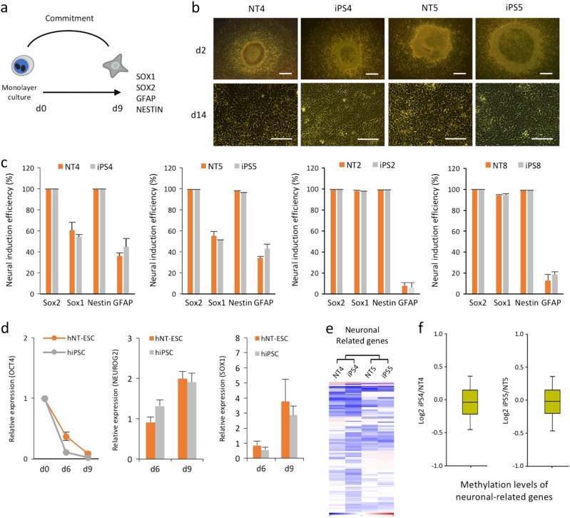
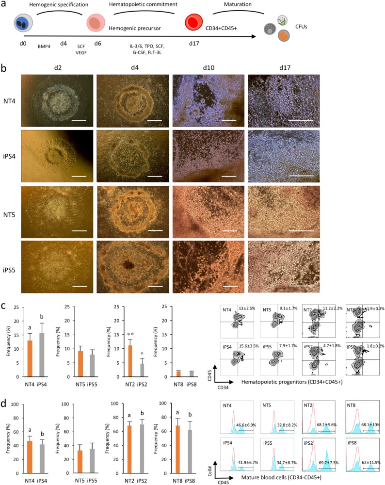
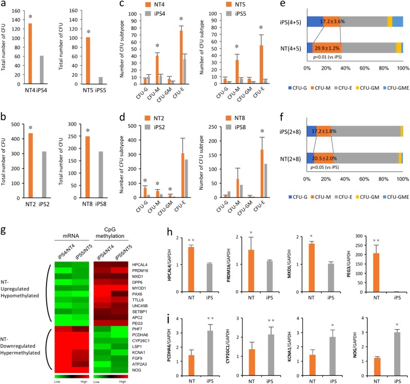
Somatic cell nuclear transfer (SCNT) or the forced expression of transcription factors can be used to generate autologous pluripotent stem cells (PSCs). Although transcriptomic and epigenomic comparisons of isogenic human NT-embryonic stem cells (NT-ESCs) and induced PSCs (iPSCs) in the undifferentiated state have been reported, their functional similarities and differentiation potentials have not been fully elucidated. Our study showed that NT-ESCs and iPSCs derived from the same donors generally displayed similar in vitro commitment capacity toward three germ layer lineages as well as proliferative activity and clonogenic capacity. However, the maturation capacity of NT-ESC-derived hematopoietic progenitors was significantly greater than the corresponding capacity of isogenic iPSC-derived progenitors. Additionally, donor-dependent variations in hematopoietic specification and commitment capacity were observed. Transcriptome and methylome analyses in undifferentiated NT-ESCs and iPSCs revealed a set of genes that may influence variations in hematopoietic commitment and maturation between PSC lines derived using different reprogramming methods. Here, we suggest that genetically identical iPSCs and NT-ESCs could be functionally unequal due to differential transcription and methylation levels acquired during reprogramming. Our proof-of-concept study indicates that reprogramming mechanisms and genetic background could contribute to diverse functionalities between PSCs.


Conflict of interest statement

The authors declare no conflict of interest.

Figures

Fig. 1
Fig. 1. Isogenic iPSCs and NT-ESCs exhibit similar proliferative and clonogenic potentials.
a A schematic diagram of the study design. Genetically matched iPSCs and NT-ESCs were derived from the dermal fibroblasts of four different donors. b Graphic representation of the cell cycle compartments in unfractionated iPSC, NT-ESC, and ESC cultures using BrdU incorporation assay. c Flow cytometry analysis of BrdU incorporation gated on the SSEA-3(+) and SSEA-3(−) subpopulations within the iPSC and NT-ESC cultures. *p < 0.05. d Representative images of the colonies that formed from dissociated single cells and the regenerated colonies counted 7 days post seed are shown. Scale bar, 100 µm. e Quantitative comparison of the clonogenic capacity of the iPSCs and NT-ESCs. f The percentages of E-cadherin in the undifferentiated iPSC and NT-ESC cultures determined by flow cytometry. g Heat maps representing the mRNA expression levels of genes associated with proliferation, cell adhesion, and pluripotency in the isogenic iPSCs and NT-ESCs. h Boxplots representing the degree of promoter gene methylation within each category. All bars indicate the mean±SD from three independent experiments
Fig. 2
Fig. 2. Isogenic iPSCs and NT-ESCs show similar alveolar epithelial cell (AEC) differentiation potentials.
a Schematic diagram of serum- and feeder-free multistep AEC differentiation from human iPSCs and NT-ESCs. DE definitive endoderm, AFE anterior foregut endoderm. b Immunofluorescence staining for CPM, NKX2.1, and EPCAM in AECs on day 14 of differentiation. Scale bar, 200 μm. c, d Flow cytometry analysis of cells harvested on days 14 and 25 showing the frequencies of specific markers for VAFE and ADAE cells, respectively. The induction efficiency of VAFE and ADAE cells was determined by measuring the percentages of CPM(+), NKX2.1(+), EPCAM(+), and SFTP-B(+) cells by flow cytometry on days 14 and 25, respectively. e AEC differentiation was measured by gradually reducing OCT4 and increasing the lineage specific markers (GATA6 and NKX2.1). The relative expression levels of target genes are normalized to GAPDH in each well. Values are relative to day 0 (day 0 = 1). f Heat maps representing the RNA expression patterns of genes associated with lung development in isogenic iPSCs and NT-ESCs. g Boxplots representing the mean lung development-related gene promoter methylation levels in each isogenic pair. All bars indicate the mean±SD from three independent experiments
Fig. 3
Fig. 3. Isogenic iPSCs and NT-ESCs exhibit similar neuronal differentiation capacities.
a A schematic of serum- and feeder-free NPC differentiation from human iPSCs and NT-ESCs. b Representative images of neural differentiation. Scale bar, 100 μm. c Flow cytometry analysis of cells harvested on day 9 showing the frequencies of specific NPC markers. The induction efficiency of neuronal cells determined by measuring the percentages of SOX2(+), SOX1(+), NESTIN(+), and GFAP(+) cells by flow cytometry on day 9. d NPC differentiation measured by gradually reducing OCT4 and increasing the neuronal lineage markers (NEUROG2 and SOX1). The relative expression levels of OCT4 are normalized to GAPDH in each well. Values are relative to day 0 (day 0 = 1). e Heat maps representing the RNA expression patterns of genes associated with neuronal differentiation in both isogenic iPSCs and NT-ESCs. f Boxplots representing the mean neuronal-related gene promoter methylation levels in each isogenic pair. All bars indicate the mean±SD from three independent experiments
Fig. 4
Fig. 4. Donor-dependent variations in hematopoietic commitment capacity between human PSC lines.
a Schematic diagram of serum- and feeder-free stepwise hematopoietic induction of human iPSCs and NT-ESCs. Flow cytometry analysis of committed hematopoietic progenitors (CD34+CD45+) and mature blood (CD34−CD45+) cells on day 17. b Representative images at different stages of hematopoietic differentiation. Scale bar, 100 μm. c, d Flow cytometry analysis of cells harvested on day 17 showing the frequencies of hematopoietic progenitor and mature blood cells. ap < 0.05 (vs NT5), bp < 0.05 (vs iPS5), *p < 0.05 (vs iPS8), **p < 0.01 (vs NT8). Both floating and attached cells were harvested to measure the efficiency if hematopoietic differentiation. All bars indicate the mean±SD from three independent experiments
Fig. 5
Fig. 5. Differential transcription and methylation levels in genes related to hematopoietic development during reprogramming may influence the maturation capacity of isogenic NT-ESCs and iPSCs.
a–f Assessment of hematopoietic progenitor capacity by counting the number (a–d) and distribution of CFU subtypes (e, f). CFU-E, erythroid; CFU-M, macrophage; and CFU-G, granulocytes; CFU-GME, granulocytes/macrophage/erythroid. *p < 0.05. g Heat maps representing up- and downregulated genes in NT lines compared with iPSC lines and the promoter methylation status of corresponding genes. h, i Verification of the transcriptomic and methylomic analysis of selected genes using qPCR. *p < 0.05, **p < 0.01. All bars indicate the mean±SD from three independent experiments

References

    1. Thomson JA, et al. Embryonic stem cell lines derived from human blastocysts. Science. 1998;282:1145–1147. doi: 10.1126/science.282.5391.1145. - DOI - PubMed
    1. Takahashi K, Yamanaka S. Induction of pluripotent stem cells from mouse embryonic and adult fibroblast cultures by defined factors. Cell. 2006;126:663–676. doi: 10.1016/j.cell.2006.07.024. - DOI - PubMed
    1. Takahashi K, et al. Induction of pluripotent stem cells from adult human fibroblasts by defined factors. Cell. 2007;131:861–872. doi: 10.1016/j.cell.2007.11.019. - DOI - PubMed
    1. Chung YG, et al. Human somatic cell nuclear transfer using adult cells. Cell Stem Cell. 2014;14:777–780. doi: 10.1016/j.stem.2014.03.015. - DOI - PubMed
    1. Tachibana M, et al. Human embryonic stem cells derived by somatic cell nuclear transfer. Cell. 2013;153:1228–1238. doi: 10.1016/j.cell.2013.05.006. - DOI - PMC - PubMed

Publication types

MeSH terms