. 2018 Oct 24;8(1):15749.
doi: 10.1038/s41598-018-34033-2.

Angiogenic and Osteogenic Synergy of Human Mesenchymal Stem Cells and Human Umbilical Vein Endothelial Cells Cocultured on a Nanomatrix

Affiliations

Angiogenic and Osteogenic Synergy of Human Mesenchymal Stem Cells and Human Umbilical Vein Endothelial Cells Cocultured on a Nanomatrix

Jun Chen et al. Sci Rep. .

Abstract

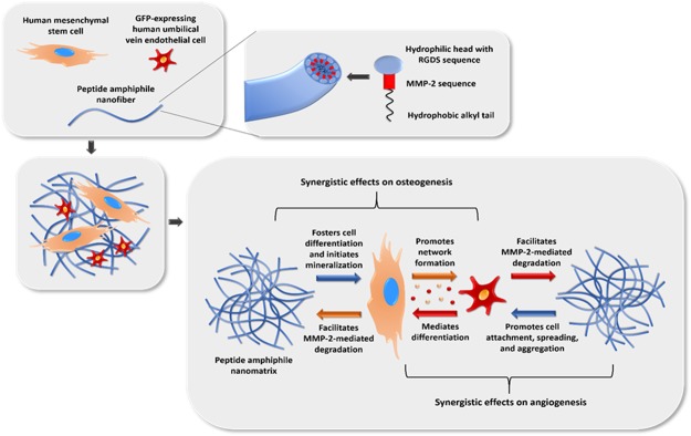
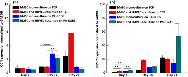
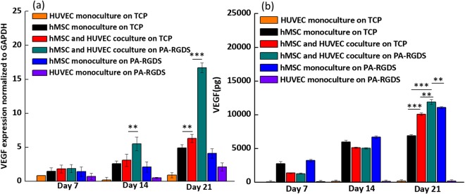
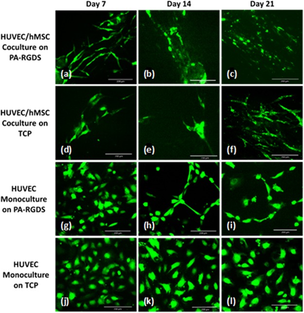
To date, bone tissue regeneration strategies lack an approach that effectively provides an osteogenic and angiogenic environment conducive to bone growth. In the current study, we evaluated the osteogenic and angiogenic response of human mesenchymal stem cells (hMSCs) and green fluorescent protein-expressing human umbilical vein endothelial cells (GFP-HUVECs) cocultured on a self-assembled, peptide amphiphile nanomatrix functionalized with the cell adhesive ligand RGDS (PA-RGDS). Analysis of alkaline phosphatase activity, von Kossa staining, Alizarin Red quantification, and osteogenic gene expression, indicates a significant synergistic effect between the PA-RGDS nanomatrix and coculture that promoted hMSC osteogenesis. In addition, coculturing on PA-RGDS resulted in enhanced HUVEC network formation and upregulated vascular endothelial growth factor gene and protein expression. Though PA-RGDS and coculturing hMSCs with HUVECs were each previously reported to individually enhance hMSC osteogenesis, this study is the first to demonstrate a synergistic promotion of HUVEC angiogenesis and hMSC osteogenesis by integrating coculturing with the PA-RGDS nanomatrix. We believe that using the combination of hMSC/HUVEC coculture and PA-RGDS substrate is an efficient method for promoting osteogenesis and angiogenesis, which has immense potential as an efficacious, engineered platform for bone tissue regeneration.


Conflict of interest statement

The authors declare no competing interests.

Figures

Figure 1
Figure 1
Schematic of the experimental strategy. Included are the expected synergistic effects on osteogenesis and angiogenesis of hMSCs and GFP-HUVECs cocultured on a peptide amphiphile nanomatrix functionalized with the RGDS motif.
Figure 2
Figure 2
ALP activity at days 1 and 7 (a) and days 14 and 28 (b) post cell-seeding. Samples were normalized by total DNA content observed in the Picogreen assay. Values are expressed as a mean ± standard error of measurement (**p = 0.01).
Figure 3
Figure 3
Day 21 Alizarin Red Quantification (a). Day 28 von Kossa staining. Row 1: PA-RGDS (bd), and Row 2: TCP (eg). Column 1: hMSCs (b,e), Column 2: HUVECs (c,f), Column 3: Coculture (d,g). Red staining shows locations of calcium deposits.
Figure 4
Figure 4
ALP (a) and Runx2 (b) gene expression. Gene expression is shown for monocultures and cocultures on TCPs and PA-RGDS at days 7, 14, and 21, as expressed as fold ratios relative to gene expression of hMSC monocultures on TCPs at day 7. Data, provided as mean ± standard deviation, are normalized to GAPDH gene expression (**p < 0.05 and ***p < 0.005).
Figure 5
Figure 5
OCN (a) and BMP-2 (b) gene expression. Gene expression is shown for monocultures and cocultures on TCPs and PA-RGDS at days 7, 14, and 21 post cell-seeding, as expressed as fold ratios relative to gene expression of hMSC monocultures on TCPs at day 7. Data are normalized to GAPDH gene expression (**p < 0.05 and ***p < 0.005).
Figure 6
Figure 6
VEGF gene expression (a) and accumulative VEGF secretion (b). Gene expression is displayed for cultures on days 7, 14, and 21 as a fold ratio relative to HUVEC monocultures on TCP at day 7. Data are expressed as mean ± standard deviation and are normalized to GAPDH gene expression (**p < 0.05 and ***p < 0.005).
Figure 7
Figure 7
GFP-expressing HUVECs, indicating extent of network formation. Images are shown for HUVEC/hMSC cocultures on PA-RGDS (ac) and on TCPs (df) and for HUVEC monocultures on PA-RGDS (gi) and on TCPs (jl). Fluorescent images show progression of vascular morphogenesis at days 7 (a,d,g,j), 14 (b,e,h,k), and 21 (c,f,i,l) post cell-seeding.

Similar articles

Cited by

References

    1. Parfitt, A. In Physiology and pharmacology of bone 1–65 (Springer, 1993).
    1. Conget PA, Minguell JJ. Phenotypical and functional properties of human bone marrow mesenchymal progenitor cells. Journal of cellular physiology. 1999;181:67–73. doi: 10.1002/(SICI)1097-4652(199910)181:1<67::AID-JCP7>3.0.CO;2-C. - DOI - PubMed
    1. Spangrude GJ, Heimfeld S, Weissman IL. Purification and characterization of mouse hematopoietic stem cells. Science. 1988;241:58. doi: 10.1126/science.2898810. - DOI - PubMed
    1. Dawson JI, Oreffo RO. Bridging the regeneration gap: stem cells, biomaterials and clinical translation in bone tissue engineering. Archives of biochemistry and biophysics. 2008;473:124–131. doi: 10.1016/j.abb.2008.03.024. - DOI - PubMed
    1. Burg KJ, Porter S, Kellam JF. Biomaterial developments for bone tissue engineering. Biomaterials. 2000;21:2347–2359. doi: 10.1016/S0142-9612(00)00102-2. - DOI - PubMed

Publication types

MeSH terms

Substances