Review
. 2019 Jan;20(1):39-50.
doi: 10.1038/s41576-018-0063-5.

Coaching from the sidelines: the nuclear periphery in genome regulation

Affiliations
Review

Coaching from the sidelines: the nuclear periphery in genome regulation

Abigail Buchwalter et al. Nat Rev Genet. 2019 Jan.

Abstract

The genome is packaged and organized nonrandomly within the 3D space of the nucleus to promote efficient gene expression and to faithfully maintain silencing of heterochromatin. The genome is enclosed within the nucleus by the nuclear envelope membrane, which contains a set of proteins that actively participate in chromatin organization and gene regulation. Technological advances are providing views of genome organization at unprecedented resolution and are beginning to reveal the ways that cells co-opt the structures of the nuclear periphery for nuclear organization and gene regulation. These genome regulatory roles of proteins of the nuclear periphery have important influences on development, disease and ageing.


Figures

Fig. 1 ∣
Fig. 1 ∣. Chromatin organization within the nucleus.
a ∣ Cross-sections through 3D models of genome structures generated from single cell Hi-C, coloured according to individual chromosomes (left panel), whether the sequence is in the A or B compartment (middle) and whether the sequence is part of a lamina-associated domain (LAD) (yellow) or contains highly expressed genes (blue) (right). b ∣ ChromEMT (chromatin electron microscopy tomography) depicts the organization of chromatin within the nucleus. Chromatin density is greatest at the nuclear periphery and decreases within the nuclear interior, with the prominent exception of decondensed euchromatin underneath nuclear pore complexes (NPCs). Gene-rich euchromatin that is transcriptionally active and enriched in histone 3 lysine 4 trimethylation (H3K4me3) marks (green) is located within the nucleoplasm, and gene-poor heterochromatin that is transcriptionally repressed and enriched for H3K9 dimethylation (H3K9me2) and H3K9me3 marks (red) is located at the nuclear periphery. ER, endoplasmic reticulum. Part a adapted from REF., Springer Nature Limited. EM image in part b adapted with permission from REF., Ou, H. D. et al. ChromEMT: visualizing 3D chromatin structure and compaction in interphase and mitotic cells. Science 357, eaag0025 (2017) https://doi.org/10.1126/science.aag0025. Reprinted with permission from AAAS.
Fig. 2 ∣
Fig. 2 ∣. NPC structure and dynamics.
The nuclear pore complex (NPC) is the major gateway for transport between the nucleus and the cytoplasm. Transport through NPCs involves the nucleoporin proteins (NUPs), the GTPase Ran, importin and exportin transport receptors and specialized factors that promote transport of Large protein and RNA complexes (not shown). Whereas core structural elements of the NPC are very stable, other NUPs shuttle dynamically on and off the NPC. Average NUP dynamics are represented for each subcomplex on the basis of their residence times at NPCs; subcomplexes range from highly dynamic (red) to fairly stable (blue). Individual NUPs within the NPC are Labelled, and NUPs in bold contain repeat domains rich in phenylalanine and glycine amino acids (FG repeats). GP210, nuclear pore membrane glycoprotein 210; POM121, nuclear envelope pore membrane protein POM121; RAE1, ribonucleic acid export 1.
Fig. 3 ∣
Fig. 3 ∣. NPC-mediated gene regulation.
a ∣ Nuclear pore complexes (NPCs) bind enhancers at the nuclear periphery to facilitate enhancer–promoter looping for gene activation. NPCs also recruit specific transcription factors (TFs) to the nuclear periphery to control gene profiles involved in cell fate. b ∣ NPCs recruit chromatin modifiers to the nuclear periphery to mediate gene repression at specific genomic loci, including ribosomal, subtelomeric and early differentiation genes. c ∣ Nucleoporin proteins (NUPs) bind the genome within the nucleus and interact with an array of proteins, including histone modifiers and RNA helicases, to regulate gene expression. d ∣ NUPs can regulate gene expression through post-transcriptional processes, such as mRNA splicing and stability. ATP-dependent RNA helicase A (DHX9) has been shown to facilitate NUP-mediated mRNA splicing, but other proteins involved in these processes still remain to be identified. m7G, 7-methylguanosine.
Fig. 4 ∣
Fig. 4 ∣. Nuclear lamina-mediated genome organization.
The nuclear lamina underlies the inner nuclear membrane (INM) and is connected to the genome through lamin-associated proteins. Lamin-associated domains (LADs) are large, gene-poor regions of heterochromatin that interact with the lamina at the nuclear periphery and are often enriched in repressive histone modifications, such as histone 3 lysine 9 methylation (H3K9me) and histone 3 lysine 27 acetylation (H3K27ac) (red). Histone modifiers and transcriptional regulators have been suggested to cooperate with INM proteins to maintain LADs. A-type lamins, in association with lamina-associated polypeptide 2α (LAP2α), can also interact with regions of euchromatin within the nucleus that are enriched in active histone modifications, including H3K4me and H3K9ac (green). BAF, barrier-to-autointegration factor; H3K9me2, H3K9 dimethylation; H3K9me3, H3K9 trimethylation; HP1, heterochromatin protein 1; LBR, lamin B receptor.
Fig. 5 ∣
Fig. 5 ∣. Nuclear decline over cellular ageing.
a ∣ Proliferating cells eventually exit the cell cycle and senesce as cellular ageing progresses. These senescent cells exhibit a disrupted lamina meshwork and altered chromatin organization. b ∣ Postmitotic cells do not divide and must maintain their function over cellular ageing. However, the identification of proteins that undergo limited turnover in non-dividing cells suggests that these proteins accumulate damage that leads to nuclear decline. Two prominent characteristics of long-lived postmitotic cells are the loss of nucleocytoplasmic compartmentalization and increased transcriptional noise.

References

    1. Laubichler MD & Davidson EH Boveri’s long experiment: sea urchin merogones and the establishment of the role of nuclear chromosomes in development. Dev. Biol 314, 1–11 (2008). - PMC - PubMed
    1. Cremer T & Cremer C Rise, fall and resurrection of chromosome territories: a historical perspective. Part I. The rise of chromosome territories. Eur. J. Histochem 50, 161–176 (2006). - PubMed
    1. Croft JA et al. Differences in the localization and morphology of chromosomes in the human nucleus. J. Cell Biol 145, 1119–1131 (1999). - PMC - PubMed
    1. Sati S & Cavalli G Chromosome conformation capture technologies and their impact in understanding genome function. Chromosoma 126, 33–44 (2017). - PubMed
    1. Lieberman-Aiden E et al. Comprehensive mapping of long-range interactions reveals folding principles of the human genome. Science 326, 289–293 (2009). - PMC - PubMed
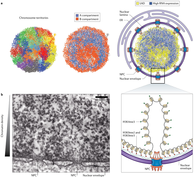
    2. This article provides the first demonstration of the HiC method for probing genome conformation indicating global properties of chromatin folding — that chromosomes occupy distinct territories and that chromatin separates into megabase-scale A and B compartments on the basis of chromatin activity and gene density.

Publication types

Substances