. 2018 Oct 24;8(1):15720.
doi: 10.1038/s41598-018-34105-3.

Locating human splenic capillary sheaths in virtual reality

Affiliations

Locating human splenic capillary sheaths in virtual reality

B S Steiniger et al. Sci Rep. .

Abstract

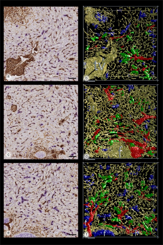
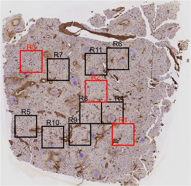
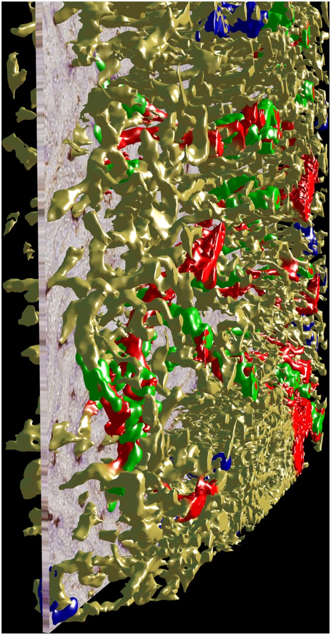
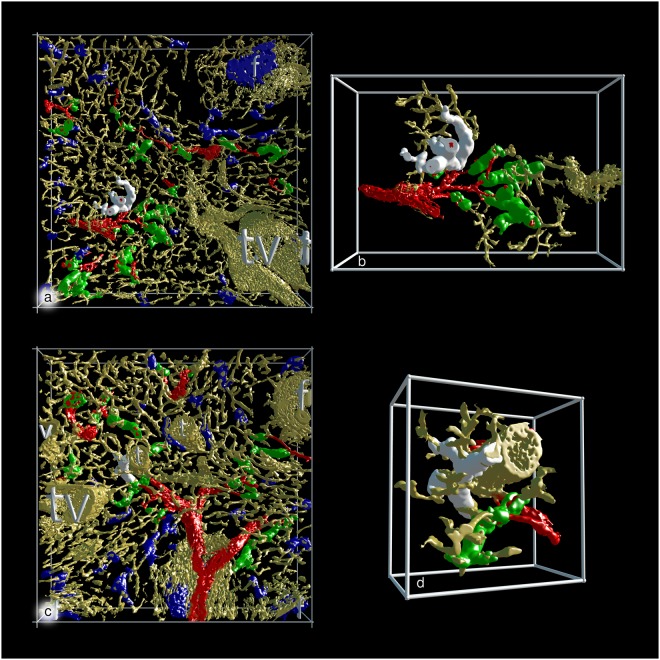
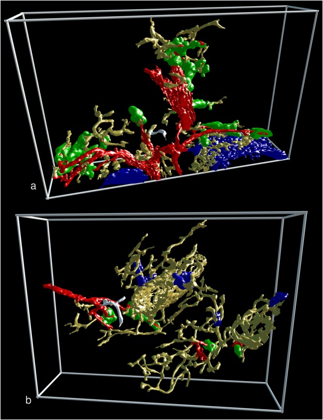
Stromal capillary sheath cells in human spleens strongly express CD271, the low affinity nerve growth factor receptor p75. Serial sections of a representative adult human spleen were double-stained for CD271 versus smooth muscle alpha actin (SMA) plus CD34 to visualise capillary sheaths, the arterial tree and endothelial cells by transmitted light. Preliminary three-dimensional (3D) reconstructions of single regions were inspected in virtual reality (VR). This method showed that a large number of CD271+ sheaths occur in a post-arteriolar position often surrounding capillaries located close to divisions of arterioles. The length and diameter of capillary sheaths are rather heterogeneous. Long sheaths were observed to accompany one or two generations of capillary branches. We hypothesise that human splenic capillary sheaths may attract recirculating B-lymphocytes from the open circulation of the red pulp to start their migration into white pulp follicles along branches of the arterial tree. In addition, they may provide sites of interaction among sheath macrophages and B-lymphocytes. Our innovative approach allows stringent quality control by inserting the original immunostained serial sections into the 3D model for viewing and annotation in VR. Longer series of sections will allow to unequivocally localise most of the capillary sheaths in a given volume.


Conflict of interest statement

The authors declare no competing interests.

Figures

Figure 1
Figure 1
Immunostained first sections and 3D models of R1 to R3 showing the location of capillary sheaths First section of R1 (a), R2 (c) and R3 (e) stained for CD34 plus SMA in brown and CD271 in blue. The first sections are compared to the 3D-model of R1 (b), R2 (d) and R3 (f) showing staining for CD34 plus SMA in yellow, for post-arteriolar CD271+ capillary sheaths in green and for capillary sheaths of undetectable location in blue. CD271+ FDCs in follicles are also blue. The direct connections of arterial vessels to green sheaths were manually marked in red. The iso-values were chosen to exclude weakly CD34+ perifollicular sinus endothelia and weakly CD271+ interstitial fibroblasts without compromising microvesssel continuity. As a consequence of this, the diameter of microvessels differs among the ROIs. Scale bars = 100 µm, f = follicle, t = trabecula, tv = trabecular vein.
Figure 2
Figure 2
Overview of all 11 regions (R) visualised in 3D.
Figure 3
Figure 3
A serial section blended into the 3D model of R2 showing staining for CD34 plus SMA in yellow, for post-arteriolar CD271+ capillary sheaths in green and for capillary sheaths of undetectable location in blue. The direct connections of arterial vessels to green sheaths were manually marked in red.
Figure 4
Figure 4
3D visualisation of the two largest sheaths found in all regions investigated. (a) The largest sheath with a demonstrable connection to an artery highlighted in white colour in R1 (view from last section of the series). (b) Same sheath as in (a) at higher magnification and after removal of non-connected structures. (c) The largest sheath without demonstrable connection to an artery highlighted in white colour in R10 (view from last section of the series). (d) Same sheath as in (c) at higher magnification and after removal of non-connected structures. (ad) show staining for CD34 plus SMA in yellow, for post-arteriolar CD271+ capillary sheaths in green and for capillary sheaths of undetectable location in blue. In (a) and (c) CD271+ FDCs in a follicle are also blue. The direct connections of arterial vessels to green sheaths were manually marked in red. Length of the horizontal part of the bounding box = 1 mm in (a) and (c); 596 µm in (b) and 293 µm in (d). f = follicle, t = trabecula, tv = trabecular vein, v = vein.
Figure 5
Figure 5
Two non-sheathed capillaries arising from an arterial vessel proximal to a capillary sheath. The non-sheathed capillaries are marked in white colour. All non-connected structures were removed. (a) Part of R3 seen from the last section in the series. (b) Part of R8 seen from the first section in the series. (a) and (b) show staining for CD34 plus SMA in yellow, for post-arteriolar CD271+ capillary sheaths in green and for capillary sheaths of undetectable location in blue. In (a) the blue structure in the lower right part corresponds to CD271+ FDCs in follicles. The direct connections of arterial vessels to green sheaths were manually marked in red. Length of the horizontal part of the bounding box = 882 µm in (a) and 709 µm in (b).

References

    1. Schweigger-Seidel F. Untersuchungen über die Milz. Virchows Arch. (Pathol. Anat) 1863;27:460–504. doi: 10.1007/BF01877431. - DOI
    1. Solnitzky O. The Schweigger-Seidel sheath (ellipsoid) of the spleen. Anat. Rec. 1937;69:55–75. doi: 10.1002/ar.1090690107. - DOI
    1. Buyssens N, Paulus G, Bourgeois N. Ellipsoids in the human spleen. Virchows Arch. (Pathol. Anat.) 1984;403:27–40. doi: 10.1007/BF00689336. - DOI - PubMed
    1. Steiniger BS, Seiler A, Lampp K, Wilhelmi V, Stachniss V. B lymphocyte compartments in the human splenic red pulp: capillary sheaths and periarteriolar regions. Histochem. Cell Biol. 2014;141:507–518. doi: 10.1007/s00418-013-1172-z. - DOI - PubMed
    1. Steiniger BS. Human spleen microanatomy: Why mice do not suffice. Immunology. 2015;145:334–346. doi: 10.1111/imm.12469. - DOI - PMC - PubMed

Substances