. 2018 Nov;563(7731):354-359.
doi: 10.1038/s41586-018-0645-6. Epub 2018 Oct 24.

De novo NAD+ synthesis enhances mitochondrial function and improves health

Affiliations

De novo NAD+ synthesis enhances mitochondrial function and improves health

Elena Katsyuba et al. Nature. 2018 Nov.

Abstract

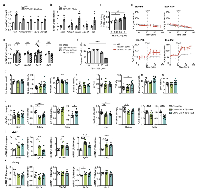
Nicotinamide adenine dinucleotide (NAD+) is a co-substrate for several enzymes, including the sirtuin family of NAD+-dependent protein deacylases. Beneficial effects of increased NAD+ levels and sirtuin activation on mitochondrial homeostasis, organismal metabolism and lifespan have been established across species. Here we show that α-amino-β-carboxymuconate-ε-semialdehyde decarboxylase (ACMSD), the enzyme that limits spontaneous cyclization of α-amino-β-carboxymuconate-ε-semialdehyde in the de novo NAD+ synthesis pathway, controls cellular NAD+ levels via an evolutionarily conserved mechanism in Caenorhabditis elegans and mouse. Genetic and pharmacological inhibition of ACMSD boosts de novo NAD+ synthesis and sirtuin 1 activity, ultimately enhancing mitochondrial function. We also characterize two potent and selective inhibitors of ACMSD. Because expression of ACMSD is largely restricted to kidney and liver, these inhibitors may have therapeutic potential for protection of these tissues from injury. In summary, we identify ACMSD as a key modulator of cellular NAD+ levels, sirtuin activity and mitochondrial homeostasis in kidney and liver.


Conflict of interest statement

Competing Financial Interests

JA, RP and NR are inventors on a patent application describing ACMSD inhibitors (application No: 14/839,209; applicant - TES Pharma S.r.l., Corciano, Italy). Granted US patent 9,708,272 (18 July, 2017). The patent application covers the results obtained with the compounds TES-991 and TES-1025 described in Figures 3-5 RP, FDF, NG and PL are employed by TES Pharma.

Figures

Extended Data Figure 1
Extended Data Figure 1. acsd-1 LOF improves NAD+ levels, mitochondrial function, and lifespan through de novo synthesis in C. elegans
a. De novo synthesis of NAD+ from tryptophan. Names of the worm’s orthologs are in blue. b. acsd-1 expression pattern across different developmental stages in wild-type worms expressing extrachromosomal array of acsd-1::gfp transgene. Scale bar=100 μm. c. acsd-1 expression pattern in adult wild-type worms expressing extrachromosomal array of acsd-1::gfp transgene; i = intestine, m = muscle, p = pharynx, v = vulva. d. acsd-1 mRNA levels in wild type and rrf-3(pk1426) mutants (n=6, each n represents a pool of ~600 worms). e. ACSD-1 activity in control (empty vector) vs acsd-1 RNAi fed worms quantified both wild type and rrf-3 mutants (n=3, where each n represents a pool of ~3600 worms) with compensation for negative controls. f. QPRT-like activity can be detected in both wild type and rrf-3 mutants (n=3, each n represents a pool of ~3600 worms). g-h. Effects of acsd-1 knockdown throughout the entire life on N2 (g) and rrf-3 mutant (h) worm lifespan. i. Lifespan of rrf-3(pk1426) mutants exposed to control or acsd-1 RNAi upon tryptophan supplementation. P*: ctrl vs ctrl+Trp 50 μM; P^: ctrl vs acsd-1 RNAi; P°: ctrl+Trp 50 μM vs acsd-1 RNAi+Trp 50 μM. j. Quantification of GFP signal in ges-1::mito::gfp reporter strain, expressing mitochondria-targeted GFP in the intestine at day 1 and 3 of adulthood (n=4, each n represents a pool of 20 worms). k. Blue native PAGE on mitochondria extracted from rrf-3 mutant worms fed with either empty vector or acsd-1 RNAi bacteria at day 2 of adulthood (n=3, each n represents mitochondria extracted from a pool of ~10’000 worms). l. Mitochondrial morphology in the pmyo-3::mito::GFP reporter strain fed with control or acsd-1 RNAi. Stars represent nuclei. Scoring includes the total perimeter of the mitochondrial network, its total area, the area occupied by the mitochondria within the cell and the circularity assessment, where 1=perfect circle and 0=line (n=6 worms). m. Epistasis between RNAi for acsd-1 and the UPRmt regulator, ubl-5. P*: ctrl vs ctrl/acsd-1 RNAi; P°: ctrl/ubl-5 RNAi vs ubl-5/acsd-1 RNAi. n. Quantification of the GFP signal in hsp-4::gfp reporter strain (n=4, each n represents a pool of 20 worms) at day 1 and 3 of adulthood. o. Quantification of the GFP signal in hsp-16.2::gfp reporter strain (n=4, each n represents a pool of 20 worms). After the first time-point sampled at 20°C, worms were exposed to 37°C, and the measurement was repeated every hour for 6 hours. p. Expression of UPRmt genes in worms at day 2 of adulthood fed with control or acsd-1 RNAi (n=6, each n represents a pool of ~600 worms). q. Expression of sod-3 mRNA at day 1 of adulthood in control or acsd-1 RNAi fed worms (n=3, each n represents a pool of ~600 worms). r. Survival of wild type (N2) worms exposed to 4 mM paraquat starting from the L4 stage, in which the knockdown of acsd-1 was performed at different life stages,. P*: ctrl vs acsd-1 RNAi whole life; P^: ctrl vs acsd-1 RNAi development; P°: ctrl vs acsd-1 RNAi adulthood. s. Epistasis between RNAi for acsd-1 and daf-16 in wild type (N2) worms exposed to 4 mM paraquat. P*: ctrl vs ctrl/acsd-1 RNAi; P^: ctrl/daf-16 RNAi vs daf-16/acsd-1 RNAi. All worm assays, except for hsp-16.2::gfp reporter strain, performed at 20°C and repeated at least once. Data are mean ± s.e.m. *P≤0.05, **P≤0.01, ***P≤0.001. P values calculated using two-tailed t-test (d, e, j, l, n-q) or Logrank test (g-i, m, r, s). For individual P values, see Source Data. For lifespan values, see Extended Data Table 1.
Extended Data Figure 2
Extended Data Figure 2. Pathways activated by Acmsd knockdown in worms are conserved in mammalian cells
a. Acmsd transcript levels reflected by the Ct values in different hepatic and renal cells and cell lines (n=4). b. Efficiency of Acmsd shRNA in mouse primary hepatocytes 48h post adenoviral transduction (n=6). c. NAD+ levels in mitochondria of AML-12 cells transduced with either shRNA control or shRNA against Acmsd (n=5). d-e. Blue native PAGE followed by in-gel activity for Complex II (blue) (d) and Complex I (purple) + IV (brown) (e) on mitochondria extracted from mouse primary hepatocytes transduced with either shRNA control or shRNA against Acmsd for 48h. The experiment was performed once. f. Primary hepatocytes extracted from a Sirt1L2/L2 mouse were transduced either with an adenovirus encoding GFP (WT condition) or the CRE-recombinase to generate Sirt1 KO primary hepatocytes. These hepatocytes were exposed to an shRNA targeting a random sequence or shRNA targeting Acmsd. Transcript levels of Acmsd and Sirt1 (n=3). g. FOXO1 acetylation levels in mouse primary hepatocytes transduced with either shRNA control or shRNA against Acmsd for 48h. The experiment was independently performed twice. Data are mean ± s.e.m.; each n represents a biologically independent sample. *P≤0.05, **P≤0.01, ***P≤0.001. P values calculated using two-tailed t-test. For gel source images see Supplementary Fig. 1. For individual P values, see Source Data.
Extended Data Figure 3
Extended Data Figure 3. Pharmacological inhibition of ACMSD has similar effects as its genetic downregulation
a-b. mRNA levels of mitochondrial genes in mouse primary hepatocytes treated for 24h with DMSO or TES-1025 (a) or TES-991 (b), at the indicated concentrations (n=3). c. SOD2 activity in mouse primary hepatocytes treated for 24h with DMSO or TES-1025, at indicated concentrations (n=4). d. Fatty acid oxidation assessed in mouse primary hepatocytes treated with DMSO or TES-991 for 24h at the indicated concentrations (n=5). FCCP (2 μM) was used as an uncoupler to reach maximal respiration. e. mRNA levels of mitochondrial genes in HK-2 cells after 24h of treatment with TES-1025 or TES-1025 in combination with SIRT1 inhibitor, EX527, at the indicated concentrations (n=5-8). f. Apoptosis rate in HK-2 cells assessed 16h after addition of 50 μM cisplatin by caspase 3/7 activity. TES-1025 was added simultaneously with the cisplatin. g. Biochemical analysis of plasma from mice fed with chow diet or chow diet supplemented with TES-991 or TES-1025 at the dose of 15 mg/kg body weight/day (ctrl and TES-991: n=10; TES-1025: n=9 mice). h-i. Quinolinic acid (QA) (h) and Nicotinic acid (NA) (i) levels in livers (n=9), kidneys (ctrl: n=10; TES-991 and TES-1025: n=9 mice) and brains (ctrl: n=11; TES-991 and TES-1025: n=12 mice). from mice fed with control chow diet or chow diet supplemented with TES-991 or TES-1025 at the dose of 15 mg/kg body weight/day. j-k. mRNA levels of β-oxidation, mitochondrial and oxidative stress defence genes in livers (j) and kidneys (k) of mice described in (g) (n=6 mice). Data are mean ± s.e.m. *P≤0.05, **P≤0.01, ***P≤0.001. P values calculated using two-tailed t-test (a-d, g-k) or one-way ANOVA (e, f). For individual P values, see Source Data.
Extended Data Figure 4
Extended Data Figure 4. ACMSD inhibitors protect hepatic function from MCD diet-induced NAFLD
a. Plasma AST levels in 16-week old C57BL/6J male mice fed for 2.5 weeks with control diet, MCD diet, and MCD diet supplemented with 15 mg/kg/day of TES-991 (n=8 mice). b. Representative photomicrographs of liver tissues stained with hematoxylin and eosin (H&E) and Oil red O from the mouse cohorts described in (a). The experiment was performed twice in an independent way. c. Representative photomicrographs of liver tissues from the mouse cohorts described in (a) stained with CD45 and the corresponding negative control. The experiment was performed twice in an independent way. d. Hepatic SOD2 activity in mouse cohorts described in (a) (CD: n=8; MCD: n=7; MCD+TES-991: n=6 mice). e-h. Liver NAD+ (e), triglyceride (TGs) content (f), plasma ALT (g), AST (h) levels in congenic C57BL/6J Sirt1hep-/- mice that match the mouse cohorts described in (a) related to age, gender and treatment duration (CD: n=8; MCD and MCD+TES-991: n=10 mice). i. Representative photomicrographs of liver tissues stained with hematoxylin and eosin (H&E) from the Sirt1hep-/- mice described in (e-h). The experiment was performed once. j. Hepatic SOD2 activity in congenic C57BL/6J Sirt1hep-/- mice described in (e-h) (CD: n=8; MCD: and MCD+TES-991: n=9 mice). k. mRNA levels of oxidative stress defence, mitochondrial, β-oxidation, inflammatory and fibrosis genes in livers of Sirt1hep-/- mice (n=8 mice). Data are mean ± s.e.m. *P≤0.05, **P≤0.01, ***P≤0.001. P values calculated using two-tailed t-test. For individual P values, see Source Data.
Extended Data Figure 5
Extended Data Figure 5. ACMSD inhibitors protect renal function in two different models of AKI
a. Schematic timeline of the cisplatin-induced AKI study. AKI was induced at day 10 after the beginning of the study in male C57BL/6J mice by a single intraperitoneal dose of cisplatin (20mg/kg body weight). Mice in the sham control group were injected with a saline solution. GFR was measured non-invasively 52h post-cisplatin administration. b-d. Representative photomicrographs of H&E stained kidney sections and the histopathological scoring for tubular necrosis (c), and inflammatory cell infiltration (d) of mouse cohorts described in (a) (Sham ctrl, Cis-AKI+TES-1025: n=6; Cis-AKI: n=5 mice). e. Schematic timeline of the IR-induced AKI study. AKI was induced at day 10 after the beginning of the study in anesthetised male C57BL/6J mice by a dorsal surgical incision and bilateral occlusion of the renal pedicles for 25 min. Mice in the sham control group underwent the same surgical procedure without application of the occluding clamp on the renal pedicles. f-j. Representative photomicrographs of H&E stained kidney sections (f) and histopathological scoring for cumulative score (g), tubular necrosis (h), tubular dilation (i), and cast formation (j) of mouse cohorts described in (e) (n=5 mice). Tubular cell necrosis (arrows), tubular dilation and casts (asterisk) and interstitial edema (crescent moon) are indicated on the pictures by the corresponding symbols in brackets. k-m. GSH protein levels (k), MPO activity (l) and NAD+ content (m) in kidneys of the IR-AKI cohorts described in (e) (n=5 mice). n. Protein expression of the respiratory complex subunits in kidneys from the mouse cohorts described in (e). The experiment was independently performed twice. Data are mean ± s.e.m. *P≤0.05, **P≤0.01, ***P≤0.001. P values calculated using two-tailed t-test. For gel source images see Supplementary Fig. 1. For individual P values, see Source Data. The histopathological scoring performed by two pathologists in a blinded and independent way (b-d, f-j).
Figure 1
Figure 1. acsd-1 LOF improves NAD+ levels, mitochondrial function, and lifespan through de novo synthesis in C. elegans
a-b. NAD+ upon tryptophan supplementation (Trp 0 and 5 μM: n=4; Trp 50 μM: n=5) (a) and feeding with control (empty vector) or acsd-1 RNAi (n=14) (b), each n represents a pool of ~1000 worms. c. Epistasis of acsd-1 and sir-2.1 RNAi. P*: ctrl vs ctrl/acsd-1 RNAi; P°: ctrl/sir-2.1 RNAi vs sir-2.1/acsd-1 RNAi. d. GFP signal in the reporter strain, expressing a mitochondria-targeted GFP in the muscle at day 1 and 3 of adulthood (n=4, each n represents a pool of 20 worms). e. mtDNA/nDNA ratio in wild type (N2) and sir-2.1(ok434) mutant worms (n=12 worms) upon control or acsd-1 RNAi. f-g. mRNA levels encoding mitochondrial proteins (n=6, each n represents a pool of ~600 worms) (f) and oxygen consumption rate in basal and uncoupled conditions (n=14, each n represents a pool of 10 worms) (g), in worms fed with control or acsd-1 RNAi. h. ATP content in rrf-3(pk1426) and N2 worms upon control or acsd-1 RNAi (n=4 and 7 respectively, each n represents a pool of ~100 worms). i. Altered ratio between nDNA- (ATP5A) and mtDNA- (MTCO1) encoded OXPHOS subunits upon acsd-1 RNAi. Each lane represents an individual pool of ~600 worms. j. Epistasis of acsd-1 with atfs-1 RNAi. P*: ctrl vs ctrl/acsd-1 RNAi; P°: ctrl/atfs-1 RNAi vs atfs-1/acsd-1 RNAi. k. GFP signal in hsp-6::GFP reporter strain fed with control or acsd-1 RNAi at day 1 and 3 of adulthood (day 1: n=3; day 3: n=4, each n represents a pool of 20 worms). l. DAF-16 nuclear translocation. Arrowheads indicate DAF-16 accumulation within nuclei. The graph represents the distribution of control and acsd-1 RNAi treated worms with DAF-16-translocated nuclei (n=25 worms). m. Epistasis of acsd-1 with daf-16 RNAi. P*: ctrl vs ctrl/acsd-1 RNAi; P°: ctrl/daf-16 RNAi vs daf-16/acsd-1 RNAi. n. GFP signal in control and acsd-1 RNAi treated sod-3::GFP reporter worms at day 1 and 3 of adulthood (n=4, each n represents a pool of 20 worms). o. ROS content in worms exposed to control or acsd-1 RNAi (n=4, each n represents a pool of 20 worms). p-q. Mobility (p) and survival (q) in N2 exposed to 4 mM paraquat starting from L4 stage, treated with control or acsd-1 RNAi throughout the entire life (n=100 worms). All worm assays performed at 20°C and repeated at least once. Data are mean ± s.e.m. *P≤0.05, **P≤0.01, ***P≤0.001. P values calculated using one-way ANOVA (a), two-tailed t-test (b, d-h, k, n-p) or Logrank test (c, j, m, q). For gel source images, see Supplementary Fig. 1. For individual P values, see Source Data. For lifespan values, see Extended Data Table 1.
Figure 2
Figure 2. Pathways activated by Acmsd knockdown in worms are conserved in mammalian cells
a-f. Mouse primary hepatocytes obtained from C57BL/6J mice were transduced with an adenovirus encoding either control shRNA or shRNA against Acmsd for 48h. NAD+ levels (n=6) (a), mtDNA/nDNA ratio (n=4) (b), citrate synthase activity (n=3) (c), expression of OXPHOS and UPRmt genes (n=3) (d), protein expression of the respiratory complex subunits (e), oxygen consumption in basal and uncoupled (FCCP 2 μM) conditions (n=15) (f). g. ATP content in control vs Acmsd shRNA conditions, in mouse primary hepatocytes grown in both medium with high glucose (HG) and low glucose (LG) supplemented with 1% oleate (n=15). h. Apoptosis evaluated by caspase 3/7 activity after 24h exposure of mouse primary hepatocytes to 0.75 mM palmitate (palmitate- condition: n=3, palmitate+ condition: n=6). i. Triglyceride (TGs) content after 24h exposure to 0.5 mM oleate in AML12 cells transduced with an adenovirus encoding either control or Acmsd shRNA (n=9). j-k. Primary hepatocytes extracted from a Sirt1L2/L2 mouse transduced with an adenovirus either encoding GFP (WT condition) or the CRE-recombinase to generate Sirt1 KO primary hepatocytes. (j) Steatosis quantification in hepatocytes treated with control or Acmsd shRNA after 24h exposure to 0.75 mM palmitate (palmitate- condition: n=3; palmitate+ condition: n=6). (k) ROS content in hepatocytes exposed to control shRNA versus Acmsd shRNA. The positive control treated with 550 μM of H2O2 (H2O2- conditions: n=3; H2O2+ condition: n=6). Data are mean ± s.e.m.; each n represents a biologically independent sample. All experiments performed independently at least twice. *P≤0.05, **P≤0.01, ***P≤0.001. P values calculated using two-tailed t-test (a-d, f-i) or two-way ANOVA (j-k). For gel source images, see Supplementary Fig. 1. For individual P values, see Source Data.
Figure 3
Figure 3. Pharmacological inhibition of ACMSD has similar effects as its genetic downregulation
a-b. NAD+ levels in mouse primary hepatocytes (PH) treated for 24h with vehicle (DMSO), TES-1025 (n=3) (a) or TES-991 (n=6) (b). c. SIRT1 activity in PH treated for 24h with vehicle or the ACMSD inhibitors (500nM) (n=3). d-f. SOD2 activity (n=3) (d), oxygen consumption in basal and uncoupled conditions (ctrl: n=6; TES-991: n=7) (e), and apoptosis rate after 36h exposure to 0.75 mM palmitate (ctrl: n=6; TES-991: n=5) (f) in PH treated for 24h with vehicle or TES-991. g. mRNA levels of fatty acid oxidation (FAO) genes in PH treated with vehicle or TES-991 (500 nM) after 6h exposure to 0.33 mM palmitate and 0.66 mM oleate (n=6). h-i. PH from a Sirt1L2/L2 mouse transduced with an adenovirus encoding GFP (Sirt1 WT) or CRE-recombinase (Sirt1 KO). FAO after 24h treatment with vehicle or TES-991 under basal (h) and uncoupled (i) conditions (n=5). j-m. Changes in NAD+ (n=4) (j), mRNA levels of mitochondrial and oxidative stress defence genes (n=6) (k), OXPHOS subunits (l) and ATP content (ctrl: n=12; TES-1025: n=13) (m) in HK-2 cells upon treatment with TES-1025 (100 μM) and EX527 (10 μM), for 24h. n. Apoptosis in HK-2 cells 16h after addition of cisplatin (50 μM). TES-1025 added 1h prior to cisplatin (n=3). o. NAD+ in livers (ctrl: n=6; TES-991: n=7; TES-1025: n=9 mice), kidneys (ctrl, TES-1025: n=8; TES-991: n=9 mice) and brains (ctrl: n=11; TES-991, TES-1025: n=12 mice) of mice fed with normal chow diet or supplemented with ACMSD inhibitors (15mg/kg body weight/day). Data are mean ± s.e.m.; each n represents a biologically independent sample. All experiments performed independently at least twice (a-n). *P≤0.05, **P≤0.01, ***P≤0.001. P values calculated using two-tailed t-test (a-c, f-g, j-k, m-o), one-way (d-e) or two-way ANOVA (h-i). For gel source images, see Supplementary Fig. 1. For individual P values, see Source Data.
Figure 4
Figure 4. ACMSD inhibitors protect hepatic function from MCD diet-induced NAFLD
a. Comparison of gross liver morphology in representative 16-week old C57BL/6J male mice fed for 2.5 weeks with control diet, MCD diet, and MCD diet supplemented with 15 mg/kg/day of TES-991. In vivo MCD diet study was performed once. b-e. Liver TGs (b), plasma ALT (c), liver NAD+ (d) and ATP (e) levels in the mouse cohorts described in (a) (CD, MCD: n=8; MCD+TES-991: n=7 mice). f. mRNA levels of oxidative stress defence, mitochondrial, β-oxidation, inflammatory and fibrosis genes in livers of the mice described in (a) (n=8 mice). Data are mean ± s.e.m. *P≤0.05, **P≤0.01, ***P≤0.001. P values calculated using two-tailed t-test. For individual P values, see Source Data.
Figure 5
Figure 5. ACMSD inhibitors protect renal function in two different models of AKI
a-c. Plasma creatinine (a), BUN (b) and renal KIM1 protein levels (c) in 12-week old C57BL/6J male mice 72h post cisplatin administration. AKI was induced at day 10 after the beginning of the study by a single intraperitoneal injection of cisplatin (20 mg/kg body weight). Sham controls injected with saline solution. TES-1025 administered at 15 mg/kg/day (n=6 mice). d. Glomerular filtration rate (GFR) in mice 52h after cisplatin or saline administration (Sham ctrl: n=7; Cis-AKI: n=6; Cis-AKI+TES-1025: n=5 mice). e. NAD+ levels in kidneys of the cisplatin-AKI cohorts (Sham ctrl: n=7; Cis-AKI, Cis-AKI+TES-1025: n=5 mice). f. Histopathological scoring of H&E stained kidney sections from mice described in (a), evaluating tubular necrosis, tubular dilation, inflammation, edema and cast (Sham ctrl, Cis-AKI+TES-1025: n=6; Cis-AKI: n=5 mice). The scoring performed by two pathologists in a blinded and independent way. g. Protein expression of the respiratory complex subunits in kidneys from mouse cohorts described in (a). The experiment was performed twice. h-i. AKI was induced in 12-week old C57BL/6J male mice by a dorsal surgical incision and bilateral occlusion of the renal pedicles for 25 min; a simple dorsal incision was performed for the Sham controls. TES-1025 administered at 15 mg/kg/day. Plasma creatinine (h) and BUN (i) in mice 48h post-surgery (n=5 mice). Data are mean ± s.e.m. *P≤0.05, **P≤0.01, ***P≤0.001. P values calculated using two-tailed t-test. For gel source images, see Supplementary Fig. 1. For individual P values, see Source Data.

Comment in

References

    1. Houtkooper RH, Pirinen E, Auwerx J. Sirtuins as regulators of metabolism and healthspan. Nat Rev Mol Cell Biol. 2012;13:225–238. doi: 10.1038/nrm3293. - DOI - PMC - PubMed
    1. Imai S, Guarente L. It takes two to tango: NAD+ and sirtuins in aging/longevity control. npj Aging and Mechanisms of Disease. 2016;2 doi: 10.1038/npjamd.2016.17. - DOI - PMC - PubMed
    1. Belenky P, Bogan KL, Brenner C. NAD+ metabolism in health and disease. Trends in biochemical sciences. 2007;32:12–19. doi: 10.1016/j.tibs.2006.11.006. - DOI - PubMed
    1. Yang Y, Sauve AA. NAD+ metabolism: Bioenergetics, signaling and manipulation for therapy. Biochim Biophys Acta. 2016;1864:1787–1800. doi: 10.1016/j.bbapap.2016.06.014. - DOI - PMC - PubMed
    1. Katsyuba E, Auwerx J. Modulating NAD(+) metabolism, from bench to bedside. The EMBO journal. 2017;36:2670–2683. doi: 10.15252/embj.201797135. - DOI - PMC - PubMed

Publication types

MeSH terms