

Development and biological evaluation of Ti6Al7Nb scaffold implants coated with gentamycin-saturated bacterial cellulose biomaterial
- PMID: 30356274
- PMCID: PMC6200220
- DOI: 10.1371/journal.pone.0205205
Development and biological evaluation of Ti6Al7Nb scaffold implants coated with gentamycin-saturated bacterial cellulose biomaterial
Abstract
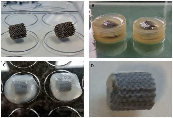
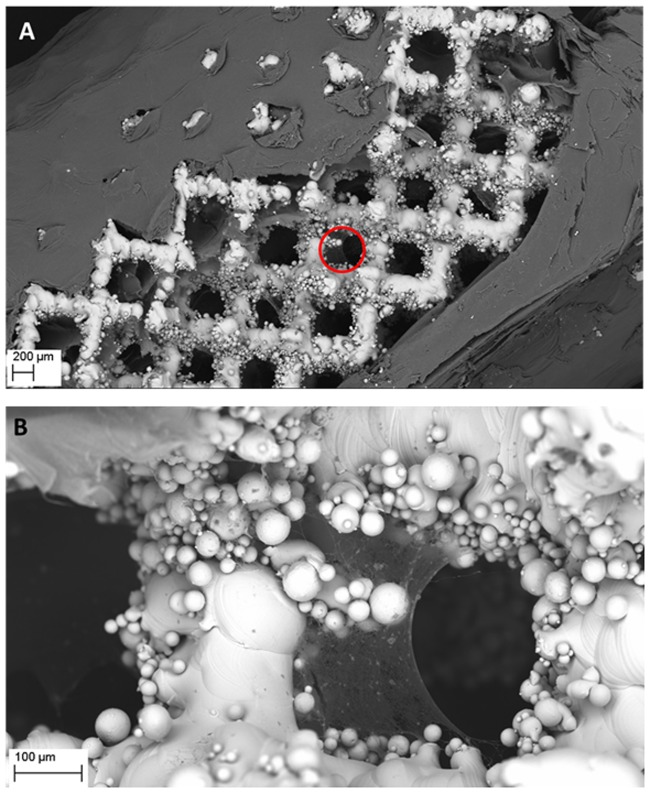
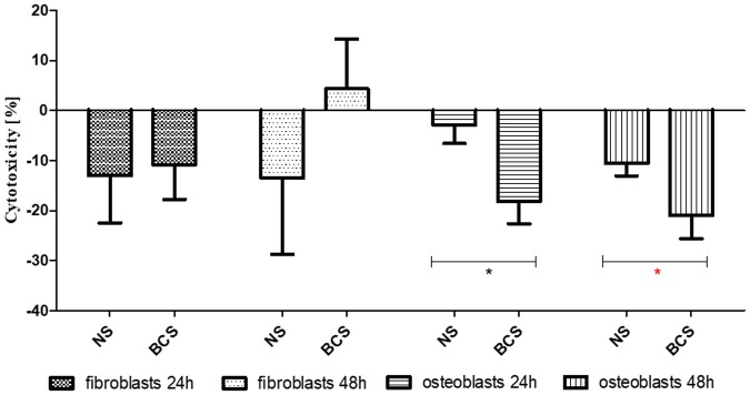
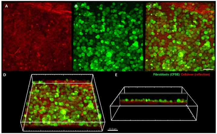
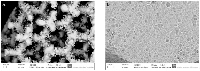
Herein we present an innovative method of coating the surface of Titanium-Aluminium-Niobium bone scaffold implants with bacterial cellulose (BC) polymer saturated with antibiotic. Customized Ti6Al7Nb scaffolds manufactured using Selective Laser Melting were immersed in a suspension of Komagataeibacter xylinus bacteria which displays an ability to produce a 3-dimensional structure of bio-cellulose polymer. The process of complete implant coating with BC took on average 7 days. Subsequently, the BC matrix was cleansed by means of alkaline lysis and saturated with gentamycin. Scanning electron microscopy revealed that BC adheres and penetrates into the implant scaffold structure. The viability and development of the cellular layer on BC micro-structure were visualized by means of confocal microscopy. The BC-coated implants displayed a significantly lower cytotoxicity against osteoblast and fibroblast cell cultures in vitro in comparison to non-coated implants. It was also noted that gentamycin released from BC-coated implants inhibited the growth of Staphylococcus aureus cultures in vitro, confirming the suitability of such implant modification for preventing hostile microbial colonization. As demonstrated using digital microscopy, the procedure used for implant coating and BC chemical cleansing did not flaw the biomaterial structure. The results presented herein are of high translational value with regard to future use of customized, BC-coated and antibiotic-saturated implants designed for use in orthopedic applications to speed up recovery and to reduce the risk of musculoskeletal infections.
Conflict of interest statement
The authors have declared that no competing interests exist.
Figures
Similar articles
-
Preparation of gentamicin-loaded electrospun coating on titanium implants and a study of their properties in vitro.Arch Orthop Trauma Surg. 2012 Jun;132(6):897-903. doi: 10.1007/s00402-012-1490-y. Epub 2012 Feb 29. Arch Orthop Trauma Surg. 2012. PMID: 22373914
-
Application of bacterial cellulose experimental dressings saturated with gentamycin for management of bone biofilm in vitro and ex vivo.J Biomed Mater Res B Appl Biomater. 2020 Jan;108(1):30-37. doi: 10.1002/jbm.b.34362. Epub 2019 Mar 18. J Biomed Mater Res B Appl Biomater. 2020. PMID: 30883023
-
Advanced biopolymer-coated drug-releasing titania nanotubes (TNTs) implants with simultaneously enhanced osteoblast adhesion and antibacterial properties.Colloids Surf B Biointerfaces. 2015 Jun 1;130:255-63. doi: 10.1016/j.colsurfb.2015.04.021. Epub 2015 Apr 18. Colloids Surf B Biointerfaces. 2015. PMID: 25944564
-
Review of titanium surface modification techniques and coatings for antibacterial applications.Acta Biomater. 2019 Jan 1;83:37-54. doi: 10.1016/j.actbio.2018.10.036. Epub 2018 Oct 26. Acta Biomater. 2019. PMID: 30541702 Review.
-
Prophylaxis and treatment of implant-related infections by antibiotic-coated implants: a review.Injury. 2006 May;37 Suppl 2:S105-12. doi: 10.1016/j.injury.2006.04.016. Injury. 2006. PMID: 16651063 Review.
Cited by
-
Antibiofilm and Antimicrobial-Enhancing Activity of Chelidonium majus and Corydalis cheilanthifolia Extracts against Multidrug-Resistant Helicobacter pylori.Pathogens. 2021 Aug 16;10(8):1033. doi: 10.3390/pathogens10081033. Pathogens. 2021. PMID: 34451497 Free PMC article.
-
The Nanofication and Functionalization of Bacterial Cellulose and Its Applications.Nanomaterials (Basel). 2020 Feb 25;10(3):406. doi: 10.3390/nano10030406. Nanomaterials (Basel). 2020. PMID: 32106515 Free PMC article. Review.
-
Evaluation of Different Methods for Cultivating Gluconacetobacter hansenii for Bacterial Cellulose and Montmorillonite Biocomposite Production: Wound-Dressing Applications.Polymers (Basel). 2020 Jan 26;12(2):267. doi: 10.3390/polym12020267. Polymers (Basel). 2020. PMID: 31991906 Free PMC article.
-
Efficacy of Bacterial Nanocellulose in Hard Tissue Regeneration: A Review.Materials (Basel). 2021 Aug 24;14(17):4777. doi: 10.3390/ma14174777. Materials (Basel). 2021. PMID: 34500866 Free PMC article. Review.
-
In Vitro Efficacy of Bacterial Cellulose Dressings Chemisorbed with Antiseptics against Biofilm Formed by Pathogens Isolated from Chronic Wounds.Int J Mol Sci. 2021 Apr 13;22(8):3996. doi: 10.3390/ijms22083996. Int J Mol Sci. 2021. PMID: 33924416 Free PMC article.
References
-
- Fux C, Stoodley P, Hall-Stoodley L, Costerton JW. Costerton, Bacterial biofilms: diagnostic and therapeutic challenge. Expert Rev Anti Infect Ther. 2003;4,667–683. - PubMed
-
- Zimmermann KA, LeBlanc JM, Sheets KT, Fox RW, Gatenholm P. Biomimetic design of a bacterial cellulose/hydroxyapatite nanocomposite for bone healing applications. Mater Sci Eng C. 2011;31(1),43–49.
-
- Chlebus E, Kuźnicka B, Kurzynowski T, Dybała B. Microstructure and mechanical behaviour of Ti-6al-7Nb alloy produced by selective laser melting. Mater Charact. 2011;62,488–495.
Publication types
MeSH terms
Substances
LinkOut - more resources
Full Text Sources
Research Materials
