. 2018 Oct 24;13(10):e0204898.
doi: 10.1371/journal.pone.0204898. eCollection 2018.

The E3 ubiquitin ligase MARCH1 regulates glucose-tolerance and lipid storage in a sex-specific manner

Affiliations

The E3 ubiquitin ligase MARCH1 regulates glucose-tolerance and lipid storage in a sex-specific manner

Candida Bhagwandin et al. PLoS One. .

Abstract

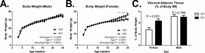
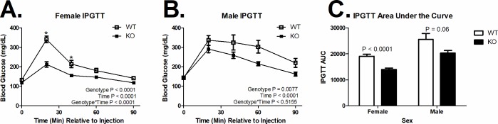
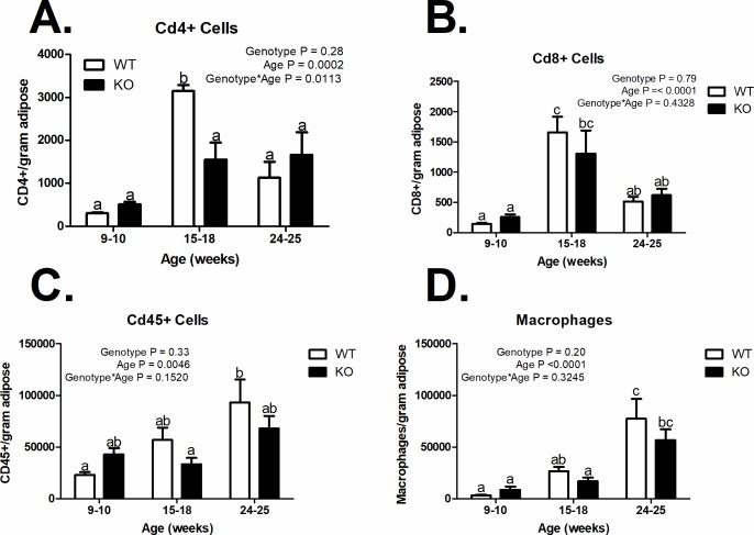
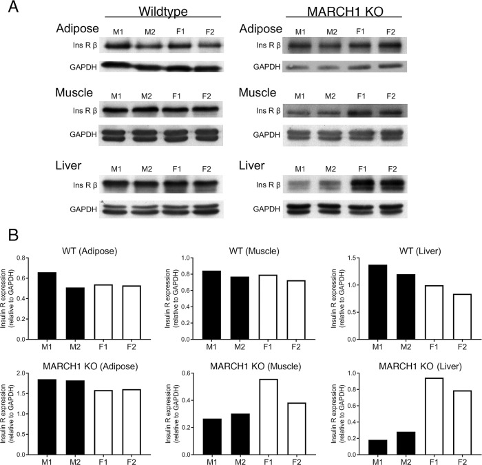
Type 2 diabetes is typified by insulin-resistance in adipose tissue, skeletal muscle, and liver, leading to chronic hyperglycemia. Additionally, obesity and type 2 diabetes are characterized by chronic low-grade inflammation. Membrane-associated RING-CH-1 (MARCH1) is an E3 ubiquitin ligase best known for suppression of antigen presentation by dendritic and B cells. MARCH1 was recently found to negatively regulate the cell surface levels of the insulin receptor via ubiquitination. This, in turn, impaired insulin sensitivity in mouse models. Here, we report that MARCH1-deficient (knockout; KO) female mice exhibit excessive weight gain and excessive visceral adiposity when reared on standard chow diet, without increased inflammatory cell infiltration of adipose tissue. By contrast, male MARCH1 KO mice had similar weight gain and visceral adiposity to wildtype (WT) male mice. MARCH1 KO mice of both sexes were more glucose tolerant than WT mice. The levels of insulin receptor were generally higher in insulin-responsive tissues (especially the liver) from female MARCH1 KO mice compared to males, with the potential to account in part for the differences between male and female MARCH1 KO mice. We also explored a potential role for MARCH1 in human type 2 diabetes risk through genetic association testing in publicly-available datasets, and found evidence suggestive of association. Collectively, our data indicate an additional link between immune function and diabetes, specifically implicating MARCH1 as a regulator of lipid metabolism and glucose tolerance, whose function is modified by sex-specific factors.


Conflict of interest statement

The authors have declared that no competing interests exist.

Figures

Fig 1
Fig 1. Body weight and visceral adiposity.
(A) Increased body weight in female mice MARCH1-deficient mice (KO) compared to wildtype mice (WT) on a chow diet after 20 weeks of age (WT n = 10–33; KO n = 12–41). (B) MARCH1-deficiency did not affect the age-related increase in body weight in male mice (WT n = 8–19; KO n = 7–28). (C) Increased visceral adiposity in female, but not male mice KO mice. Number (n) is indicated within bars). *indicates significant difference P < 0.05. Data are plotted at the mean +/- SEM.
Fig 2
Fig 2. Glucose tolerance.
(A) MARCH1 deletion (KO) decreased glucose clearance in female (WT n = 10, KO n = 12) and (B) male (WT n = 8, KO n = 7) mice. (C) Area under the glucose tolerance curve by sex and genotype (n is indicated within bars). *Indicates significant difference at that time point (P < 0.05). Data are plotted at the mean +/- SEM.
Fig 3
Fig 3. Immune cell infiltration of visceral adipose tissue.
(A) CD4+ cells (n = 3), (B) CD8+ cells (n = 3), (C) CDd45+ cells (n = 3–6), and (D) Macrophages/gram of visceral adipose tissue (n = 3–6). a, b, c = bars that share a common superscript do not differ significantly (P > 0.05). The numbers are based on flow cytometric analyses using the gating and scheme shown in S1 Fig. Data are plotted at the mean +/- SEM.
Fig 4
Fig 4. Comparison of insulin receptor levels in female versus male mice.
Proteins from adipose tissue, skeletal muscle, and liver from either WT or MARCH1 KO mice were isolated, separated by SDS-PAGE, and immunoblotted using antibodies recognizing insulin receptor β-chain and GAPDH (as a loading control). Each group consisted of two male and two female littermates. (A) Immunoblots showing the expression levels of insulin receptor β-chain (Ins R β) and GAPDH present in each sample. (B) Immunoblots were scanned by laser densitometry and band intensity was quantified. The expression level of insulin receptor β-chain was plotted relative to the amount of GAPDH present in each sample.

References

    1. Johnson Andrew MF, Olefsky Jerrold M. The Origins and Drivers of Insulin Resistance. Cell. 2013;152(4):673–84. 10.1016/j.cell.2013.01.041 - DOI - PubMed
    1. Muoio DM, Newgard CB. Mechanisms of disease: molecular and metabolic mechanisms of insulin resistance and beta-cell failure in type 2 diabetes. Nat Rev Mol Cell Biol. 2008;9(3):193–205. Epub 2008/01/18. 10.1038/nrm2327 . - DOI - PubMed
    1. Basu A, Dalla Man C, Basu R, Toffolo G, Cobelli C, Rizza RA. Effects of type 2 diabetes on insulin secretion, insulin action, glucose effectiveness, and postprandial glucose metabolism. Diabetes care. 2009;32(5):866–72. Epub 2009/02/07. 10.2337/dc08-1826 ; PubMed Central PMCID: PMCPmc2671126. - DOI - PMC - PubMed
    1. He L, Sabet A, Djedjos S, Miller R, Sun X, Hussain MA, et al. Metformin and insulin suppress hepatic gluconeogenesis through phosphorylation of CREB binding protein. Cell. 2009;137(4):635–46. Epub 2009/05/20. 10.1016/j.cell.2009.03.016 ; PubMed Central PMCID: PMCPmc2775562. - DOI - PMC - PubMed
    1. Ashcroft FM, Rorsman P. Diabetes mellitus and the beta cell: the last ten years. Cell. 2012;148(6):1160–71. Epub 2012/03/20. 10.1016/j.cell.2012.02.010 . - DOI - PMC - PubMed

Publication types

Substances