. 2018 Sep 25:2018:3649391.
doi: 10.1155/2018/3649391. eCollection 2018.

Airflow and Particle Deposition in Acinar Models with Interalveolar Septal Walls and Different Alveolar Numbers

Affiliations

Airflow and Particle Deposition in Acinar Models with Interalveolar Septal Walls and Different Alveolar Numbers

Jinxiang Xi et al. Comput Math Methods Med. .

Abstract

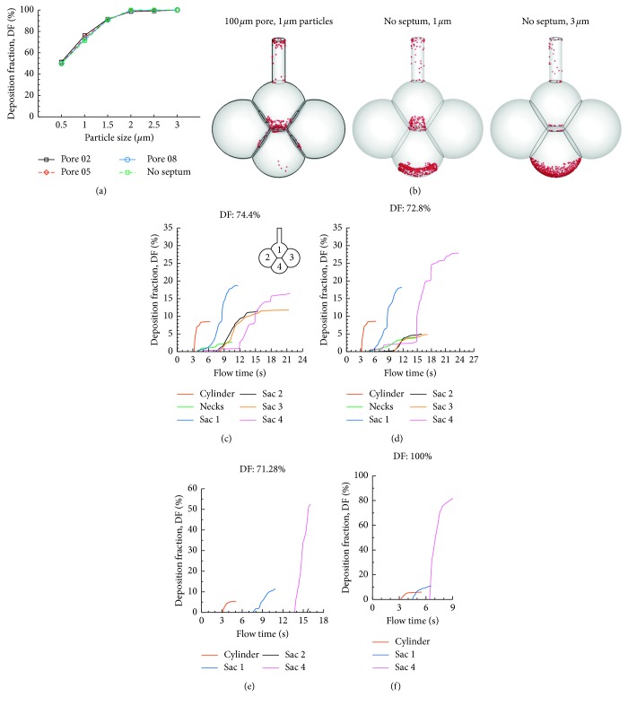
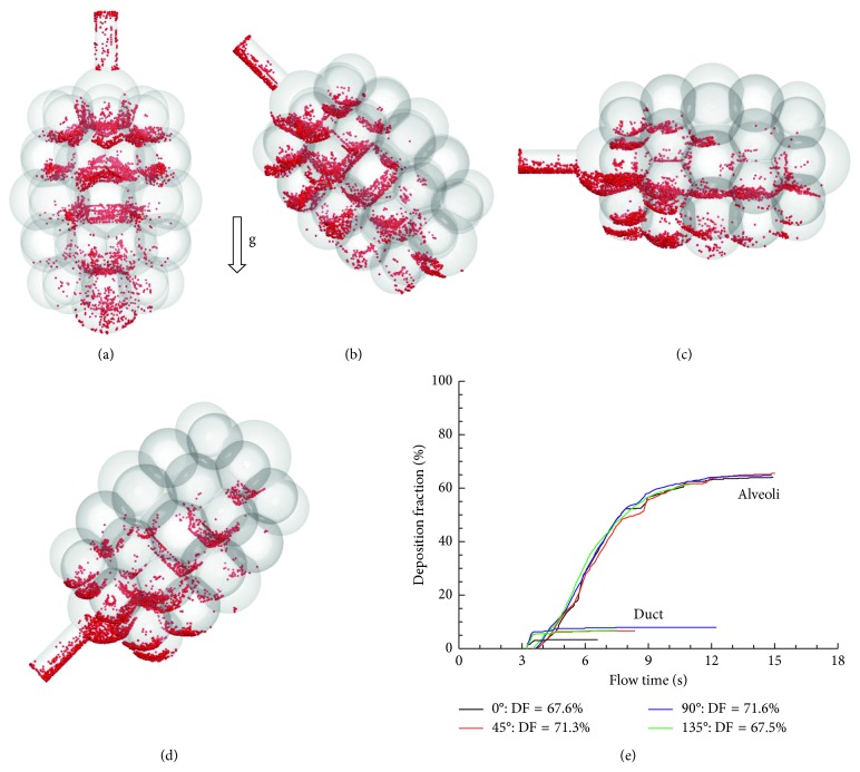
Unique features exist in acinar units such as multiple alveoli, interalveolar septal walls, and pores of Kohn. However, the effects of such features on airflow and particle deposition remain not well quantified due to their structural complexity. This study aims to numerically investigate particle dynamics in acinar models with interalveolar septal walls and pores of Kohn. A simplified 4-alveoli model with well-defined geometries and a physiologically realistic 45-alveoli model was developed. A well-validated Lagrangian tracking model was used to simulate particle trajectories in the acinar models with rhythmically expanding and contracting wall motions. Both spatial and temporal dosimetries in the acinar models were analyzed. Results show that collateral ventilation exists among alveoli due to pressure imbalance. The size of interalveolar septal aperture significantly alters the spatial deposition pattern, while it has an insignificant effect on the total deposition rate. Surprisingly, the deposition rate in the 45-alveoli model is lower than that in the 4-alveoli model, indicating a stronger particle dispersion in more complex models. The gravity orientation angle has a decreasing effect on acinar deposition rates with an increasing number of alveoli retained in the model; such an effect is nearly negligible in the 45-alveoli model. Breath-holding increased particle deposition in the acinar region, which was most significant in the alveoli proximal to the duct. Increasing inhalation depth only slightly increases the fraction of deposited particles over particles entering the alveolar model but has a large influence on dispensing particles to the peripheral alveoli. Results of this study indicate that an empirical correlation for acinar deposition can be developed based on alveolar models with reduced complexity; however, what level of geometry complexity would be sufficient is yet to be determined.


Figures

Figure 1
Figure 1
Simplified and complex multiple-alveoli models with septa and pores: (a) simplified 4-alveoli (or 4-sac) model with pores (septal apertures) of 40 μm in diameter; (b) 4-alveoli model with 100 μm pores; (c) 4-alveoli model with 160 μm pores; (d) 4-alveoli model with no spectrum; and (e) 45-alveoli model with 40 μm pores.
Figure 2
Figure 2
Airflow contour and stream traces in the 4-alveoli models with a pore size of (A) 40 µm, (B) 160 µm, and (C) no septal wall during (a) inhalation and (b) exhalation.
Figure 3
Figure 3
Instantaneous snapshots of particle positions in the 4-alveoli model during the first cycle after particle inhalation. Particles were 1 µm in diameter. Due to particle dispersion, some particles exited the geometry with expiratory airflow. (a) T = 0.25 s. (b) T = 0.50 s. (c) T = 0.75 s. (d) T = 1.00 s. (e) T = 1.25 s. (f) T = 1.75 s. (g) T = 2.00 s. (h) T = 2.25 s. (i) T = 2.50 s. (j) T = 2.75 s.
Figure 4
Figure 4
Instantaneous snapshots of particle positions in the 4-alveoli model during the second cycle after particle inhalation. All the remaining particles will deposit eventually due to particle interception or gravitational sedimentation. (a) T = 3.25 s. (b) T = 3.50 s. (c) T = 3.75 s. (d) T = 4.00 s. (e) T = 4.25 s. (f) T = 4.75 s. (g) T = 5.00 s. (h) T = 5.25 s. (i) T = 5.50 s. (j) T = 5.75 s.
Figure 5
Figure 5
Surface deposition of 1 µm particles in the 4-alveoli model with a pore size of 40 µm: (a) side view and (b) bottom view.
Figure 6
Figure 6
Cumulative deposition rate with time for 1 µm particles in the 4-alveoli model: (a) dynamic model with rhythmic wall motions and (b) static model. Each respiration cycle has a period of 3.0 s.
Figure 7
Figure 7
Comparison of particle deposition between different particle sizes in the 4-alveoli model: (a) total deposition fractions for 0.5–3 µm particles and the time resolution of sectional cumulative deposition for (b) 0.5 µm, (c) 1 µm, and (d) 2 µm. Deposition was quantified in each section of the model, that is, cylinder, the four alveoli (sacs), and the interalveolar pores (necks).
Figure 8
Figure 8
Comparison of the cumulative deposition with time for different gravity orientation angles in the 4-alveoli model: (a) 0°, (b) 45°, (c) 90°, and (d) 135° counterclockwise from the gravity.
Figure 9
Figure 9
Comparison of total deposition rates for different breath-holding periods in the 4-alveoli model: (a) total deposition and (b) temporal deposition for 4 s breath-holding.
Figure 10
Figure 10
Comparison of particle deposition rates between different inhalation depths in the 4-alveoli model: (a) total deposition at different tidal volumes (VT) and the time resolution of sectional cumulative deposition for (b) 2 standard VT (i.e., volume expansion factor: 0.466), (c) 3 standard VT (i.e., volume expansion factor: 0.699), and (d) 4 VT (i.e., volume expansion factor: 0.932). Each respiration cycle has a period of 3.0 s.
Figure 11
Figure 11
Pore size effects on alveolar deposition in the 4-alveoli model: (a) total deposition rates in the alveoli with different pore sizes, (b) surface deposition in different alveoli models, and the time resolution of sectional cumulative deposition for (c) pore size: 100 µm, particles: 1 µm, (d) pore size: 160 µm, particles: 1 µm, (e) no septum, particles: 1 µm, and (f) no septum, particles: 3 µm.
Figure 12
Figure 12
Airflow and particle deposition in the 45-alveoli model: (a) airflow, (b) surface deposition, (c) comparison of deposition rate between the 45-alveoli model and 4-alveoli and single-alveolus models, and (d) deposition with time.
Figure 13
Figure 13
Comparison of the cumulative deposition with time for different gravity orientation angles in the 45-alveoli model: (a) 0°, (b) 45°, (c) 90°, and (d) 135° counterclockwise from the gravity, and (e) deposition.
Figure 14
Figure 14
Comparison of particle deposition with different breath depths in the 45-alveoli model: (a) total deposition vs. inhalation depth and the time resolution of sectional cumulative deposition for (b) 0.5 standard VT (i.e., volume expansion factor: 0.117), (c) 0.75 standard VT (i.e., volume expansion factor: 0.175), (d) 1.05 standard VT (i.e., volume expansion factor: 0.245), and (e) 1.1 standard VT (i.e., volume expansion factor: 0.256).

Similar articles

Cited by

References

    1. Ochs M., Nyengaard J. R., Jung A., et al. The number of alveoli in the human lung. American Journal of Respiratory and Critical Care Medicine. 2004;169(1):120–124. doi: 10.1164/rccm.200308-1107oc. - DOI - PubMed
    1. Desplechain C., Foliguet B., Barrat E., Grignon G., Touati F. The pores of Kohn in pulmonary alveoli. Bulletin Européen de Physiopathologie Respiratoire. 1983;19(1):59–68. - PubMed
    1. Terry P. B., Traystman R. J. The clinical significance of collateral ventilation. Annals of the American Thoracic Society. 2016;13(12):2251–2257. doi: 10.1513/annalsats.201606-448fr. - DOI - PMC - PubMed
    1. Namati E., Thiesse J., de Ryk J., McLennan G. Alveolar dynamics during respiration: are the pores of Kohn a pathway to recruitment? American Journal of Respiratory Cell and Molecular Biology. 2008;38(5):572–578. doi: 10.1165/rcmb.2007-0120oc. - DOI - PMC - PubMed
    1. Nagai A., Inano H., Matsuba K., Thurlbeck W. M. Scanning electronmicroscopic morphometry of emphysema in humans. American Journal of Respiratory and Critical Care Medicine. 1994;150(5):1411–1415. doi: 10.1164/ajrccm.150.5.7952569. - DOI - PubMed

LinkOut - more resources