. 2018 Oct 9:9:1383.
doi: 10.3389/fphys.2018.01383. eCollection 2018.

Profibrotic, Electrical, and Calcium-Handling Remodeling of the Atria in Heart Failure Patients With and Without Atrial Fibrillation

Affiliations

Profibrotic, Electrical, and Calcium-Handling Remodeling of the Atria in Heart Failure Patients With and Without Atrial Fibrillation

Cristina E Molina et al. Front Physiol. .

Abstract

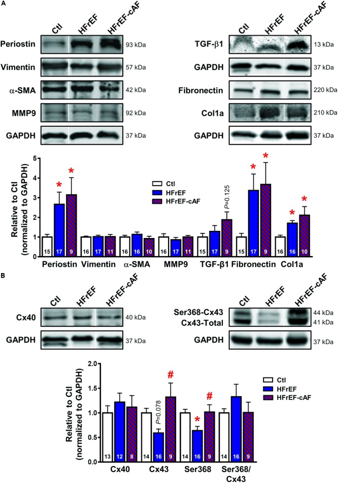
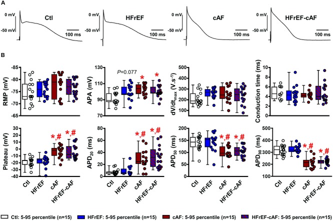
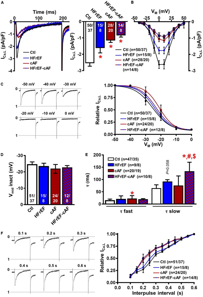
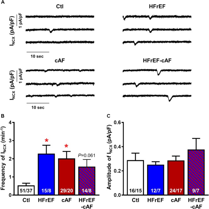
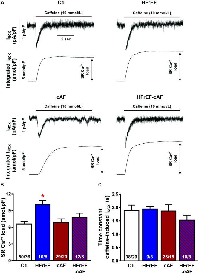
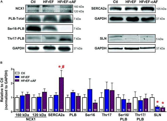
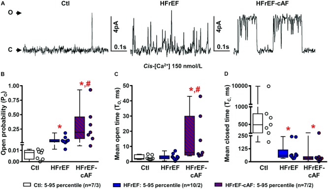
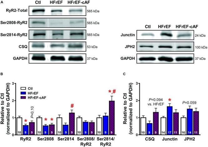
Atrial fibrillation (AF) and heart failure (HF) are common cardiovascular diseases that often co-exist. Animal models have suggested complex AF-promoting atrial structural, electrical, and Ca2+-handling remodeling in the setting of HF, but data in human samples are scarce, particularly regarding Ca2+-handling remodeling. Here, we evaluated atrial remodeling in patients with severe left ventricular (LV) dysfunction (HFrEF), long-standing persistent ('chronic') AF (cAF) or both (HFrEF-cAF), and sinus rhythm controls with normal LV function (Ctl) using western blot in right-atrial tissue, sharp-electrode action potential (AP) measurements in atrial trabeculae and voltage-clamp experiments in isolated right-atrial cardiomyocytes. Compared to Ctl, expression of profibrotic markers (collagen-1a, fibronectin, periostin) was higher in HFrEF and HFrEF-cAF patients, indicative of structural remodeling. Connexin-43 expression was reduced in HFrEF patients, but not HFrEF-cAF patients. AP characteristics were unchanged in HFrEF, but showed classical indices of electrical remodeling in cAF and HFrEF-cAF (prolonged AP duration at 20% and shorter AP duration at 50% and 90% repolarization). L-type Ca2+ current (ICa,L) was significantly reduced in HFrEF, cAF and HFrEF-cAF, without changes in voltage-dependence. Potentially proarrhythmic spontaneous transient-inward currents were significantly more frequent in HFrEF and HFrEF-cAF compared to Ctl, likely resulting from increased sarcoplasmic reticulum (SR) Ca2+ load (integrated caffeine-induced current) in HFrEF and increased ryanodine-receptor (RyR2) single-channel open probability in HFrEF and HFrEF-cAF. Although expression and phosphorylation of the SR Ca2+-ATPase type-2a (SERCA2a) regulator phospholamban were unchanged in HFrEF and HFrEF-cAF patients, protein levels of SERCA2a were increased in HFrEF-cAF and sarcolipin expression was decreased in both HFrEF and HFrEF-cAF, likely increasing SR Ca2+ uptake and load. RyR2 protein levels were decreased in HFrEF and HFrEF-cAF patients, but junctin levels were higher in HFrEF and relative Ser2814-RyR2 phosphorylation levels were increased in HFrEF-cAF, both potentially contributing to the greater RyR2 open probability. These novel insights into the molecular substrate for atrial arrhythmias in HF-patients position Ca2+-handling abnormalities as a likely trigger of AF in HF patients, which subsequently produces electrical remodeling that promotes the maintenance of the arrhythmia. Our new findings may have important implications for the development of novel treatment options for AF in the context of HF.

Keywords: Ca2+ handling; atrial fibrillation; heart failure; heart failure with reduced ejection fraction; human atrial cardiomyocytes.


Figures

FIGURE 1
FIGURE 1
Atrial profibrotic and connexin remodeling. (A) Representative Western blot examples (top) and quantification of protein expression (bottom; mean ± SEM) of periostin, vimentin, α-smooth muscle actin (α-SMA), matrix metallopeptidase 9 (MMP9), transforming growth factor-β1 (TGF-β1), fibronectin and collagen 1α (col1a) in right-atrial tissue homogenates from Ctl (white bars), HFrEF (blue bars) or HFrEF-cAF (red/blue-striped bars) patients. Vertical white lines separate non-adjacent lanes on the same blot. (B) Representative Western blot examples (top) and quantification of protein expression (bottom; mean ± SEM) of total connexin-40 (Cx40), total and Ser368-phosphorylated connexin-43 (Cx43). GAPDH was used as loading control and is shown for the samples used for Western blots of periostin, vimentin, α-SMA and MMP9, for TGF-β1, and for fibronectin and col1a. Numbers in bars indicate number of patients. indicates P < 0.05 vs. Ctl, # indicates P < 0.05 vs. HFrEF.
FIGURE 2
FIGURE 2
Electrical remodeling in multicellular human atrial trabeculae. (A) Representative action potential (AP) recordings in multicellular preparations from Ctl, HFrEF, cAF or HFrEF-cAF patients (from left to right). (B) Quantification of AP parameters in Ctl (white bars), HFrEF (blue bars), cAF (red bars) or HFrEF-cAF (red/blue-striped bars) patients. Top row shows (from left to right): resting membrane potential (RMP), AP amplitude (APA), maximal AP upstroke velocity (dV/dtmax) and conduction time (note different n-numbers). Bottom row shows (from left to right): level of AP plateau and AP duration (APD) at 20%, 50%, and 90% of repolarization. indicates P < 0.05 vs. Ctl, # indicates P < 0.05 vs. HFrEF. Numbers indicate number of patients.
FIGURE 3
FIGURE 3
L-type Ca2+ current (ICa,L) measurements in isolated human atrial cardiomyocytes. (A) Representative ICa,L recording during a 200-ms depolarizing pulse to +10 mV in a Ctl (black), HFrEF (blue), cAF (red) or HFrEF-cAF (blue/red) atrial cardiomyocyte (left) and quantification of ICaL amplitude (right). (B) Current-voltage relationship of peak ICa,L. (C) Representative examples of ICa,L inactivation protocol (left) and voltage dependence of inactivation (right) (D) midpoint of inactivation in Ctl, HFrEF, cAF and HFrEF-cAF patients. (E) Fast and slow time constants of inactivation during a depolarization to +10 mV in Ctl, HFrEF, cAF and HFrEF-cAF patients. (F) Representative examples (left) and quantification (right) of ICa,L recovery from inactivation with various interpulse intervals. Numbers in bars indicate number of cells/number of patients. indicates P < 0.05 vs. Ctl, # indicates P < 0.05 vs. HFrEF, $ indicates P < 0.05 vs. cAF.
FIGURE 4
FIGURE 4
Spontaneous Ca2+-release events in isolated human atrial cardiomyocytes. (A) Representative recordings of NCX-mediated transient-inward current (INCX) in atrial cardiomyocytes from Ctl, HFrEF, cAF or HFrEF-cAF patients. (B,C) Frequency (B) and amplitude (C) of spontaneous INCX in the four groups. Numbers in bars indicate number of cells/number of patients. indicates P < 0.05 vs. Ctl.
FIGURE 5
FIGURE 5
Sarcoplasmic reticulum (SR) Ca2+ load in isolated human atrial cardiomyocytes. (A) Representative recordings of NCX-mediated inward current (INCX) induced by caffeine (10 mmol/l) application in atrial cardiomyocytes from Ctl, HFrEF, cAF or HFrEF-cAF patients (top panels). The bottom panels show the integral of membrane current over time, the amplitude of which is an accepted index of SR Ca2+ load. (B,C) SR Ca2+ load (B) and time constant of caffeine-induced INCX (an indicator of NCX function) in the four groups. Numbers in bars indicate number of cells/number of patients. indicates P < 0.05 vs. Ctl.
FIGURE 6
FIGURE 6
Molecular determinants of increased sarcoplasmic reticulum (SR) Ca2+ load. (A) Representative Western blot examples of protein expression of Na+-Ca2+-exchanger type-1 (NCX1; full-length protein of 160 kDa with proteolytic fragment at 120 kDa), SR Ca2+-ATPase type-2a (SERCA2a) and its inhibitory regulators phospholamban (PLB; total PLB as well as Ser16- and Thr17-phosphorylated PLB) and sarcolipin (SLN). GAPDH was used as loading control. Ser16-PLB of HFrEF-cAF represents an extreme example. (B) Quantification of NCX1, SERCA2a, total PLB, Ser16- and Thr17-phosphorylated PLB, Ser16/total PLB, Thr17/total PLB, and SLN in right-atrial tissue homogenates of Ctl (white bars), HFrEF (blue bars) or HFrEF-cAF (red/blue-striped bars). Numbers in bars indicate number of patients. indicates P < 0.05 vs. Ctl, # indicates P < 0.05 vs. HFrEF.
FIGURE 7
FIGURE 7
Single-channel recordings of cardiac ryanodine receptor type-2 (RyR2) channels. (A) Representative RyR2 single-channel recording in a right-atrial sample from a Ctl, HFrEF or HFrEF-cAF patient. Upward deflections reflect channel opening. (B–D) Quantification of open probability (B), mean open time (C), and mean closed time (D) of single RyR2 channels from Ctl (white bars/symbols), HFrEF (blue bars/symbols) or HFrEF-cAF (red/blue-striped bars/symbols) patients. Numbers represent number of channels/number of patients. indicates P < 0.05 vs. Ctl, # indicates P < 0.05 vs. HFrEF.
FIGURE 8
FIGURE 8
Molecular determinants of ryanodine receptor type-2 (RyR2) dysfunction. (A) Representative Western blot examples of total RyR2, Ser2808-, and Ser2814-phosphorylated RyR2 and calsequestrin (CSQ; left), as well as junctin and junctophilin-2 (JPH2; right) protein expression levels. GAPDH was used as loading control. (B) Quantification of total RyR2, Ser2808-, and Ser2814-phosphorylated RyR2, Ser2808/total RyR2, and Ser2814/total RyR2 protein expression levels in Ctl (white bars), HFrEF (blue bars) and HFrEF-cAF (red/blue-striped bars) patients. (C) Protein expression levels of the RyR2-interacting proteins CSQ, junctin, and JPH2 in Ctl, HFrEF and HFrEF-cAF patients. Data are shown relative to Ctl. Numbers in bars indicate number of patients. indicates P < 0.05 vs. Ctl, # indicates P < 0.05 vs. HFrEF.

References

    1. Akkaya M., Higuchi K., Koopmann M., Damal K., Burgon N. S., Kholmovski E., et al. (2013). Higher degree of left atrial structural remodeling in patients with atrial fibrillation and left ventricular systolic dysfunction. J. Cardiovasc. Electrophysiol. 24 485–491. 10.1111/jce.12090 - DOI - PubMed
    1. Andrade J., Khairy P., Dobrev D., Nattel S. (2014). The clinical profile and pathophysiology of atrial fibrillation: relationships among clinical features, epidemiology, and mechanisms. Circ. Res. 114 1453–1468. 10.1161/CIRCRESAHA.114.303211 - DOI - PubMed
    1. Beavers D. L., Wang W., Ather S., Voigt N., Garbino A., Dixit S. S., et al. (2013). Mutation E169K in junctophilin-2 causes atrial fibrillation due to impaired RyR2 stabilization. J. Am. Coll. Cardiol. 62 2010–2019. 10.1016/j.jacc.2013.06.052 - DOI - PMC - PubMed
    1. Benjamin E. J., Virani S. S., Callaway C. W., Chamberlain A. M., Chang A. R., Cheng S., et al. (2018). Heart disease and stroke statistics-2018 update: a report from the American heart association. Circulation 137 e67–e492. 10.1161/CIR.0000000000000558 - DOI - PubMed
    1. Brundel B. J., Henning R. H., Kampinga H. H., Van Gelder I. C., Crijns H. J. (2002). Molecular mechanisms of remodeling in human atrial fibrillation. Cardiovasc. Res. 54 315–324. - PubMed