. 2018 Oct 9:9:2248.
doi: 10.3389/fimmu.2018.02248. eCollection 2018.

Inflammatory Potential of Four Different Phases of Calcium Pyrophosphate Relies on NF-κB Activation and MAPK Pathways

Affiliations

Inflammatory Potential of Four Different Phases of Calcium Pyrophosphate Relies on NF-κB Activation and MAPK Pathways

Laure Campillo-Gimenez et al. Front Immunol. .

Abstract

Background: Calcium pyrophosphate (CPP) microcrystal deposition is associated with wide clinical phenotypes, including acute and chronic arthritis, that are interleukin 1β (IL-1β)-driven. Two CPP microcrystals, namely monoclinic and triclinic CPP dihydrates (m- and t-CPPD), have been identified in human tissues in different proportions according to clinical features. m-CPP tetrahydrate beta (m-CPPTβ) and amorphous CPP (a-CPP) phases are considered as m- and t-CPPD crystal precursors in vitro. Objectives: We aimed to decipher the inflammatory properties of the three crystalline phases and one amorphous CPP phase and the intracellular pathways involved. Methods: The four synthesized CPP phases and monosodium urate crystals (MSU, as a control) were used in vitro to stimulate the human monocytic leukemia THP-1 cell line or bone marrow-derived macrophages (BMDM) isolated from WT or NLRP3 KO mice. The gene expression of pro- and anti-inflammatory cytokines was evaluated by quantitative PCR; IL-1β, IL-6 and IL-8 production by ELISA; and mitogen-activated protein kinase (MAPK) activation by immunoblot analysis. NF-κB activation was determined in THP-1 cells containing a reporter plasmid. In vivo, the inflammatory potential of CPP phases was assessed with the murine air pouch model via cell analysis and production of IL-1β and CXCL1 in the exudate. The role of NF-κB was determined by a pharmacological approach, both in vivo and in vitro. Results:In vitro, IL-1β production induced by m- and t-CPPD and m-CPPTβ crystals was NLRP3 inflammasome dependent. m-CPPD crystals were the most inflammatory by inducing a faster and higher production and gene expression of IL-1β, IL-6, and IL-8 than t-CPPD, m-CPPTβ and MSU crystals. The a-CPP phase did not show an inflammatory property. Accordingly, m-CPPD crystals led to stronger activation of NF-κB, p38, extracellular signal-regulated kinase 1/2 (ERK1/2) and c-Jun N-terminal kinase (JNK) MAPKs. Inhibition of NF-κB completely abrogated IL-1β and IL-8 synthesis and secretion induced by all CPP crystals. Also, inhibition of JNK and ERK1/2 MAPKs decreased both IL-1β secretion and NF-κB activation induced by CPP crystals. In vivo, IL-1β and CXCL1 production and neutrophil infiltration induced by m-CPPD crystals were greatly decreased by NF-κB inhibitor treatment. Conclusion: Our results suggest that the inflammatory potential of different CPP crystals relies on their ability to activate the MAPK-dependent NF-κB pathway. Studies are ongoing to investigate the underlying mechanisms.

Keywords: CPP crystals; IL-1β; MAP kinase signaling; NF-κB pathway; NLRP3 inflammasome; macrophages; microcrystal-induced arthritis.


Figures
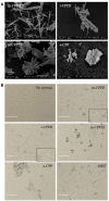
Figure 1
Figure 1
Physicochemical and morphological characteristics of the four CPP phases. (A) SEM micrographs of the four synthetic pure CPP phases tested in this study (scale bar = 2 μm). (B) Optical micrographs of THP-1/crystal culture by EVOS system microscopy (X20).
Figure 2
Figure 2
Kinetics of IL-1β, IL-8, and IL-6 production in response to CPP phases. THP-1 cells were primed the day before experiments. Cells were harvested at different times of crystal stimulation as indicated. (A) IL-1β concentration was assessed in supernatant after 6-h stimulation with different doses of crystals. (B–D) Cells were stimulated with 200 μg/ml of different crystals and, at the indicated time, supernatants were collected to quantify IL-1β (B), IL-8 (C), and IL-6 (D) concentrations (n = 3). Kruskal-Wallis test with FDR correction between crystal and PBS (#) or between CPP crystals (*). #,*p < 0.05; ##,**p < 0.01; ###,***p < 0.001.
Figure 3
Figure 3
Differential effects of CPP phases on the inflammatory response in vitro. THP-1 were primed the day before experiments. After 6 h of crystal stimulation, supernatants, and total RNA were collected. (A) IL-1β (n = 10) and IL-8 (n = 3) concentrations were quantified from cell supernatants by ELISA. Kruskal-Wallis test with FDR correction between crystals and PBS (#) or between CPP crystals (*); (B) Correlation analysis between mean of IL-1β/IL-8 concentrations measured at 6 h after crystal stimulation and specific surface area (S.S.A) of each CPP phase. Spearman t-test. (C,D) THP-1 cells were lysed for quantification of IL-1β, IL-6, IL-8, and TNF-α (n = 10) and IL-1RA, IL-10, IL-37, and TGF-β (n = 3) mRNA expression by qPCR. Kruskal-Wallis test with FDR correction between crystal and PBS (#) or between CPP phases (*). #,*p < 0.05; ##,**p < 0.01; ###,***p < 0.001.
Figure 4
Figure 4
CPP crystals differentially trigger inflammatory response in vivo. Air pouches were created by two injections of 3 ml of sterile air at day 0 and day 3. At day 6, crystals or PBS were injected into the air pouches and inflammation was assessed 6 and 24 h after. (A–C) From the air pouch lavage, number of infiltrated cells was evaluated by cell counting with Trypan blue exclusion (A); proportion of recruited neutrophils (PMN) and macrophages among total cells was determined by flow cytometry after staining with anti-Ly6G-PE and F4/80-APC monoclonal antibodies (mAbs), respectively (B); IL-1β and CXCL1 concentrations were quantified by ELISA (C). Two-way ANOVA with FDR correction between crystal and PBS at 6 h (#) or m-CPPD and other crystals ($) or between 6 and 24 h (*). #, $,*p < 0.05; ##, $$,**p < 0.01; ###, $$$,***p < 0.001. (D) Correlation analysis between cell count and IL-1β (Left) or CXCL1 level (Middle) or between IL-1β and CXCL1 levels (Right), at 6 h of crystal stimulation. Spearman test, *p < 0.05; **p < 0.01; ***p < 0.001.
Figure 5
Figure 5
CPP crystal-induced IL-1β production is NLRP3-dependent. (A) Human IL-1β concentration from PMA-primed THP-1 cell cultures after 6 h stimulation by different crystals in absence (PBS) or presence of caspase 1 inhibitor (iCasp1, 10 μM, n = 5). Two-way ANOVA test with FDR correlation between PBS and iCasp1. (B) Mouse IL-1β and CXCL-1 concentrations from wild type (wt, Left) and nlrp3−/− (Right) BMDM cultures (n = 6) after 6 h stimulation by different crystals. 2-way ANOVA test with FDR correlation between crystal and PBS (#) or between CPP crystals (*). #,*p < 0.05; ##,**p < 0.01; ###,***p < 0.001. (C) mRNA quantification of NLRP3, ASC and pro-Casp1 gene expression from PMA-primed THP-1 cells stimulated by different crystals. Kruskal-Wallis test with FDR correlation between crystals and PBS (#) or between CPP crystals (*). #,*p < 0.05; ##,**p < 0.01; ###,***p < 0.001. (D) Immunoblot analysis of NLRP3 protein expression from primed THP-1 cells or BMDM isolated from wt or nlrp3−/− mice (box in B) after 6 h of crystal stimulation; one representative of 3 independent experiments.
Figure 6
Figure 6
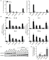
NF-κB regulates CPPD microcrystal-induced inflammation. Cells were primed the day before experiments. (A–C) 6 h after crystal stimulation of THP-1 cells, in the absence (PBS) or presence of NF-κB inhibitor (BAY-11-7085 [iNF-κB], 10 μM), supernatants were collected to quantify IL-1β (n = 5) and IL-8 (n = 3) concentrations (A), and cells were lysed for analyzing IL-1β, IL-6, IL-8, and TNF-α gene expression by qPCR (n = 4) (B) or proIL-1β protein expression by western blot analysis (one experiment representative of three) (C). Two-way ANOVA test with FDR correction: *p < 0.05; **p < 0.01; ***p < 0.001 (iNF-κB compared to PBS). (D) 8 h after crystal stimulation, supernatants of THP-1-Lucia cells were collected to determine the activity of secreted luciferase. Results are reported as relative NF-κB activity (n = 8); Kruskal-Wallis test with FDR correction between crystal and PBS (#) or between CPP crystals (*). #*p < 0.05; ##**p < 0.01; ###***p < 0.001. HKLM, heat-killed Listeria monocytogenes.
Figure 7
Figure 7
NF-κB regulates m-CPPD microcrystal-induced inflammation in vivo. Six hour after PBS or m-CPPD injection into the air pouches, in absence (PBS) or in presence of iNF-κB, the air pouch exudates were collected for analysis. (A) The number of total infiltrated cells was evaluated by cell counting with Trypan blue exclusion. (B,C) the absolute number (Left) and proportion (Right) of recruited neutrophils (PMN) and macrophages (Mø) among total cells was determined by flow cytometry after staining with anti-Ly6G-PE and F4/80-APC mAbs, respectively. (D) IL-1β and CXCL1 levels were quantified by ELISA. Kruskal-Wallis test with FDR correction between crystal and PBS (#) or between CPP crystals (*). #*p < 0.05; ##**p < 0.01; ###***p < 0.001.
Figure 8
Figure 8
CPPD microcrystals activate NF-κB and induce inflammation via MAPK pathways. Cells were primed the day before experiments. (A) THP-1 cells were incubated for 30 min with crystals then lysed for immunoblot analysis of p38, p42/44, and JNK MAPK phosphorylation (one experiment representative of three). (B–D) Cells were stimulated with crystals in the absence (PBS) or presence of p38 (SB203580 (ip38), 10 μM), JNK (SP600125 (iJNK), 10 μM) or p42/44 (PD98059 (ip42/44), 100 μM) inhibitors. (B) THP-1-Lucia cell supernatants were collected at 8 h for determining luciferase activity (n = 4). HKLM, heat-killed Listeria monocytogenes. (C,D) THP-1 supernatants were collected at 6 h (C: ip42/44 and iJNK) or at 2 h (D: ip38) to quantify IL-1β and IL-8 levels by ELISA (n = 4). Multiple t-test with FDR correction PBS and MAPK inhibitors (*). *p < 0.05; **p < 0.01; ***p < 0.001.
Figure 9
Figure 9
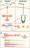
Schematic representation of the inflammatory potential of CPP crystals relying on both NF-κB (priming signal) and NLRP3 inflammasome (maturation signal) activation. (A) In our in vitro model of macrophages (THP-1 or BMDMs), uncoated CPP crystals act in synergy (red arrows) with PMA or LPS used for cell differentiation and activation. We showed that CPP crystals amplify NF-κB activation through a MAPK-dependent signaling pathways leading to an increased production of pro-inflammatory cytokines including TNF-α, IL-8, IL-6, and pro-IL-1β (A, left: priming signal). CPP crystals also enhance NLRP3 production and activation that induces ASC and pro-caspase-1 oligomerization (NLRP3 assembly) leading to caspase-1 maturation and subsequently cleavage of pro-IL-1β into active IL-1β (A, right: maturation signal). (B) CPP crystals possess different inflammatory properties with m-CPPD crystals being the most phlogistic ones followed by t-CPPD and m-CPPTβ, while a-CPP phase does not harbor inflammatory response. The inflammatory potential of each crystal depends on their ability to differentially activate the MAPK/NF-κB pathways and NLRP3 inflammasome complex. How CPP crystals differentially modulate NF-κB and NLRP3 remains unknown.

References

    1. Richette P, Bardin T, Doherty M. An update on the epidemiology of calcium pyrophosphate dihydrate crystal deposition disease. Rheumatology (2009) 48:711–5. 10.1093/rheumatology/kep081 - DOI - PubMed
    1. Ryu K, Iriuchishima T, Oshida M, Kato Y, Saito A, Imada M, et al. . The prevalence of and factors related to calcium pyrophosphate dihydrate crystal deposition in the knee joint. Osteoarthr Cartil. (2014) 22:975–9. 10.1016/j.joca.2014.04.022 - DOI - PubMed
    1. Balić-Žunić T, Christoffersen MR, Christoffersen J. Structure of the β form of calcium pyrophosphate tetrahydrate. Acta Crystallogr. B (2000) 56:953–8. 10.1107/S0108768100009447 - DOI - PubMed
    1. Gras P, Rey C, Marsan O, Sarda S, Combes C. Synthesis and characterisation of hydrated calcium pyrophosphate phases of biological interest. Eur J Inorg Chem. (2013) 2013:5886–95. 10.1002/ejic.201300955 - DOI
    1. Zhang W, Doherty M, Bardin T, Barskova V, Guerne P-A, Jansen TL, et al. . European League Against Rheumatism recommendations for calcium pyrophosphate deposition. Part I: terminology and diagnosis. Ann Rheum Dis. (2011) 70:563–70. 10.1136/ard.2010.139105 - DOI - PubMed

Publication types

MeSH terms