. 2018 Oct 2:9:1388.
doi: 10.3389/fphys.2018.01388. eCollection 2018.

A Theoretical Model: Elastic Analysis of the Evolution of the Crypt Opening Between the Fundic Gland and the Pyloric Gland

Affiliations

A Theoretical Model: Elastic Analysis of the Evolution of the Crypt Opening Between the Fundic Gland and the Pyloric Gland

Fei Xiong et al. Front Physiol. .

Abstract

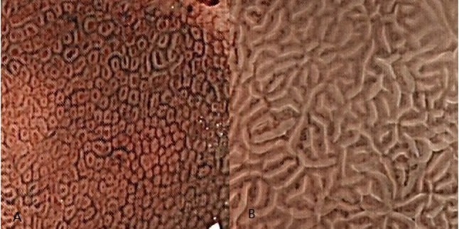
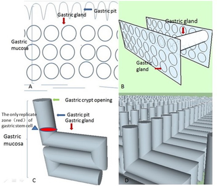
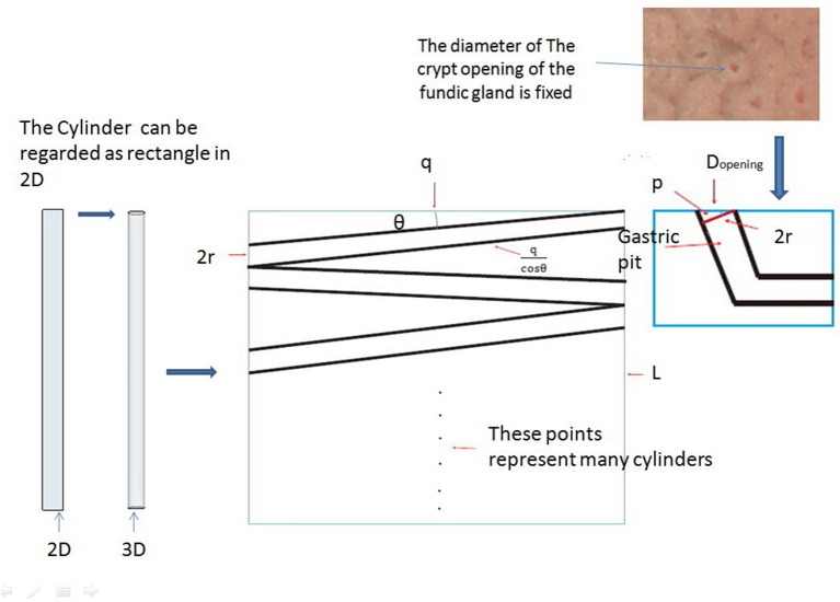
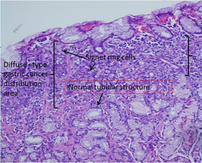
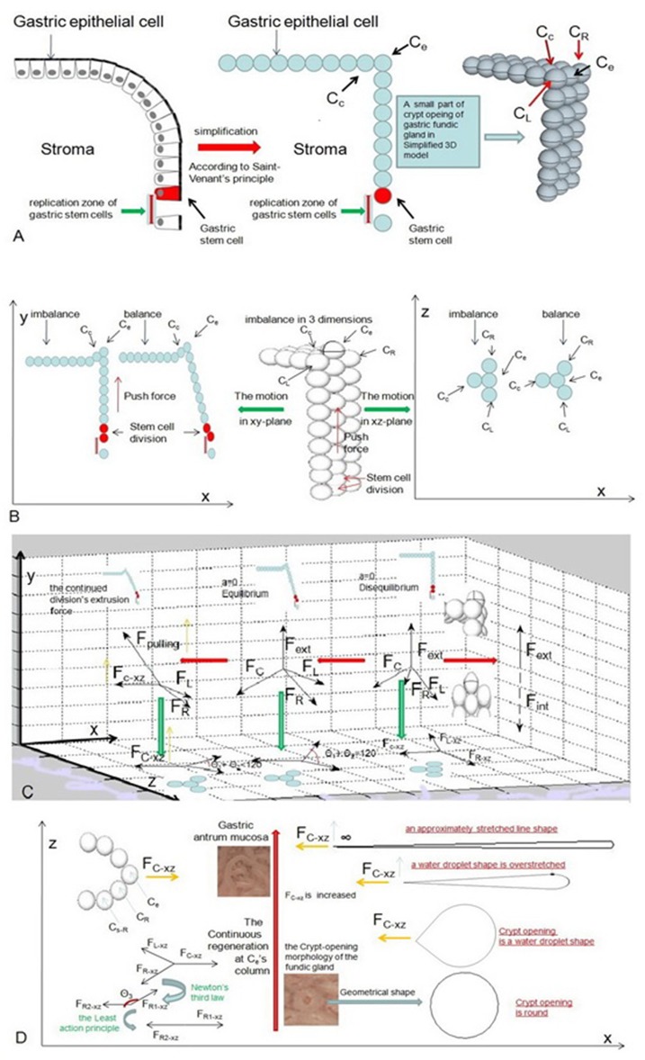
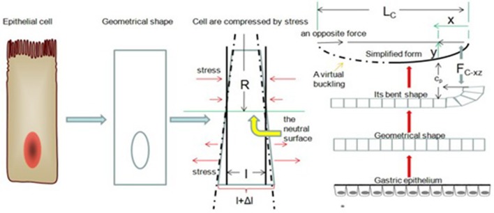
In recent years, with the development of magnified endoscopic technology, the microstructure of the gastric mucosa surface has been widely studied. However, it is unclear why the crypt opening shape of the fundic gland is different from that of the pyloric gland. We attempted to explain the problem by means of physical concepts, mathematical tools and some pathological perspectives. We first constructed an "L" type tubular structure on the basis of the pathology of the gastric mucosa and some geometric principles and then analyzed the distortion of marginal crypt epithelia after we added cells in the model via the mechanism of continuous regeneration. Finally, we determined that the crypt opening shape of the pyloric gland is derived mathematically from that of the fundic gland with the help of the idea of the Riemann sum. According to the derivation of the Euler force, it is possible that the epithelial-mesenchymal transition (EMT) protects the integrity of the gastric mucosa. Our model suggests that the evolution of the fundic gland and the pyloric gland triggers the EMT via elastic deformation. The basic logic of our model is the principle of least action.

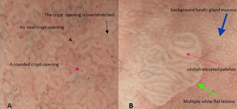
Keywords: crypt opening; epithelial-mesenchymal transition; fundic gland; multiple white flat lesions; the Euler force.


Figures

Figure 1
Figure 1
The Crypt opening morphology of the gastric mucosa. These images were taken using magnified endoscopic technology with narrow-band imaging (NBI-ME). (A) The subepithelial capillary network (SECN) looks like a honeycomb in the gastric body, and we can see many approximate round or oval dots in the middle of the honeycombs. These black dots are the crypt openings of fundic glands. (B) Although we cannot see the crypt opening in the gastric antrum mucosa, gastric crypt openings are approximately linear or have a reticular groove based on NBI-ME and pathological examination. These openings are hidden between marginal crypt epithelia (white stripes).
Figure 2
Figure 2
These four conceptual diagrams show the derivation process of these stereoscopic structures of gastric glands in an ideal state based on the principle of least action. (A) According to isoperimetric inequality, we delineated an ideal geometrical morphology of the gastric gland in 2D space. (B) Let us suppose there are many similar 2D spaces (like Figure 2A) overlapping along a straight line that is parallel to the gastric mucosal surface, which gives us many cylinders. From the least action principle, these cylinders are parallel to the gastric mucosal surface. (C) When we unite the geometric shapes of the gastric body's gastric pits with these cylinders, we can obtain an “L” type tubular structure of the gastric gland in the ideal state. (D) The “L” type tubular structure of the gastric gland fills the whole mucosa in the ideal state. Its real state is disorganized and chaotic. The neat rows of these “L” type tubular structures are only easy to observe (the distribution of these structures does not contribute to the final result). A gastric unit consists of several glands that open into a common pit. However, if we apply that definition, our model becomes more complex in what was already a very complex environment. Our aim is to find the mechanism for the formation of the crypt opening of the gastric mucosa, not the stereoscopic structures of the gastric gland. For simplification, we assume that a single pit is connected to a single gland.
Figure 3
Figure 3
We can think of cylinders as rectangles in a 2D plane. The middle part of the picture shows the rectangle plane distribution at any angle θk (k = 1, 2, 3 … n), and we can demonstrate that these rectangles are parallel to q based on the principle of least action with the help of some properties of triangles and the definition of area.
Figure 4
Figure 4
These two pictures show the mucosal surface structure of the fundus of the stomach with the help of magnified endoscopy with narrow-band imaging (NBI-ME). (A) There are some different morphological characteristics of gastric crypt openings. (B) This image shows the morphological characteristics of a flat white lesion. There are huge differences in the epithelial structures between background fundic gland mucosa and multiple white flat lesions.
Figure 5
Figure 5
Gastric signet ring cell carcinoma confined to the mucosal layer in the picture. If we carefully observe the diffuse-type gastric cancer distribution area, the lateral length of the distribution area is far longer than the longitudinal length. This characteristic suggests that diffuse-type gastric cancer spreads laterally in the pathological section. In addition, based on our model, we find many normal tubular structures below the diffuse-type gastric cancer distribution area.
Figure 6
Figure 6
These pictures show the motion of the epithelial cell in an ideal state. (A) We need to simplify these cells into roundness based on Saint-Venant's principle and suppose that cells are arranged in rows and columns. (B) When a gastric stem cell splits and forms two new gastric epithelial cells within Ce's column, these forces act acrossCe and are analyzed at the initial stage. (C) Our model shows the changes in these forces and the changes in the position of Ce for balance. (D) The crypt opening demonstrates an approximately stretched line shape when FCxz is infinite.
Figure 7
Figure 7
Buckling of the gastric epithelium. The cell components above the neutral surface are compressed, and the cell components below the neutral surface are stretched. When FCxz is increasing, the gastric epithelium can be simplified as a bending line.

Similar articles

References

    1. Ambros V. (2004). The functions of animal microRNAs. Nature 431, 350–355. 10.1038/nature02871 - DOI - PubMed
    1. Beasley D. E., Koltz A. M., Lambert J. E., Fierer N., Dunn R. R. (2015). The evolution of stomach acidity and its relevance to the human microbiome. PLoS ONE 10:e0134116. 10.1371/journal.pone.0134116 - DOI - PMC - PubMed
    1. Boeriu A., Boeriu C., Drasovean S., Pascarenco O., Mocan S., Stoian M., et al. . (2015). Narrow-band imaging with magnifying endoscopy for the evaluation of gastrointestinal lesions. World J. Gastrointest. Endosc. 7:10. 10.4253/wjge.v7.i2.110 - DOI - PMC - PubMed
    1. Brannan J. R., Boyce W. E. (2015). Differential Equations: An Introduction to Modern Methods and Applications Textbook Solutions. Hoboken, NJ: Wiley; & Sons.
    1. Broadbelt N. V., Stahl P. J., Chen J., Mizrahi M., Lal A., Bozkurt A., et al. . (2007). Early upregulation of iNOS mRNA expression and increase in NO metabolites in pressurized renal epithelial cells. Am. J. Physiol. Renal. Physiol. 293, F1877–1888. 10.1152/ajprenal.00238.2007 - DOI - PubMed

LinkOut - more resources