Motor Exit Point (MEP) Glia: Novel Myelinating Glia That Bridge CNS and PNS Myelin
- PMID: 30356886
- PMCID: PMC6190867
- DOI: 10.3389/fncel.2018.00333
Motor Exit Point (MEP) Glia: Novel Myelinating Glia That Bridge CNS and PNS Myelin
Abstract
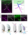
Oligodendrocytes (OLs) and Schwann cells (SCs) have traditionally been thought of as the exclusive myelinating glial cells of the central and peripheral nervous systems (CNS and PNS), respectively, for a little over a century. However, recent studies demonstrate the existence of a novel, centrally-derived peripheral glial population called motor exit point (MEP) glia, which myelinate spinal motor root axons in the periphery. Until recently, the boundaries that exist between the CNS and PNS, and the cells permitted to cross them, were mostly described based on fixed histological collections and static lineage tracing. Recent work in zebrafish using in vivo, time-lapse imaging has shed light on glial cell interactions at the MEP transition zone and reveals a more complex picture of myelination both centrally and peripherally.
Keywords: boundary cap cell; motor exit point glia; myelin; oligodendrocyte; schwann cell; zebrafish.
Figures


Similar articles
-
Spinal cord precursors utilize neural crest cell mechanisms to generate hybrid peripheral myelinating glia.Elife. 2021 Feb 8;10:e64267. doi: 10.7554/eLife.64267. Elife. 2021. PMID: 33554855 Free PMC article.
-
Contact-mediated inhibition between oligodendrocyte progenitor cells and motor exit point glia establishes the spinal cord transition zone.PLoS Biol. 2014 Sep 30;12(9):e1001961. doi: 10.1371/journal.pbio.1001961. eCollection 2014 Sep. PLoS Biol. 2014. PMID: 25268888 Free PMC article.
-
Radial glia inhibit peripheral glial infiltration into the spinal cord at motor exit point transition zones.Glia. 2016 Jul;64(7):1138-53. doi: 10.1002/glia.22987. Epub 2016 Mar 31. Glia. 2016. PMID: 27029762 Free PMC article.
-
Glial plasticity at nervous system transition zones.Biol Open. 2023 Oct 15;12(10):bio060037. doi: 10.1242/bio.060037. Epub 2023 Oct 3. Biol Open. 2023. PMID: 37787575 Free PMC article. Review.
-
The CNS-PNS transitional zone of the rat. Morphometric studies at cranial and spinal levels.Prog Neurobiol. 1992;38(3):261-316. doi: 10.1016/0301-0082(92)90022-7. Prog Neurobiol. 1992. PMID: 1546164 Review.
Cited by
-
Spinal cord precursors utilize neural crest cell mechanisms to generate hybrid peripheral myelinating glia.Elife. 2021 Feb 8;10:e64267. doi: 10.7554/eLife.64267. Elife. 2021. PMID: 33554855 Free PMC article.
-
Evolutionary Origins of the Oligodendrocyte Cell Type and Adaptive Myelination.Front Neurosci. 2021 Dec 1;15:757360. doi: 10.3389/fnins.2021.757360. eCollection 2021. Front Neurosci. 2021. PMID: 34924932 Free PMC article. Review.
-
More Than Mortar: Glia as Architects of Nervous System Development and Disease.Front Cell Dev Biol. 2020 Dec 14;8:611269. doi: 10.3389/fcell.2020.611269. eCollection 2020. Front Cell Dev Biol. 2020. PMID: 33381506 Free PMC article. Review.
-
Liver X Receptors and Their Implications in the Physiology and Pathology of the Peripheral Nervous System.Int J Mol Sci. 2019 Aug 27;20(17):4192. doi: 10.3390/ijms20174192. Int J Mol Sci. 2019. PMID: 31461876 Free PMC article. Review.
-
Schwann Cell Role in Selectivity of Nerve Regeneration.Cells. 2020 Sep 20;9(9):2131. doi: 10.3390/cells9092131. Cells. 2020. PMID: 32962230 Free PMC article. Review.
References
Publication types
Grants and funding
LinkOut - more resources
Full Text Sources
Molecular Biology Databases
Miscellaneous

