. 2018 Jul 30;5(10):1800614.
doi: 10.1002/advs.201800614. eCollection 2018 Oct.

Simple and Low-Cost Sampling of Cell-Free Nucleic Acids from Blood Plasma for Rapid and Sensitive Detection of Circulating Tumor DNA

Affiliations

Simple and Low-Cost Sampling of Cell-Free Nucleic Acids from Blood Plasma for Rapid and Sensitive Detection of Circulating Tumor DNA

Choong Eun Jin et al. Adv Sci (Weinh). .

Abstract

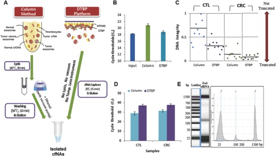
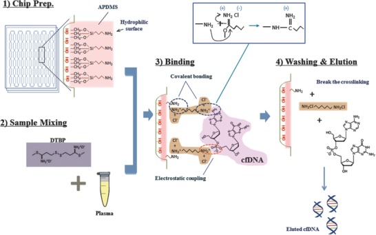
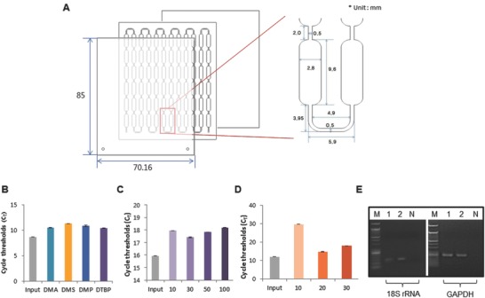
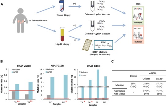
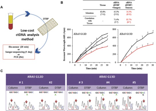
Cell-free nucleic acids (cfNAs) are emerging diagnostic biomarkers for monitoring the treatment and recurrence of cancers. In particular, the biological role and clinical usefulness of cfNAs obtained from the plasma of patients with various cancers are popular and still intensely explored, yet most studies are limited by technical problems during cfNA isolation. A dimethyl dithiobispropionimidate (DTBP)-based microchannel platform that enables spontaneous cfNA capture in 15 min with minimal cellular background and no requirements for use of bulky instruments is reported first. This platform identified KRAS and BRAF hot-spot mutations following cfDNA isolation from the blood plasma and tissues obtained from 30 colorectal cancer patients. The correlation of mutations between the primary tissues and plasma from the patients was high using this platform with whole genome sequencing compared to the spin-column method. This platform can also be combined with various detection approaches (biooptical sensor, Sanger sequencing, and polymerase chain reaction (PCR)) for rapid, simple, low-cost, and sensitive circulating tumor DNA detection in blood plasma. The efficiency and versatility of this platform in isolating cfNAs from liquid biopsies has applications in cancer treatment and precision medicine.

Keywords: biooptical sensors; cell‐free nucleic acids; circulating tumor DNA; liquid biopsy; microfluidics platforms.


Figures

Figure 1
Figure 1
Simple and low‐cost cell‐free nucleic acid (cfNA) sampling for blood‐based testing. A) Schematic representation of the principle of cfNA isolation using the DTBP platform. Workflow of the column‐based method for cfNA isolation with a cell lysis step, high temperatures, and instruments (centrifuge and vacuum pump) (left). The DTBP platform can directly capture cfNA from plasma within 15 min without the requirements of a cell lysis step, high temperatures, or instruments (right). B) Comparison of the capture efficiency with the Alu element amplicon using the column‐based and DTBP platform. The error bars indicate standard deviations from the mean, based on at least three independent experiments. C) The integrity of isolated cfDNA using the column‐based method and the DTBP platform (CTL: 10 healthy control samples, CRC: 14 colorectal cancer samples). D) Real‐time PCR fluorescence signals for the amplified Actin gene (400 bp) with the isolated cfDNA using the column‐based method and the DTBP platform for checking the cellular DNA background. The error bars indicate the standard deviation from the mean, based on at least three independent experiments. E) Electrophoreogram of the isolated cfDNA using the DTBP platform. The lower peak is 25 bp and the upper peak is 1500 bp for size reference.
Figure 2
Figure 2
Operation principles of a cfNA isolation based microfluidic system with DTBP. 1) Chip preparation: assembling the microfluidic platform and inner surface modification with APDMS for binding the amine group of DTBP. 2) Sample mixing: blood plasma samples were mixed with DTBP solution (30 mg mL−1) and injected into the platform. 3) Binding: DTBP binds to the amine group of both APDMS and nucleic acids by covalent bonding and electrostatic coupling. 4) Washing and elution: after washing with PBS, elution buffer leads to the breakage of the cross‐linking, thus eluting cfDNA (or cfRNA).
Figure 3
Figure 3
Characterization of the DTBP platform for simple and low‐cost cfNA isolation. A) The DTBP platform consists of several microwells in a single microfluidic chip. The width, depth, and length were optimized for cfNA isolation. B) The amplification efficiency of this platform is dependent on the type of homobifunctional imidoester reagents (dimethyl adipimidate (DMA), dimethyl suberimidate (DMS), dimethyl pimelimidate (DMP), and DTBP). The cfDNA capture rate is dependent on the C) DTBP concentration (mg mL−1) and D) APDMS concentration (µL mL−1). The error bars indicate the standard deviation from the mean, based on at least three independent experiments. E) cfRNA isolation from blood plasma using the DTBP platform. (M: size marker, 1: plasma #1, 2: plasma #2, N: negative control).
Figure 4
Figure 4
Application of the DTBP platform for cfDNA isolation and ctDNA analysis. A) Workflow scheme of 14 clinical samples, including primary tissues and blood plasma from colorectal cancer patients. 1) Mutation screening using the column‐based method for extraction from tissue biopsies and whole exome sequencing (WES) for detection. 2) Mutation screening using the column‐based method for extraction from blood plasma and WES for detection. 3) Mutation screening using the DTBP platform for extraction from blood plasma and WES for detection. B) Mutation ratio of the isolated ctDNA using the column‐based method (gray) and the DTBP platform (sky blue) for detecting BRAF V600E (left), KRAS G12D (middle), and KRAS G13D (right) mutations. The red line represents the cut‐off (criterion) for reporting a sample once the mutation (positive/negative) was detected. Two asterisks (*) represent samples from which the mutation was detected in only cfDNA and not in tissue DNA. Dual asterisks (**) represent samples from which mutations were detected only in cfDNA using the DTBP platform but not using the column‐based method. C) Correlation between WES results of primary tissues and plasma among 14 clinical samples with the ctDNA isolated using the column‐based method and the DTBP platform.
Figure 5
Figure 5
Simple and low‐cost ctDNA analysis for clinical diagnosis. A) Combination of the DTBP platform and Sanger sequencing for low‐cost ctDNA analysis. B) ctDNA was isolated from 11 plasma samples of colorectal cancer patients using the DTBP platform, and followed by the use of the biosensor for ctDNA analysis. The correlation between the primary tissues (OncoPanel result) and blood plasma (with the Sanger sequencing and the biooptical sensor) (up) was analyzed. Resonant wavelength shift using the biooptical sensor with either the G12D (down‐left) or the G13D mutant primers (down‐right). The error bars indicate the standard deviation of the mean, based on at least three independent experiments. C) Validation of this simple and low‐cost ctDNA analysis using five plasma samples in which mutations were not identified in the column‐based method.

References

    1. Diaz L. A., Bardelli A., J. Clin. Oncol. 2014, 21, 579. - PMC - PubMed
    1. Schwarzenbach H., Hoon D. S. B., Pantel K., Nat. Rev. Cancer 2011, 11, 426. - PubMed
    1. Wan J. C. M., Massie C., Garcia‐Corbacho J., Mouliere F., Brenton J. D., Caldas C., Pacey S., Baird R., Rosenfeld N., Nat. Rev. Cancer 2017, 17, 223. - PubMed
    1. Cohen J. D., Li L., Wang Y., Thoburn C., Afsari B., Danilova L., Douville C., Javed A. A., Wong F., Mattox A., Hruban R. H., Wolfgang C. L., Goggins M. G., Molin M., Wang T., Roden R., Klein A. P., Ptak J., Dobbyn L., Schaefer J., Silliman N., Popoli M., Vogelstein J. T., Browne J. D., Schoen R. E., Brand R. E., Tie J., Gibbs P., Wong H., Mansfield A. S., Jen J., Hanash S. M., Falconi M., Allen P. J., Zhou S., Bettegowda C., Diaz L., Tomasetti C., Kinzler K. W., Vogelstein B., Lennon A. M., Papadopoulos N., Science 2018, 359, 926. - PMC - PubMed
    1. Gerlinger M., N. Engl. J. Med. 2012, 366, 883. - PMC - PubMed

LinkOut - more resources