. 2018 Aug 7;5(10):1800749.
doi: 10.1002/advs.201800749. eCollection 2018 Oct.

Zinc-Modified Sulfonated Polyetheretherketone Surface with Immunomodulatory Function for Guiding Cell Fate and Bone Regeneration

Affiliations

Zinc-Modified Sulfonated Polyetheretherketone Surface with Immunomodulatory Function for Guiding Cell Fate and Bone Regeneration

Wei Liu et al. Adv Sci (Weinh). .

Abstract

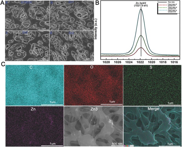
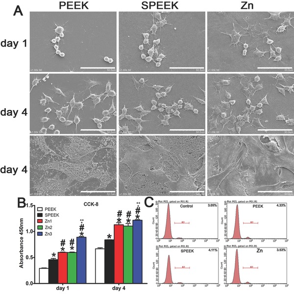
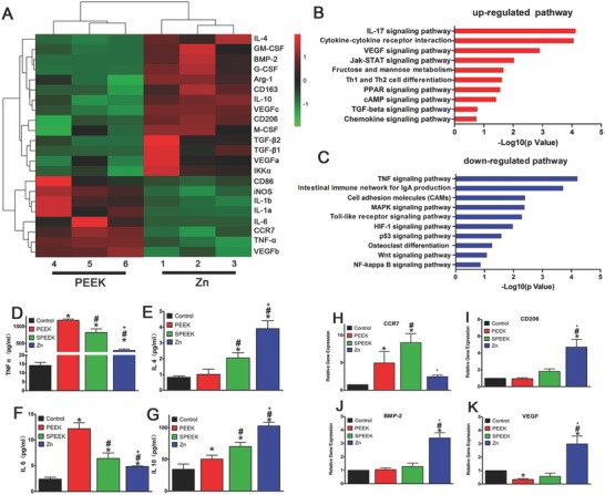
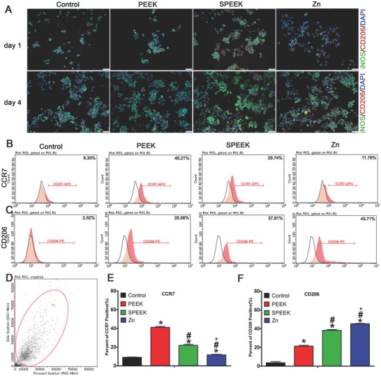
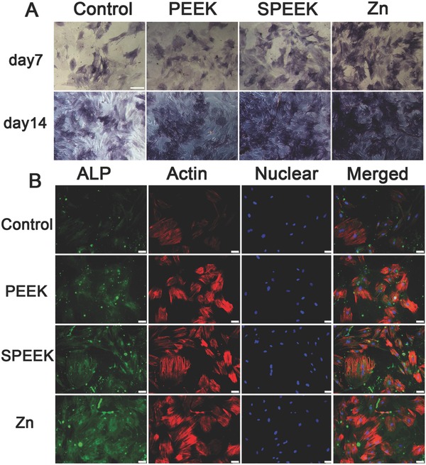
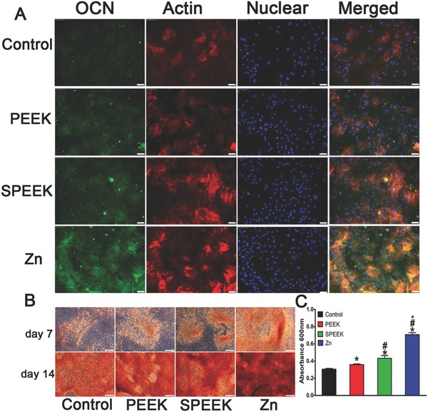
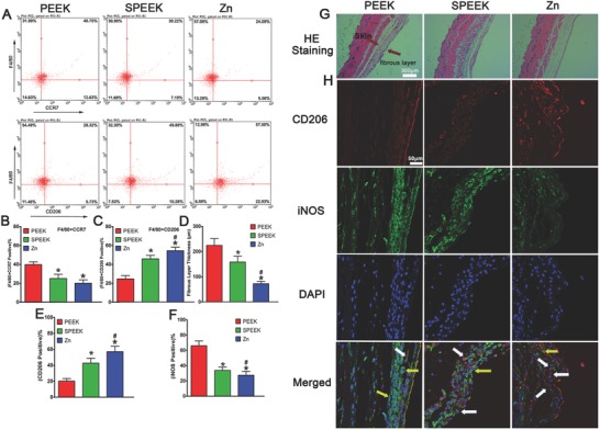
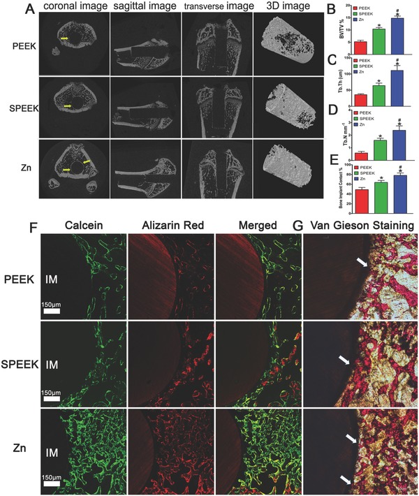
The cytokines released by immune cells are considered important factors to induce bone tissue regeneration. However, the pathway of those bone-targeting macrophage cytokines induced by biomaterial surface under tissue microenvironment is rarely reported. In this study, the immunomodulatory capability of zinc ions on macrophage polarization and its effects on osteogenic differentiation are investigated. Hence, a layer of zinc ions are incorporated on sulfonated polyetheretherketone (SPEEK) biomaterials by using a customized magnetron sputtering technique. The results reveal that the microenvironment on Zn-coated SPEEK can modulate nonactivated macrophage polarization to an anti-inflammatory phenotype and induce the secretion of anti-inflammatory and osteogenic cytokines. The osteogenic differentiation capability of bone marrow stromal cells (BMSCs) is therefore enhanced, leading to improved osteointegration between the zinc-coated SPEEK and bone tissue. This study verifies that zinc ion is a promising additive in the osteoimmunomodulation process and provides knowledge that may pave the way to develop the next generation of immunomodulatory biomaterials.

Keywords: bone formation; immunomodulation; macrophages; stem cells; zinc.


Figures

Figure 1
Figure 1
Characterization of different samples. A) Scanning electron images of SPEEK, Zn1, Zn2, and Zn3. B) XPS results of Zn1, Zn2, and Zn3. C) EDS mapping for the major elements of Zn coated SPEEK (Zn3).
Figure 2
Figure 2
A) Scanning electron images of RAW264.7 and rBMSC cells on PEEK, SPEEK and Zn, bar: 50 µm. B) CCK8 results of RAW264.7 cultured on PEEK, SPEEK and Zn for 1 and 4 d. C) Percentage of dead cells culture on materials using PI staining determined by flow cytometry. (*, #, +, and ++ represent p < 0.05 when compared with PEEK, SPEEK, Zn1, Zn2 respectively). (*, #, +, and ++ represent p < 0.05 when compared with PEEK, SPEEK, Zn1, Zn2 respectively).
Figure 3
Figure 3
Gene expression analysis of RAW264.7 cultured on samples. A) Microarray heat map depicting the fold change of selected genes expression. B–C) Representative Top 10 upregulated or downregulated pathways analyzed by KEGG pathway method. D–G) Elisa results of TNF‐ɑ, IL‐4, IL‐6, and IL‐10 respectively. H–K) RT‐PCR results of CCR7, CD206, BMP‐2, and VEGF respectively. (*, #, and + represent p < 0.05 when compared with Control, PEEK, SPEEK, respectively).
Figure 4
Figure 4
Immunofluorescent staining and surface markers of RAW264.7 cells cultured on samples. A) Immunofluorescent staining of RAW scratched from materials after cultured for 4 d. B,C) Representative dot images of surface markers (CCR7 and CD206) of RAW264.7 analyzed by flow cytometry. D) Representative gate of forward scatter (FSC) and side scatter (SSC). E,F) Percentage of CCR7 or CD206 positive cells respectively. (*, #, and + represent p < 0.05 when compared with Control, PEEK, SPEEK, respectively).
Figure 5
Figure 5
A) ALP staining of rBMSC cultured in conditioned medium for 7 and 14 d. B) ALP immunofluorescent staining of rBMSC in conditioned medium: green (ALP), red (actin), blue (DAPI).
Figure 6
Figure 6
A) OCN immunofluorescent staining of rBMSC in conditioned medium: green (osteocalcin), red(actin), blue (DAPI). B) Alizarin Red staining of rBMSC cultured in conditioned medium for 7 and 14 d. C) Quantitative analysis of Alizarin Red staining. (*, #, and + represent p < 0.05 when compared with Control, PEEK, SPEEK, respectively).
Figure 7
Figure 7
Results of mouse air pouch model. A) Representative dot images of surface markers of cells harvested from air pouches analyzed by flow cytometry. B,C) Percentage of (F4/80+CCR7) or (F4/80+CD206) positive cells respectively. D) Fibrous layer thickness of air pouches' skin. E,F) Percentage of CD206 and iNOS positive cells in the fibrous layer by immunofluorescent staining. G) HE staining of the air pouches' skin. H) Immunofluorescent staining air pouches' skin: red (CD206), green (iNOS), and blue (DAPI). (* and # represent p < 0.05 when compared with PEEK, SPEEK, respectively).
Figure 8
Figure 8
Results of the in vivo bone‐repairing model. A) Coronal, sagittal, transverse, and 3D images of Micro‐CT, yellow arrow indicates new bone. B–D) Quantitative analysis of Micro‐CT data: BV/TV%, Tb.Th, and Tb.N respectively. E) Bone implant contact measured from inset (G). F) Undecalcified sections of sequential polychrome labels for bone: red (Alizarin red), green (Calcein). G) Van Gieson staining of undecalcified sections, white arrow indicates bone implant contact. (* and # represent p < 0.05 when compared with PEEK, SPEEK respectively).

Similar articles

Cited by

References

    1. a) Chen Z., Klein T., Murray R. Z., Crawford R., Chang J., Wu C., Xiao Y., Mater. Today 2016, 19, 304;
    2. b) Takayanagi H., Nat. Rev. Immunol. 2007, 7, 292. - PubMed
    1. Einhorn T. A., Gerstenfeld L. C., Nat. Rev. Rheumatol. 2015, 11, 45. - PMC - PubMed
    1. a) Takizawa T., Nakayama N., Haniu H., Aoki K., Okamoto M., Nomura H., Tanaka M., Sobajima A., Yoshida K., Kamanaka T., Ajima K., Oishi A., Kuroda C., Ishida H., Okano S., Kobayashi S., Kato H., Saito N., Adv. Mater. 2018, 30; - PubMed
    2. b) Pina S., Oliveira J. M., Reis R. L., Adv. Mater. 2015, 27, 1143. - PubMed
    1. Franz S., Rammelt S., Scharnweber D., Simon J. C., Biomaterials 2011, 32, 6692. - PubMed
    1. a) Kohli N., Ho S., Brown S. J., Sawadkar P., Sharma V., Snow M., Garcia‐Gareta E., Bone 2018, 110, 38; - PubMed
    2. b) Anderson J. M., Rodriguez A., Chang D. T., Semin. Immunol. 2008, 20, 86. - PMC - PubMed

LinkOut - more resources