. 2018 Aug 16;5(10):1800781.
doi: 10.1002/advs.201800781. eCollection 2018 Oct.

A Broad-Spectrum ROS-Eliminating Material for Prevention of Inflammation and Drug-Induced Organ Toxicity

Affiliations

A Broad-Spectrum ROS-Eliminating Material for Prevention of Inflammation and Drug-Induced Organ Toxicity

Lanlan Li et al. Adv Sci (Weinh). .

Abstract

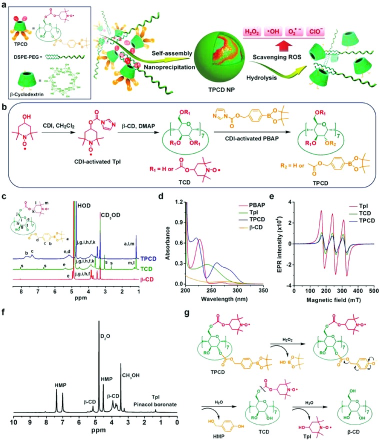
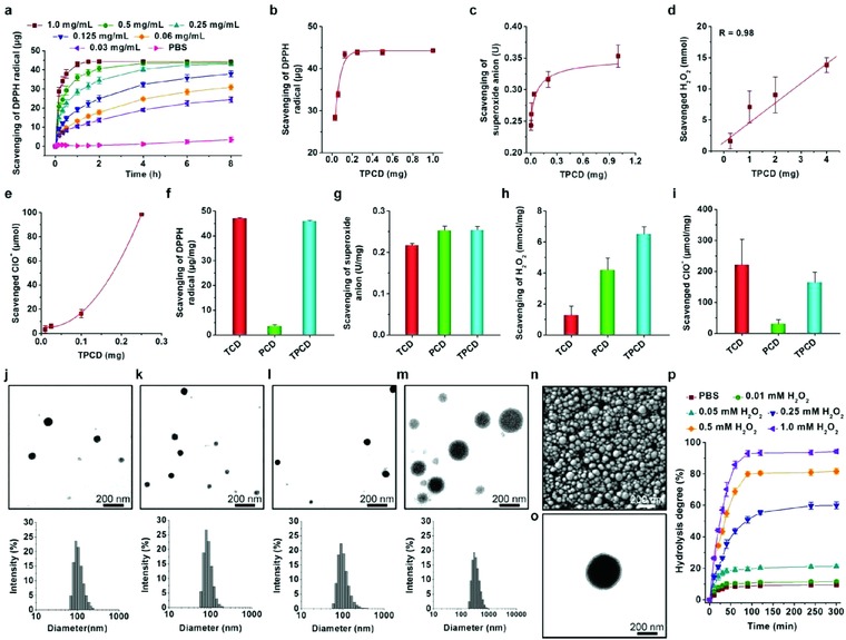
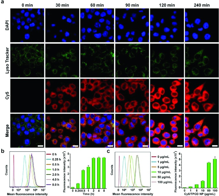
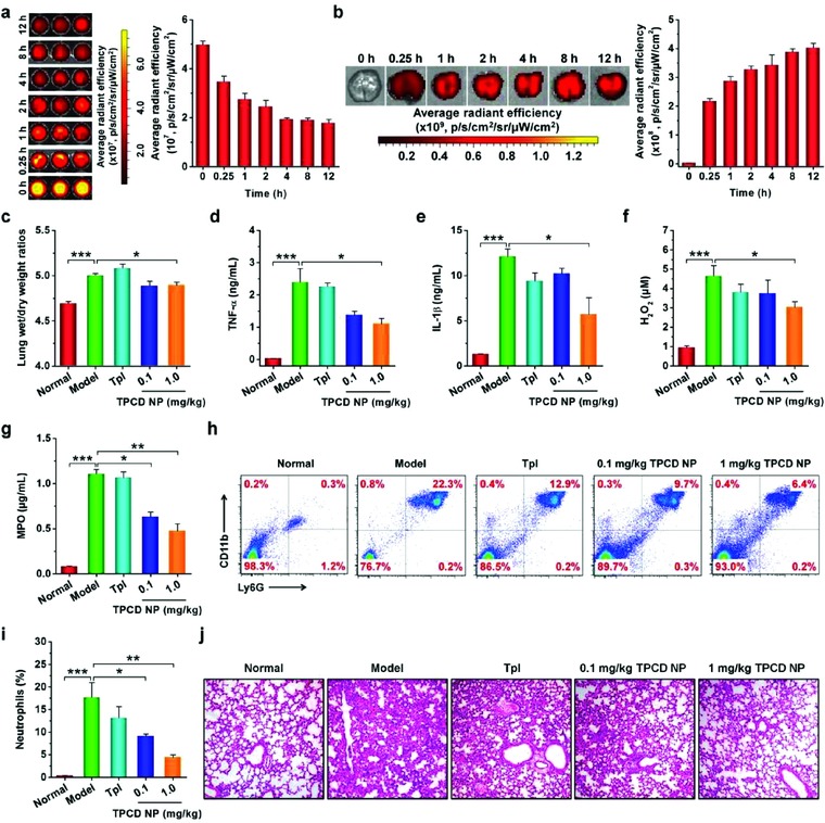
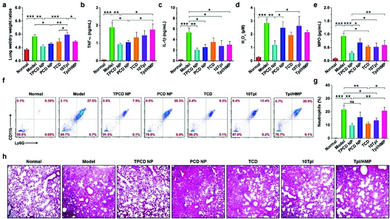
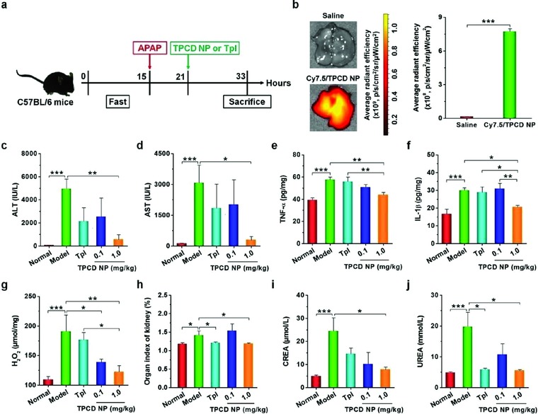
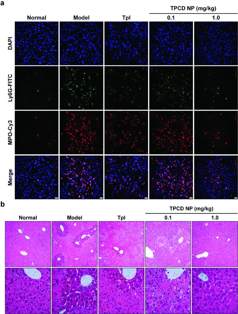
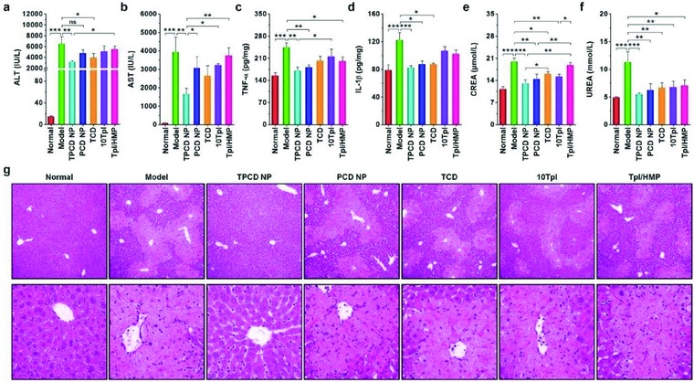
Despite the great potential of numerous antioxidants for pharmacotherapy of diseases associated with inflammation and oxidative stress, many challenges remain for their clinical translation. Herein, a superoxidase dismutase/catalase-mimetic material based on Tempol and phenylboronic acid pinacol ester simultaneously conjugated β-cyclodextrin (abbreviated as TPCD), which is capable of eliminating a broad spectrum of reactive oxygen species (ROS), is reported. TPCD can be easily synthesized by sequentially conjugating two functional moieties onto a β-cyclodextrin scaffold. The thus developed pharmacologically active material may be easily produced into antioxidant and anti-inflammatory nanoparticles, with tunable size. TPCD nanoparticles (TPCD NP) effectively protect macrophages from oxidative stress-induced apoptosis in vitro. Consistently, TPCD NP shows superior efficacies in three murine models of inflammatory diseases, with respect to attenuating inflammatory responses and mitigating oxidative stress. TPCD NP can also protect mice from drug-induced organ toxicity. Besides the passive targeting effect, the broad spectrum ROS-scavenging capability contributes to the therapeutic benefits of TPCD NP. Importantly, in vitro and in vivo preliminary experiments demonstrate the good safety profile of TPCD NP. Consequently, TPCD in its native and nanoparticle forms can be further developed as efficacious and safe therapies for treatment of inflammation and oxidative stress-associated diseases.

Keywords: antioxidants; inflammation; nanoparticles; reactive oxygen species; targeted therapy.


Figures

Figure 1
Figure 1
Design, preparation, and characterization of a ROS‐scavenging material. a) Schematic illustration of engineering of a broad spectrum ROS‐scavenging material and nanoparticle based on functionalized β‐cyclodextrin (β‐CD). b) The synthetic route of β‐CD conjugated with Tempol (Tpl) and PBAP units (TPCD). CDI, 1,1‐carbonyldiimidazole; DMAP, 4‐dimethylaminopyridine; TCD, Tpl‐conjugated β‐CD; PBAP, 4‐(hydroxymethyl) phenylboronic acid pinacol ester. c) 1H NMR spectra of different materials including β‐CD, TCD, and TPCD. The character “s” denotes the presence of trace amount of solvent impurities, such as DMSO and DMAP residuals from the precipitation process. d) UV–visible absorption spectra of different compounds. e) EPR spectra of free Tpl, TCD, and TPCD. f) A 1H NMR spectrum of the hydrolyzed TPCD sample in D2O. HMP, p‐(hydroxymethyl)phenol. g) The mechanism for the H2O2‐mediated hydrolysis of TPCD.
Figure 2
Figure 2
ROS‐scavenging capability of TPCD and characterization of TPCD nanoparticles. a) Time‐dependent scavenging of the DPPH radical by different doses of TPCD. b) DPPH radical‐scavenging efficiency of TPCD at varied doses after 20 h of incubation. c–e) Elimination of superoxide anion, H2O2, and hypochlorite by TPCD after incubation for 40 min, 24 h, and 15 min, respectively. f–i) Comparison of scavenging capabilities of different materials for radical, superoxide anion, H2O2, and hypochlorite. j–m) TEM images (the upper panel) and size distribution profiles (the lower panel) of TPCD NPs fabricated by a self‐assembly/nanoprecipitation method using methanol, methanol/acetonitrile, methanol/dimethylformamide, and methanol/tetrahydrofuran as the solvent for TPCD, respectively. For all solvent mixtures, the volume ratio of methanol to another solvent was kept at 1:2. n) A typical SEM image of TPCD NP prepared using methanol as the solvent. o) TEM image of phosphotungstic acid‐stained TPCD NP. p) Hydrolysis curves of TPCD NP (prepared using methanol as a solvent) in PBS (0.01 m, pH 7.4) containing various concentrations of H2O2. Data in (a–i, p) are mean ± SE (n = 3).
Figure 3
Figure 3
Cellular uptake of Cy5‐labeled TPCD NP in RAW264.7 macrophages. a) Fluorescence images showing time‐dependent internalization of Cy5/TPCD NP at 2 µg mL−1 of Cy5 in RAW264.7 cells. For observation by confocal microscopy, nuclei were stained with DAPI (blue), while late endosomes and lysosomes were stained with LysoTracker (green). Scale bars, 10 µm. b) Typical flow cytometric curves (left) and quantitative analysis (right) of time‐dependent cellular uptake of Cy5/TPCD NP at 2 µg mL−1 of Cy5 in RAW264.7 cells. c) Flow cytometric profiles (left) and quantification results (right) indicating cellular uptake of Cy5/TPCD NP at various doses after 2 h of incubation in RAW264.7 cells. Data are mean ± SE (n = 3).
Figure 4
Figure 4
In vitro antioxidative stress and in vivo anti‐inflammatory activities in murine models of acute inflammation. a,b) Flow cytometric profiles and quantitative data of apoptotic RAW264.7 cells after different treatments. The control, TPCD NP, and H2O2 groups were treated with fresh medium, 100 µg mL−1 of TPCD NP, and 200 × 10−6 m H2O2, respectively. The H2O2 + TPCD NP group was first incubated with 100 µg mL−1 of TPCD NP for 2 h, followed by culture with 200 × 10−6 m H2O2 for 24 h. c,d) Flow cytometric profiles and quantitative analysis of apoptotic macrophages treated with different formulations. The groups of TPCD NP, PCD NP, TCD, 10Tpl, Tpl, and Tpl/HMP were preincubated with the corresponding formulations for 2 h, and then stimulated with 200 × 10−6 m H2O2 for 24 h. The 10Tpl group was treated with Tpl at the tenfold dose of that contained in TPCD NP. e) In vivo efficacy of TPCD NP in rats with carrageen‐induced edema. At 0.5 h after challenge by i.d. injection of carrageen, a single dose of different formulations was locally administered. The dose of free Tpl was the same as that of TPCD NP at 5.0 mg kg−1. f–i) The levels of H2O2, MPO, TNF‐α, and IL‐1β in cell‐free peritoneal exudates collected from mice with zymosan‐induced peritonitis. At 1 h after zymosan induction by i.p. injection, different treatments were performed. TPCD NP at 1.0 mg kg−1 contained the same dose of Tpl as the free Tpl group. j–m) The expression of H2O2, MPO, TNF‐α, and IL‐1β in peritoneal exudates from peritonitis mice after treatment with different controls. Data are mean ± SE (b, d, n = 3; e, n = 5; f–m, n = 6). Statistical significance was assessed by one‐way ANOVA with post hoc LSD tests for data in (b, e–m). *P < 0.05, **P < 0.01, ***P < 0.001; ns, no significance. Due to heterogeneity of variance, the Kruskal–Wallis test was used for statistical analysis of data in (d). *P < 0.001, **P < 0.0001, ***P < 0.00001.
Figure 5
Figure 5
Targeted therapy of acute lung injury with TPCD NP in mice. a) Fluorescence images (left) and quantitative analysis (right) of the blood samples collected from mice with acute lung injury (ALI) at various time points after i.v. injection of Cy7.5/TPCD NP. b) Ex vivo images (left) and quantitative analysis (right) illustrating the distribution of Cy7.5/TPCD NP in the lung of ALI mice after i.v. administration for different periods of time. c) The lung wet‐to‐dry weight ratios after different treatments. d–g) The expression levels of TNF‐α, IL‐1β, H2O2, and MPO in bronchoalveolar lavage fluid from mice with LPS‐induced ALI and subjected to different treatments. h,i) Representative flow cytometric profiles and quantitative analysis of neutrophil counts in pulmonary tissues of ALI mice. j) H&E‐stained pathological sections of lung tissues. ALI in mice was induced by intratracheal (i.t.) administration of LPS. 1 h after challenge, mice were i.v. administered with different formulations. The normal group was treated with saline. The TPCD NP groups were administered with either lower (0.1 mg kg−1) or higher (1.0 mg kg−1) dose of TPCD NP, and 1.0 mg kg−1 TPCD contained the same dose of the Tpl unit as the free Tpl group. At 12 h after various treatments, bronchoalveolar lavage fluid was collected for different quantitative analyses. In separate experiments, lung tissues were isolated for additional quantitative and histological analyses. Data are mean ± SE (a,b, n = 3; c–g, i, n = 6). Statistical significance was assessed by one‐way ANOVA with post hoc LSD tests. *P < 0.05, **P < 0.01, ***P < 0.001.
Figure 6
Figure 6
Comparison of in vivo efficacies of TPCD NP with different nanotherapy and small‐molecule controls in ALI mice. a) The lung wet‐to‐dry weight ratios after treatment with different formulations. b–e) The levels of TNF‐α, IL‐1β, H2O2, and MPO in bronchoalveolar lavage fluid from mice with LPS‐induced ALI and subjected to different treatments. f,g) Flow cytometric profiles and quantitative data of neutrophil counts in pulmonary tissues of ALI mice. h) H&E‐stained sections of lung tissues. ALI in mice was induced by intratracheal (i.t.) administration of LPS. After 1 h, mice were treated with different formulations via i.v. injection. The normal group was treated with saline. The PCD NP and TCD groups were administered at the same dose of the PBAP or Tpl unit as that of 1.0 mg kg−1 of TPCD NP, respectively. In the 10Tpl group, the Tpl dose was tenfold of that contained in TPCD NP. For the Tpl/HMP group, the Tpl dose was the same as that in TPCD NP, while the HMP dose equaled to that generated after complete hydrolysis of TPCD NP. At 12 h after different treatments, bronchoalveolar lavage fluid was collected for quantitative analyses. In a separate study, lung tissues were isolated for quantitative and histological analyses. Data are mean ± SE (a–e, g, n = 6). Statistical significance was assessed by one‐way ANOVA with post hoc LSD tests. *P < 0.05, **P < 0.01, ***P < 0.001; ns, no significance.
Figure 7
Figure 7
Detoxification of acetaminophen (APAP)‐induced hepatotoxicity and renal injury by TPCD NP. a) Schematic illustration of the establishment of APAP‐induced organ toxicity and treatment regimens. b) Representative ex vivo images and quantitative analysis of Cy7.5/TPCD NP accumulation in the liver of mice at 12 h after administration. c,d) The levels of ALT and AST in serum collected from mice with APAP‐induced toxicity. e–g) The expression levels of TNF‐α, IL‐1β, and H2O2 in the hepatic tissues. h) The organ index of kidney. i,j) The serum levels of CREA and UREA. For therapy studies, at 6 h after i.p. stimulation with APAP at 200 mg kg−1, mice were treated with different formulations. Mice in the normal group were not challenged with APAP. The model group was administered with saline. TPCD NP at 1.0 mg kg−1 contained the same dose of the Tpl unit as that of the free Tpl group. At 12 h after different treatments, animals were euthanized and serum was collected for quantification of biochemical markers. Additionally, the liver tissues were isolated for quantification of inflammatory mediators. Data are mean ± SE (b, n = 3; c–j, n = 6). Statistical significance was analyzed by one‐way ANOVA with post hoc LSD tests. *P < 0.05, **P < 0.01, ***P < 0.001.
Figure 8
Figure 8
Evaluation of in vivo detoxification efficacy of TPCD NP in mice with APAP‐induced hepatotoxicity. a) Immunofluorescence images show the infiltration of neutrophils and expression of MPO in the liver. Scale bars, 20 µm. b) H&E‐stained histopathological sections of liver tissues resected from mice subjected to different treatments. The lower panel indicates high‐resolution images.
Figure 9
Figure 9
Comparison of in vivo detoxification efficacy of TPCD NP with different nanotherapy and small‐molecule controls in mice with APAP‐induced hepatotoxicity and renal injury. a,b) The levels of ALT and AST in serum collected from mice induced with APAP. c,d) The levels of TNF‐α and IL‐1β in the hepatic tissues. e,f) The serum levels of CREA and UREA. At 6 h after i.p. stimulation with APAP at 200 mg kg−1, mice were treated with different formulations. The PCD NP and TCD groups were treated at the same dose of the PBAP or Tpl unit as that of 1.0 mg kg−1 TPCD NP, respectively. In the 10Tpl group, the Tpl dose was tenfold of that contained in TPCD NP. For the Tpl/HMP group, the Tpl dose was the same as that in TPCD NP, while the HMP dose equaled to that generated after complete hydrolysis of TPCD NP. At 12 h after different treatments, animals were euthanized and serum was collected for quantification of biochemical markers. The liver tissues were isolated for quantification of inflammatory mediators. g) H&E‐stained histopathological sections of liver tissues. The lower panel indicates high‐resolution images. Data are mean ± SE (n = 6). Statistical significance was assessed by one‐way ANOVA with post hoc LSD tests for data in (a–c, e, f). *P < 0.05, **P < 0.01, ***P < 0.001; ns, no significance. Due to heterogeneity of variance, the Kruskal–Wallis test was used for statistical analysis of data in (d). *P < 0.01, ***P < 0.0001.

Similar articles

Cited by

References

    1. Nathan C., Nature 2002, 420, 846. - PubMed
    1. Fullerton J. N., Gilroy D. W., Nat. Rev. Drug Discovery 2016, 15, 551. - PubMed
    1. Buckley C. D., Gilroy D. W., Serhan C. N., Immunity 2014, 40, 315. - PMC - PubMed
    1. Tabas I., Glass C. K., Science 2013, 339, 166. - PMC - PubMed
    1. Okin D., Medzhitov R., Curr. Biol. 2012, 22, R733. - PMC - PubMed

LinkOut - more resources