. 2018 Aug 24;5(10):1800382.
doi: 10.1002/advs.201800382. eCollection 2018 Oct.

A Light-Triggered Mesenchymal Stem Cell Delivery System for Photoacoustic Imaging and Chemo-Photothermal Therapy of Triple Negative Breast Cancer

Affiliations

A Light-Triggered Mesenchymal Stem Cell Delivery System for Photoacoustic Imaging and Chemo-Photothermal Therapy of Triple Negative Breast Cancer

Chang Xu et al. Adv Sci (Weinh). .

Abstract

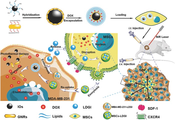
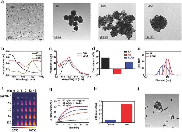
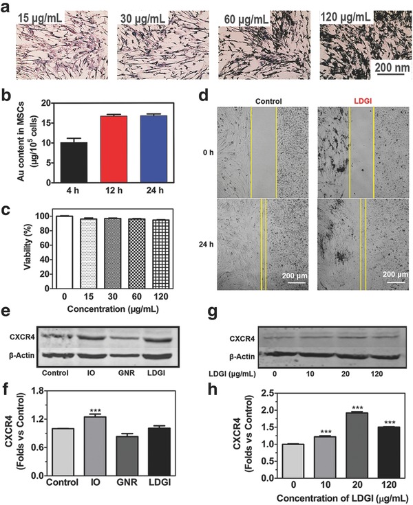
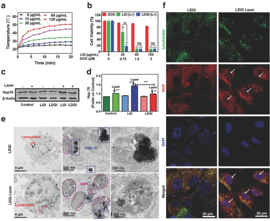
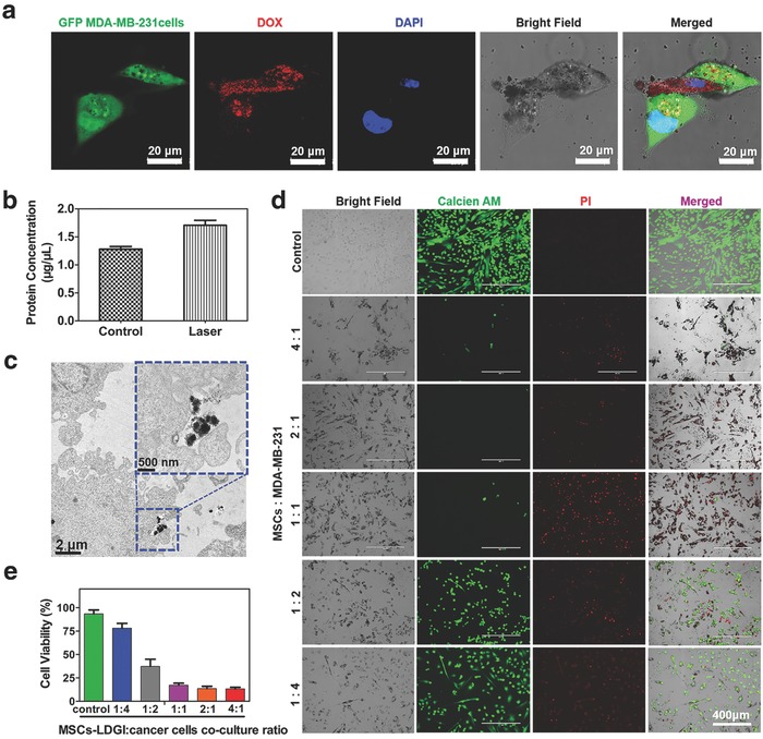
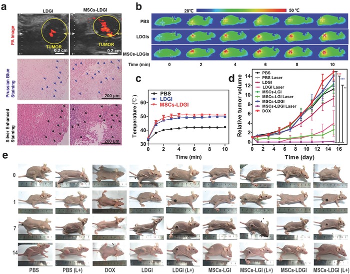
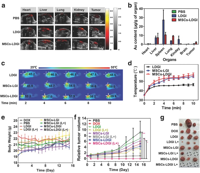
Targeted therapy is highly challenging and urgently needed for patients diagnosed with triple negative breast cancer (TNBC). Here, a synergistic treatment platform with plasmonic-magnetic hybrid nanoparticle (lipids, doxorubicin (DOX), gold nanorods, iron oxide nanocluster (LDGI))-loaded mesenchymal stem cells (MSCs) for photoacoustic imaging, targeted photothermal therapy, and chemotherapy for TNBC is developed. LDGI can be efficiently taken up into the stem cells with good biocompatibility to maintain the cellular functions. In addition, CXCR4 on the MSCs is upregulated by iron oxide nanoparticles in the LDGI. Importantly, the drug release and photothermal therapy can be simultaneously achieved upon light irradiation. The released drug can enter the cell nucleus and promote cell apoptosis. Interestingly, light irradiation can control the secretion of cellular microvehicles carrying LDGI for targeted treatment. A remarkable in vitro anticancer effect is observed in MDA-MB-231 with near-infrared laser irradiation. In vivo studies show that the MSCs-LDGI has the enhanced migration and penetration abilities in the tumor area via both intratumoral and intravenous injection approaches compared with LDGI. Subsequently, MSCs-LDGI shows the best antitumor efficacy via chemo-photothermal therapy compared to other treatment groups in the TNBC model of nude mice. Thus, MSCs-LDGI multifunctional system represents greatly synergistic potential for cancer treatment.

Keywords: light‐controlled release; photothermal therapy; plasmonic–magnetic nanoparticles; stem cells; triple negative breast cancer.


Figures

Scheme 1
Scheme 1
The scheme of LDGI‐loaded MSCs for enhanced tumor migration ability, PTT, and chemotherapy of TNBC.
Figure 1
Figure 1
Characterization of LDGI. a) Representative TEM images of GNR, IO, and LDGI. b) UV–vis spectra of IO, GNR, and LDGI. c) Fluorescence spectra of LDGI and DOX (1 µg mL−1). d) Zeta potential of IO, GNR, and LDGI in deionized water. e) Size distribution of GI and LDGI in deionized water. f) Photothermal effect of LDGI at different concentrations and g) the corresponding temperature changing curves under 808 nm laser at 3 W cm−2 for 15 min. h) Drug release behavior of the LDGI aqueous solution with laser irradiation (3 W cm−2, 3 min). i) The TEM image of LDGI post irradiation.
Figure 2
Figure 2
Loading LDGI into MSCs and the migration ability of the combination platform. a) Prussian blue stain of MSCs treated with different concentrations of LDGI (15, 30, 60, 120 µg mL−1) after 24 h incubation. b) ICP‐MS quantification of LDGI at the incubation times of 4, 12, and 24 h. c) Cell viability with different concentrations of LDGI. d) MSCs migration toward MDA‐MB‐231 cells treated with 25 µg mL−1 LDGI and without LDGI in 24 h. e) Western blot analysis of CXCR4 expression in MSCs after treatment with nanoparticles including IO, GNR, and LDGI. f) Quantification of the western blotting results in (e). g) Specific surface markers of stem cells expression on MSCs treated with different concentrations of LDGI and h) the quantification of western blotting results by measuring the band density and then normalizing it to β‐actin.
Figure 3
Figure 3
NIR light–triggered DOX release and photothermal effect of LDGI. a) Temperature rising curve of MSCs incubated with LDGI at different concentrations (0, 30, 60, 120 µg mL−1) with laser irradiation (3 W cm−2, 20 min). b) Cell viability of MSCs incubated with DOX, LGI, and LDGI nanoparticles for 24 h and treated with NIR laser (3 W cm−2, 5 min). c) Western blot analysis of Hsp70 protein expression in MSCs after treatment with nanoparticles including LGI and LDGI post laser irradiation (1.5 W cm−2, 5 min) and d) the quantification of western blotting results by measuring the band density and then normalizing according to β‐actin. e) Representative TEM images of LDGI‐loaded MSCs treated with and without laser (3 W cm−2, 5 min). f) Confocal images of DOX release from LDGI in MSCs before and after laser (3 W cm−2, 5 min). Green fluorescence represents the lysotracker green accumulated in lysosomes, red fluorescence represents DOX trapped in LDGI and free DOX, and blue fluorescence represents 4′,6‐diamidino‐2‐phenylindole (DAPI) in the cell nucleus.
Figure 4
Figure 4
The anticancer effect of MSCs‐LDGI against MDA‐MB‐231 cells. a) Confocal images of green florescence protein (GFP)‐labeled MDA‐MB‐231 cells cocultured with MSCs‐LDGI (green florescence represents MDA‐MB‐231 cells, red florescence represents DOX trapped in LDGI, and blue florescence represents the cell nucleus). b) Protein quantification of cells culture medium after laser irradiation (1.5 W cm−2, 5 min). c) TEM image of exocytosis of LDGI from MSCs‐LDGI post 24 h incubation. d) LIVE/DEAD cell viability tests of MSCs‐LDGI cocultured with MDA‐MB‐231 cells post laser irradiation (green fluorescence represents live cells stained with calcein‐AM solution and red fluorescence represents dead cells stained with propidium iodide (PI) solution). e) Cell viability of MSCs‐LDGI and MDA‐MB‐231 cells with different ratios for 24 h with and without irradiation tested by CCK‐8 kit after 24 h.
Figure 5
Figure 5
In vivo antitumor effect of MSCs‐LDGI (intratumoral injection). a) In vivo photoacoustic image, Prussian blue staining, and silver‐enhanced staining of tumor tissues of LDGI and MSCs‐LDGI, respectively. b) Infrared thermal images and c) temperature increasing curves of PBS, LDGI, MSCs‐LGI, and MSCs‐LDGI with continuous laser irradiation of 808 nm laser (1.5 W cm−2, 10 min) in vivo. d) Relative tumor volume and e) images of mice of being intratumorally injected with DOX, PBS, LDGI, MSCs‐LGI, MSCs‐LDGI with and without NIR laser irradiation within 15 days.
Figure 6
Figure 6
In vivo antitumor effect of MSCs‐LDGI via intravenous injection. a) In vivo photoacoustic image of mice organs of MSCs‐LDGI, LDGI, and PBS, respectively. b) Biodistribution of gold amount in heart, liver, spleen, lung, kidney, brain, and tumor tissues post 3 days of intravenous injection. c) In vivo infrared thermal images and d) the corresponding temperature increasing curves of LDGI, MSCs‐LGI, and MSCs‐LDGI with continuous laser irradiation of 808 nm laser (1.5 W cm−2, 10 min). e) Body weight curves of mice injected with DOX, PBS, LDGI, MSCs‐LGI, MSCs‐LDGI with and without NIR laser. f) Relative tumor volumes and g) the image of tumors from the mice treated with DOX, PBS, LDGI, MSCs‐LGI, MSCs‐LDGI with and without NIR laser irradiation post 15 days.

References

    1. Torre L. A., Siegel R. L., Ward E. M., Jemal A., Cancer Epidemiol., Biomarkers Prev. 2016, 25, 16. - PubMed
    1. Jemal A., Bray F., Center M. M., Ferlay J., Ward E., Forman D., CA ‐ Cancer J. Clin. 2011, 61, 69. - PubMed
    1. Ahn S. G., Kim S. J., Kim C., Jeong J., Int. J. Breast Cancer 2016, 19, 223. - PMC - PubMed
    1. Foulkes W. D., Smith I. E., Reis‐Filho J. S., N. Engl. J. Med. 2010, 363, 1938. - PubMed
    1. Dent R., Trudeau M., Pritchard K. I., Hanna W. M., Kahn H. K., Sawka C. A., Lickley L. A., Rawlinson E., Sun P., Narod S. A., Clin. Cancer Res. 2007, 13, 4429. - PubMed

LinkOut - more resources