

Sclerostin promotes human dental pulp cells senescence
- PMID: 30356963
- PMCID: PMC6195797
- DOI: 10.7717/peerj.5808
Sclerostin promotes human dental pulp cells senescence
Abstract
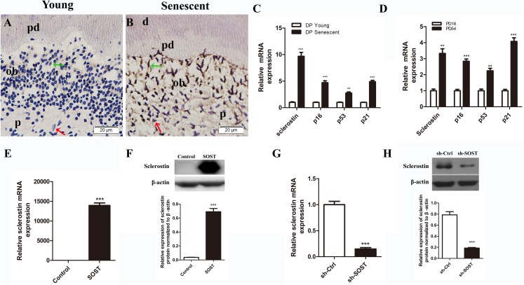
Background: Senescence-related impairment of proliferation and differentiation limits the use of dental pulp cells for tissue regeneration. Deletion of sclerostin improves the dentinogenesis regeneration, while its role in dental pulp senescence is unclear. We investigated the role of sclerostin in subculture-induced senescence of human dental pulp cells (HDPCs) and in the senescence-related decline of proliferation and odontoblastic differentiation.
Methods: Immunohistochemical staining and qRT-PCR analyses were performed to examine the expression pattern of sclerostin in young (20-30-year-old) and senescent (45-80-year-old) dental pulps. HDPCs were serially subcultured until senescence, and the expression of sclerostin was examined by qRT-PCR analysis. HDPCs with sclerostin overexpression and knockdown were constructed to investigate the role of sclerostin in HDPCs senescence and senescence-related impairment of odontoblastic differentiation potential.
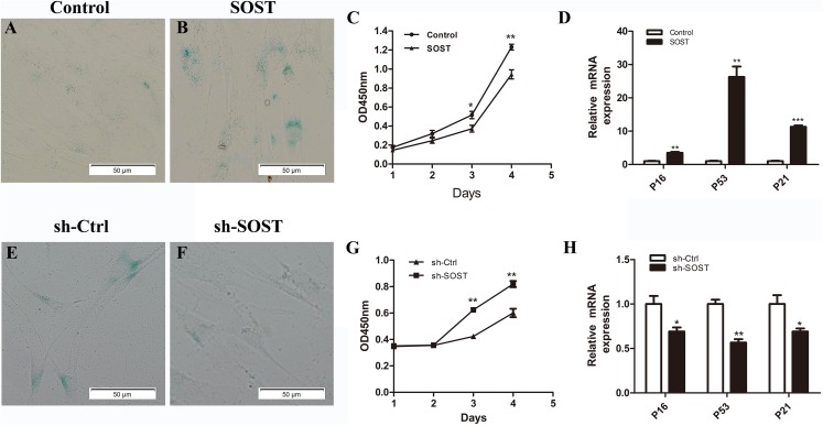
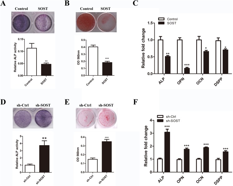
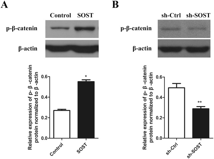
Results: By immunohistochemistry and qRT-PCR, we found a significantly increased expression level of sclerostin in senescent human dental pulp compared with that of young human dental pulp. Additionally, elevated sclerostin expression was found in subculture-induced senescent HDPCs in vitro. By sclerostin overexpression and knockdown, we found that sclerostin promoted HDPCs senescence-related decline of proliferation and odontoblastic differentiation potential with increased expression of p16, p53 and p21 and downregulation of the Wnt signaling pathway.
Discussion: The increased expression of sclerostin is responsible for the decline of proliferation and odontoblastic differentiation potential of HDPCs during cellular senescence. Anti-sclerostin treatment may be beneficial for the maintenance of the proliferation and odontoblastic differentiation potentials of HDPCs.
Keywords: Human dental pulp cell; Sclerostin; Senescence; p16; p21; p53.
Conflict of interest statement
The authors declare that they have no competing interests.
Figures
Similar articles
-
Senescence and odontoblastic differentiation of dental pulp cells.J Cell Physiol. 2018 Jan;234(1):849-859. doi: 10.1002/jcp.26905. Epub 2018 Aug 4. J Cell Physiol. 2018. PMID: 30078208
-
Calcium-sensing receptor-ERK signaling promotes odontoblastic differentiation of human dental pulp cells.Bone. 2017 Aug;101:191-201. doi: 10.1016/j.bone.2017.05.012. Epub 2017 May 12. Bone. 2017. PMID: 28506888
-
Effects of high-mobility group box 1 on the proliferation and odontoblastic differentiation of human dental pulp cells.Int Endod J. 2013 Dec;46(12):1153-63. doi: 10.1111/iej.12112. Epub 2013 Apr 19. Int Endod J. 2013. PMID: 23600680
-
The effect of SIRT6 on the odontoblastic potential of human dental pulp cells.J Endod. 2014 Mar;40(3):393-8. doi: 10.1016/j.joen.2013.11.010. Epub 2013 Dec 19. J Endod. 2014. PMID: 24565659
-
Hypoxia‑induced mitophagy regulates proliferation, migration and odontoblastic differentiation of human dental pulp cells through FUN14 domain‑containing 1.Int J Mol Med. 2022 May;49(5):72. doi: 10.3892/ijmm.2022.5128. Epub 2022 Apr 1. Int J Mol Med. 2022. PMID: 35362539 Free PMC article.
Cited by
-
The Influence of Pro-Inflammatory Factors on Sclerostin and Dickkopf-1 Production in Human Dental Pulp Cells Under Hypoxic Conditions.Front Bioeng Biotechnol. 2019 Dec 17;7:430. doi: 10.3389/fbioe.2019.00430. eCollection 2019. Front Bioeng Biotechnol. 2019. PMID: 31921831 Free PMC article.
-
Aging and Senescence of Dental Pulp and Hard Tissues of the Tooth.Front Cell Dev Biol. 2020 Nov 30;8:605996. doi: 10.3389/fcell.2020.605996. eCollection 2020. Front Cell Dev Biol. 2020. PMID: 33330507 Free PMC article. Review.
-
Multidifferentiation potential of dental-derived stem cells.World J Stem Cells. 2021 May 26;13(5):342-365. doi: 10.4252/wjsc.v13.i5.342. World J Stem Cells. 2021. PMID: 34136070 Free PMC article. Review.
-
Deep coverage and quantification of the bone proteome provides enhanced opportunities for new discoveries in skeletal biology and disease.PLoS One. 2023 Oct 10;18(10):e0292268. doi: 10.1371/journal.pone.0292268. eCollection 2023. PLoS One. 2023. PMID: 37816044 Free PMC article.
-
Modulators of Wnt Signaling Pathway Implied in Dentin Pulp Complex Engineering: A Literature Review.Int J Mol Sci. 2022 Sep 13;23(18):10582. doi: 10.3390/ijms231810582. Int J Mol Sci. 2022. PMID: 36142496 Free PMC article. Review.
References
-
- Artsi H, Cohen-Kfir E, Gurt I, Shahar R, Bajayo A, Kalish N, Bellido TM, Gabet Y, Dresner-Pollak R. The Sirtuin1 activator SRT3025 down-regulates sclerostin and rescues ovariectomy-induced bone loss and biomechanical deterioration in female mice. Endocrinology. 2014;155(9):3508–3515. doi: 10.1210/en.2014-1334. - DOI - PMC - PubMed
-
- Chan BY, Fuller ES, Russell AK, Smith SM, Smith MM, Jackson MT, Cake MA, Read RA, Bateman JF, Sambrook PN, Little CB. Increased chondrocyte sclerostin may protect against cartilage degradation in osteoarthritis. Osteoarthritis and Cartilage. 2011;19(7):874–885. doi: 10.1016/j.joca.2011.04.014. - DOI - PubMed
-
- Chen S, Rani S, Wu YM, Unterbrink A, Gu TT, Gluhak-Heinrich J, Chuang HH, MacDougall M. Differential regulation of dentin sialophosphoprotein expression by Runx2 during odontoblast cytodifferentiation. Journal of Biological Chemistry. 2005;280(33):29717–29727. doi: 10.1074/jbc.M502929200. - DOI - PubMed
LinkOut - more resources
Full Text Sources
Research Materials
Miscellaneous
