

Review
doi: 10.7759/cureus.3171.
Osteonecrosis of the Femoral Head: Etiology, Investigations, and Management
Affiliations
- PMID: 30357068
- PMCID: PMC6197539
- DOI: 10.7759/cureus.3171
Item in Clipboard
Review
Osteonecrosis of the Femoral Head: Etiology, Investigations, and Management
Cureus.
.
Abstract
Femoral head osteonecrosis is a condition caused by a compromise of the blood supply of the femoral head. The precarious blood supply of the head and its role as a major weight-bearing joint makes it one of the most common bones to be affected by osteonecrosis. We describe the etiology, clinical presentation, investigations and common management options used nowadays to treat it.
Keywords: bisphosphonates; core decompression; hyperbaric oxygen; osteonecrosis.
Conflict of interest statement
The authors have declared that no competing interests exist.
Figures
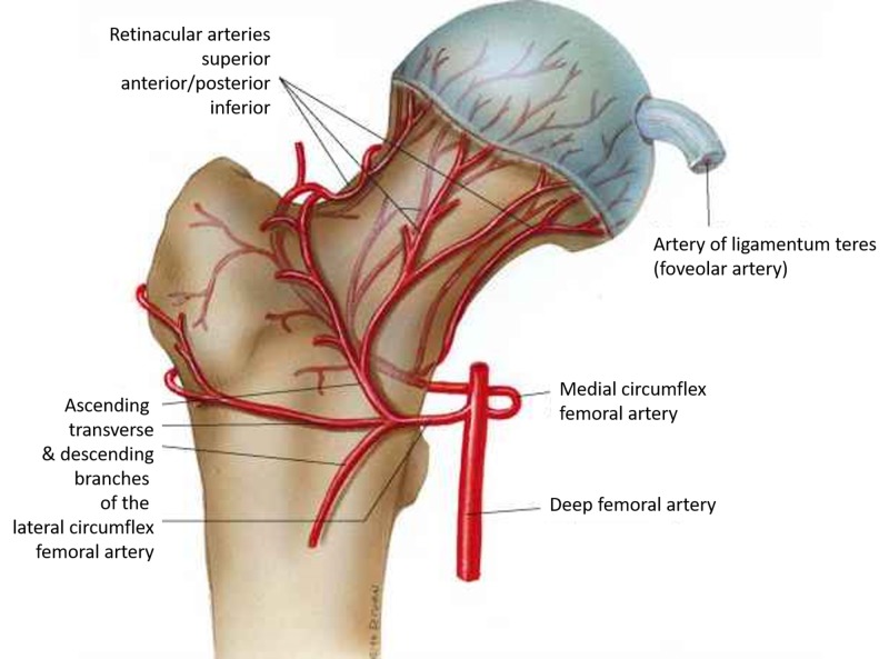
Femoral head and neck blood supply (Courtesy ALPF Medical Research)
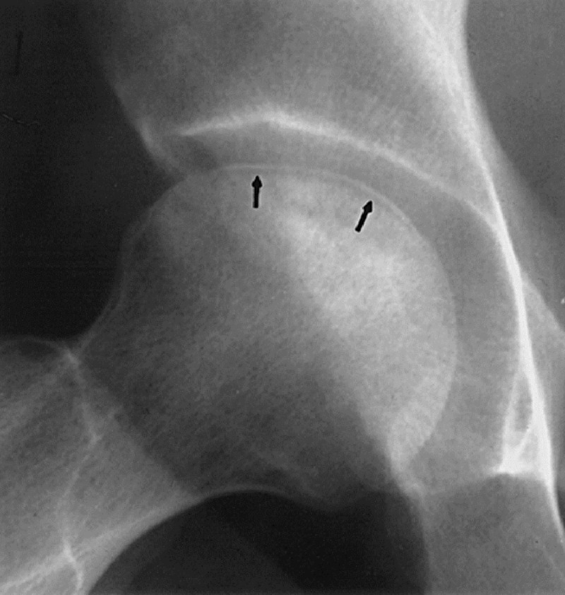
Crescent sign. Arrows showing the hypointense crescent. (Courtesy http://onradiology.blogspot.com )
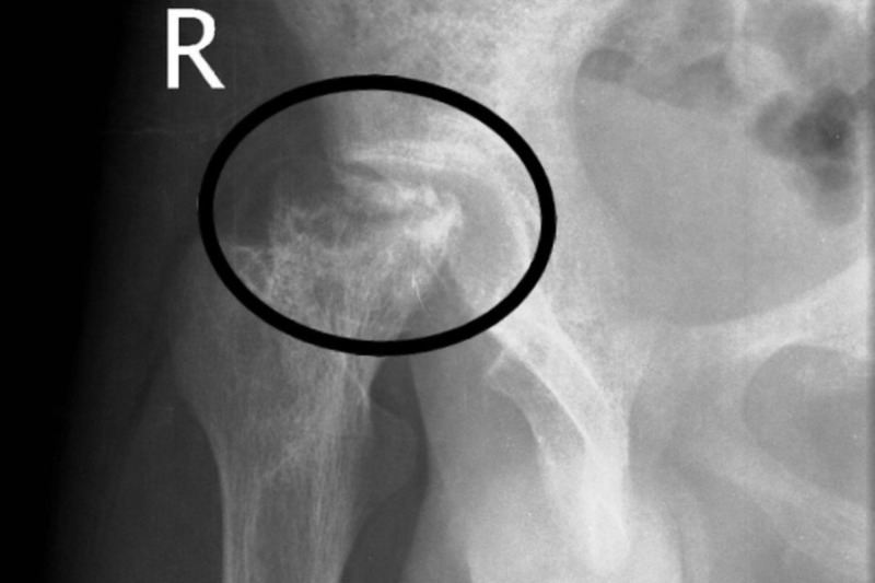
Advanced osteonecrosis shown in the circle.
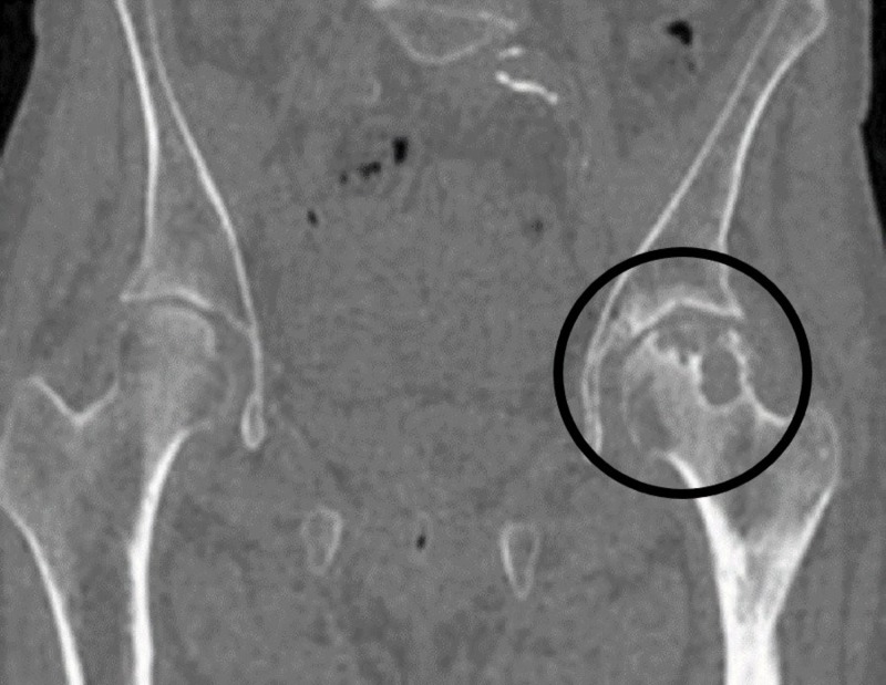
Osteonecrosis left hip encircled.
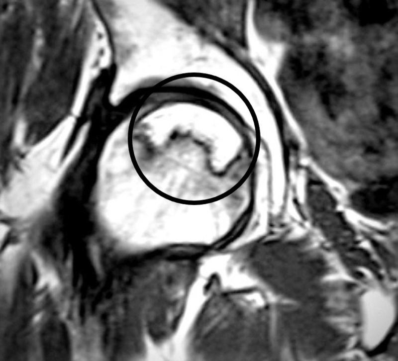
T1 MRI image encircled, showing osteonecrosis in the femoral headband-like lesion. MRI: Magnetic resonance imaging
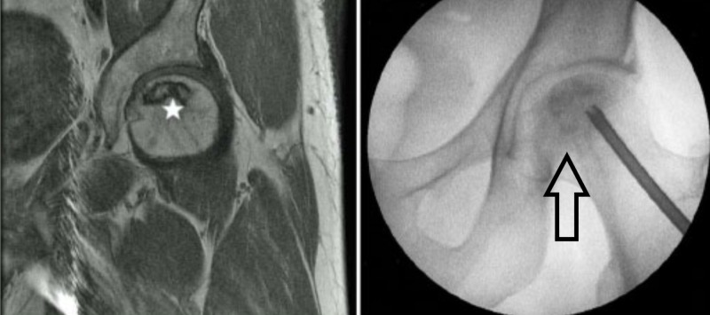
MRI with ONFH (left-sided image; star showing the osteonecrosis); X-ray left hip; arrow showing osteonecrosis and decompression. MRI: Magnetic resonance imaging; ONFH: Osteonecrosis of the femoral head

Post-decompression bone strut using a vascular fibular graft (shown in the circle). (Courtesy: Penn Medicine)
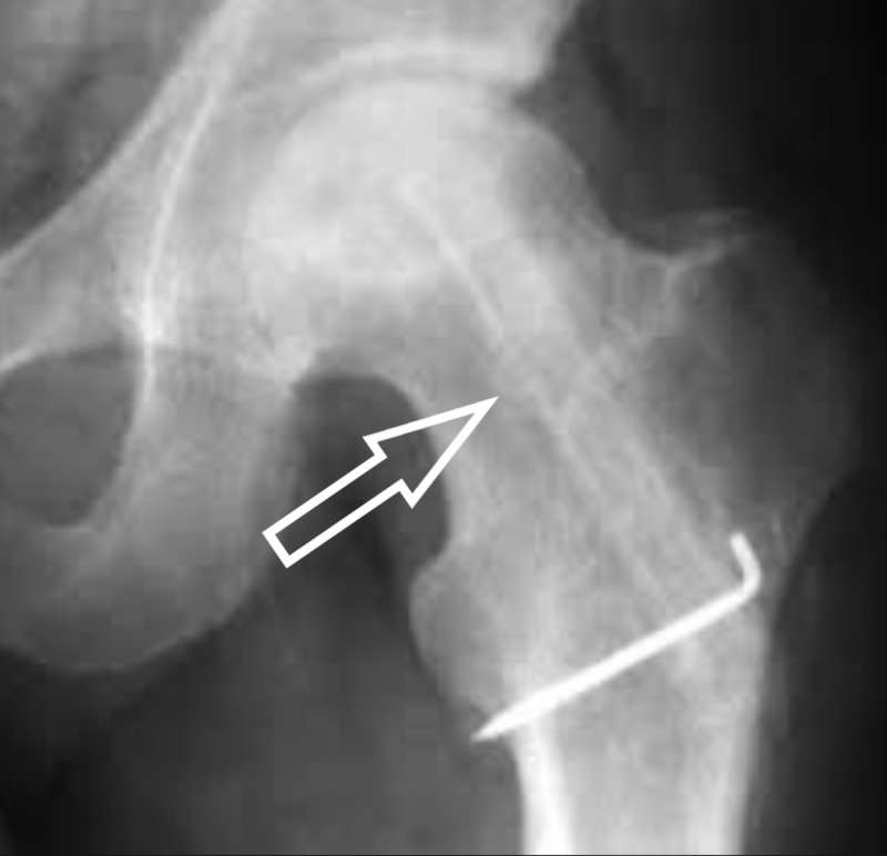
X-ray showing bone grafting after decompression with the help of k-wires (Kirschner wires).
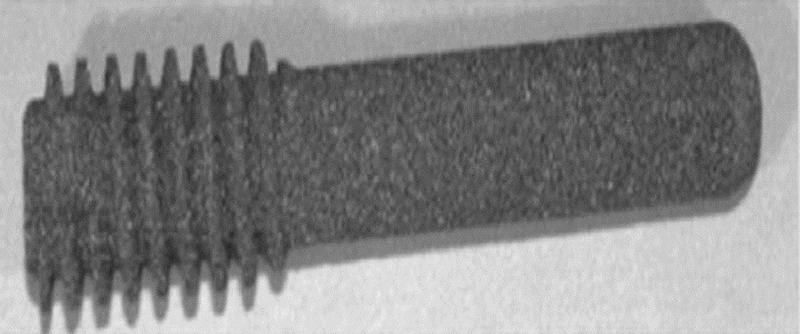
Tantalum rod in vitro.
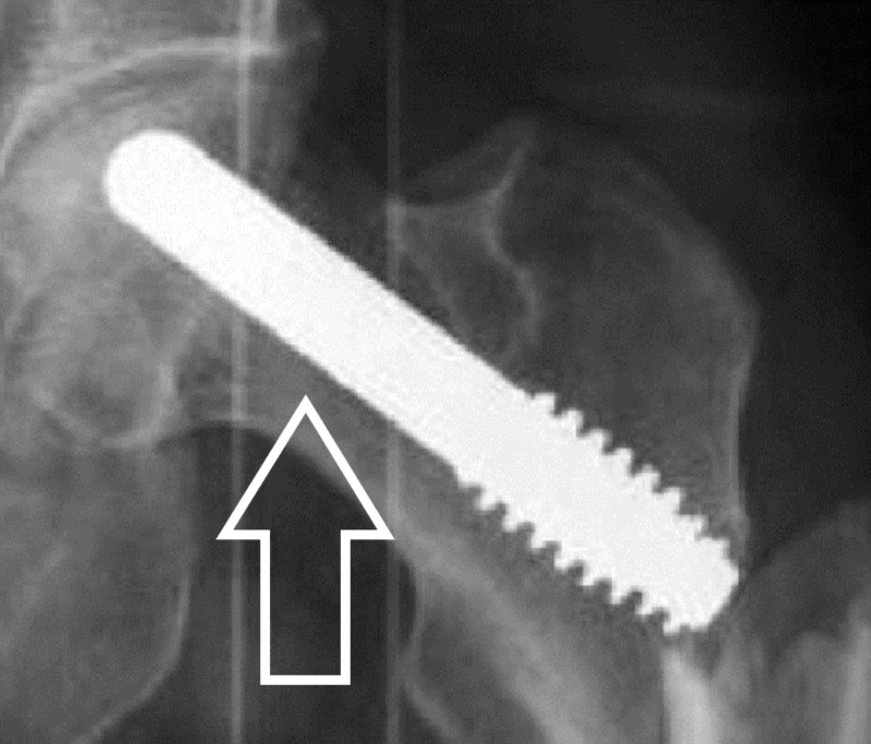
Tantalum rod in vivo highlighted by an arrow.
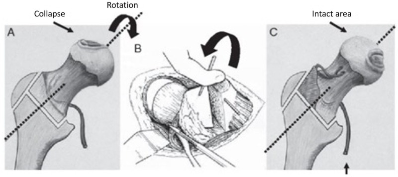
The osteotomy is to rotate the damaged weight-bearing surface, so the undamaged area becomes the weight-bearing part of the femoral head. (Courtesy: Nakashima Y, Kubota H, Yamamoto T, Mawatari T, Motomura G, Iwamoto Y: Transtrochanteric rotational osteotomy for late-onset Legg-Calve-Perthes disease. J Pediatr Orthop. 2011, 31:223-228)
References
-
- Avascular necrosis of the hip - diagnosis and treatment [Article in German] Drescher W, Pufe T, Smeets R, Eisenhart-Rothe R v, Jäger M, Tingart M. Z Orthop Unfall. 2011;149:231–240. - PubMed
-
- The surgical anatomy of the blood supply to the femoral head: description of the anastomosis between the medial femoral circumflex and inferior gluteal arteries at the hip. Grose AW, Gardner MJ, Sussmann PS, Helfet DL, Lorich DG. J Bone Joint Surg Br. 2008;90-B:1298–1303. - PubMed
Publication types
LinkOut - more resources
Full Text Sources