

Antecedents of Soft Drusen, the Specific Deposits of Age-Related Macular Degeneration, in the Biology of Human Macula
- PMID: 30357337
- PMCID: PMC6733529
- DOI: 10.1167/iovs.18-24883
Antecedents of Soft Drusen, the Specific Deposits of Age-Related Macular Degeneration, in the Biology of Human Macula
Abstract
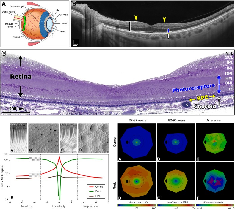
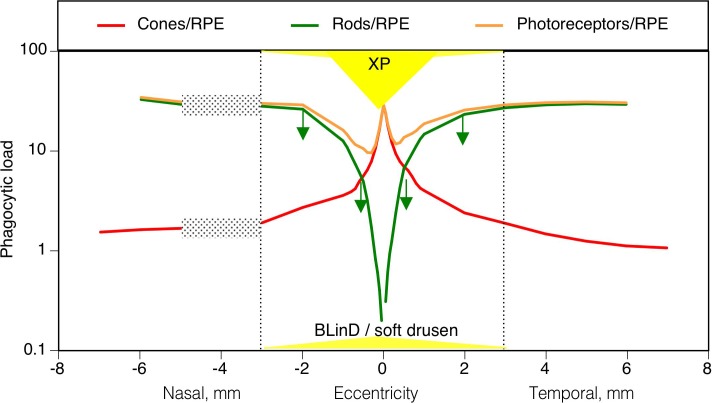
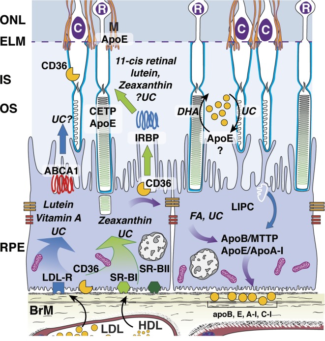
AMD pathobiology was irreversibly changed by the recent discovery of extracellular cholesterol-containing deposits in the subretinal space, between the photoreceptors and retinal pigment epithelium (RPE), called subretinal drusenoid deposits (SDDs). SDDs strikingly mirror the topography of rod photoreceptors in human macula, raising the question of whether an equivalent process results in a deposition related to foveal cones. Herein we propose that AMD's pathognomonic lesion-soft drusen and basal linear deposit (BLinD, same material, diffusely distributed)-is the leading candidate. Epidemiologic, clinical, and histologic data suggest that these deposits are most abundant in the central macula, under the fovea. Strong evidence presented in a companion article supports the idea that the dominant ultrastructural component is large apolipoprotein B,E-containing lipoproteins, constitutively secreted by RPE. Lipoprotein fatty acids are dominated by linoleate (implicating diet) rather than docosahexaenoate (implicating photoreceptors); we seek within the retina cellular relationships and dietary drivers to explain soft druse topography. The delivery of xanthophyll pigments to highly evolved and numerous Müller cells in the human fovea, through RPE, is one strong candidate, because Müller cells are the main reservoir of these pigments, which replenish from diet. We propose that the evolution of neuroglial relations and xanthophyll delivery that underlie exquisite human foveal vision came with a price, that is, soft drusen and sequela, long after our reproductive years.
Figures
References
-
- Zweifel SA, Spaide RF, Curcio CA, Malek G, Imamura Y. Reticular pseudodrusen are subretinal drusenoid deposits. Ophthalmology. 2010;117:303–312.e1. - PubMed
-
- Sarks J, Arnold J, Ho IV, Sarks S, Killingsworth M. Evolution of reticular pseudodrusen. Br J Ophthalmol. 2011;95:979–985. - PubMed
-
- Greferath U, Guymer RH, Vessey KA, Brassington K, Fletcher EL. Correlation of histologic features with in vivo imaging of reticular pseudodrusen. Ophthalmology. 2016;123:1320–1331. - PubMed
Publication types
MeSH terms
Substances
Grants and funding
LinkOut - more resources
Full Text Sources
Medical
