. 2019 Jan 8;47(D1):D315-D321.
doi: 10.1093/nar/gky952.

EncoMPASS: an online database for analyzing structure and symmetry in membrane proteins

Affiliations

EncoMPASS: an online database for analyzing structure and symmetry in membrane proteins

Edoardo Sarti et al. Nucleic Acids Res. .

Abstract

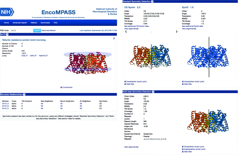
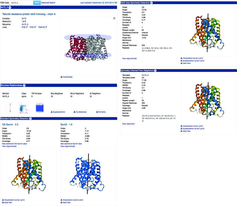
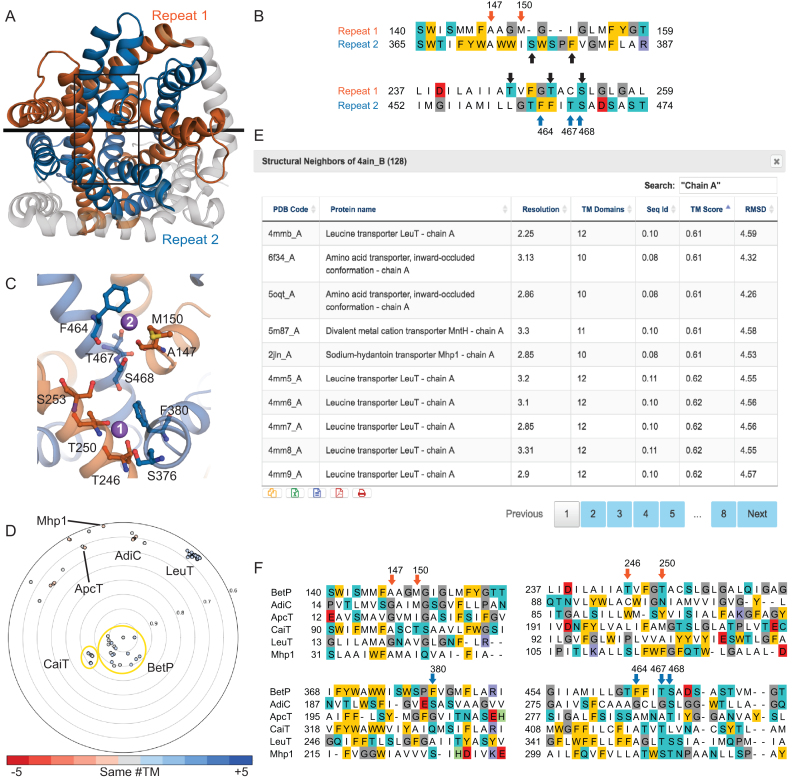
The EncoMPASS online database (http://encompass.ninds.nih.gov) collects, organizes, and presents information about membrane proteins of known structure, emphasizing their structural similarities as well as their quaternary and internal symmetries. Unlike, e.g. SCOP, the EncoMPASS database does not aim for a strict classification of membrane proteins, but instead is organized as a protein chain-centric network of sequence and structural homologues. The online server for the EncoMPASS database provides tools for comparing the structural features of its entries, making it a useful resource for homology modeling and active site identification studies. The database can also be used for inferring functionality, which for membrane proteins often involves symmetry-related mechanisms. To this end, the online database also provides a comprehensive description of both the quaternary and internal symmetries in known membrane protein structures, with a particular focus on their orientation relative to the membrane.


Figures

Figure 1.
Figure 1.
Example EncoMPASS online database complex page. Representative page for the whole structure of PDB entry 3M75 (a TehA homologue), including a brief summary of the characteristics of each of its transmembrane subunits (left) and analysis of the symmetries within the complex (right) using two standard symmetry detection algorithms as well as the multi-step symmetry detection (MSSD) method. Note that on these pages the MSSD section contains results only if quaternary symmetries (i.e., involving multiple chains) have been detected. According to all three methods (CE-Symm, SymD, and MSSD), the TehA homologue forms a three-fold circularly (C3) symmetric assembly. Note that in the molecular viewer in the SymD section, the repeats are not differentiated by color, since SymD does not report residue ranges for the symmetry-related repeats. According to the MSSD results, the axis of this C3 symmetry is perpendicular to the membrane plane, i.e., the protomers have parallel topologies.
Figure 2.
Figure 2.
Example EncoMPASS online database chain page. Representative page for chain A of PDB entry 3M75. These pages follow the same outline as for the complex in Figure 1, but contain analysis specific to each chain, including sequence and structural relationships, and any detected internal symmetries. Chain A of 3M75 has 39 sequence and structure neighbors. (Visualization of the polar or scatter plots, shown here as clickable icons, reveals that these structures are all closely related, both structurally and by sequence.) In terms of the internal symmetry, all methods agree that chain A contains a five-fold circular pseudo-symmetry (C5), although there are some differences in the details. As in the pages for complexes, the MSSD section reports the relationship of the symmetry axis to the membrane plane. The last section (‘Symmetry Inferred from Neighbors’), only available for chains, reports results based on inference from putative structural homologues, in cases where MSSD detected a more comprehensive symmetry than in the chain of interest. By inferring the symmetry information from another TehA-homologue structure, 3M72 chain A, this approach detected a symmetry relationship for 3M75 chain A with greater coverage (86% of the chain, compared to 77% when using MSSD) and a higher similarity (TM-score 0.70 rather than 0.68).
Figure 3.
Figure 3.
Examining structure-function relationships in the sodium-coupled betaine transporter BetP and its homologues using EncoMPASS. (A) Structural symmetry reported by MSSD for BetP (PDB identifier 4AIN chain B), in which residues 135–326 (repeat 1, orange) are pseudo-C2-symmetric to residues 360–542 (repeat 2, blue), and related by an axis (black line) that lies in the plane of the membrane. The protein is viewed from the extracellular side of the membrane. The protein is shown in cartoon helices; elements of the structure not related by symmetry are colored gray. Note that ligands are not included in this representation. (B) Sequence alignment between repeats extracted from the structural alignment in the regions contributing residues to the Na2 binding site. The Na1 site residues are Ala147, Met150 from repeat 1 (orange arrows) and Phe464, Thr467 and Ser468 from repeat 2 (blue arrows). Residues in a similar region of the other repeat (black arrows) are candidates for the Na1 binding site. (C) Close-up of the Na2 and putative Na1-binding site regions in the area outlined with a black box in (A). Sodium ions are shown as purple spheres, and residues discussed in the main text are shown in ball-and-stick format. (D) Structural relationships between BetP and all other structures in EncoMPASS with TM-scores <0.6. Structures of another BCCT family-member, CaiT are closely related (TM-score ∼0.85), while structures of other LeuT-fold family members are more distantly related. Example LeuT-fold family members are labelled. Each point is colored according to the difference in number of transmembrane segments from the chain in the center, according to the provided scale. (E) Clicking on the entry for ‘All Neighbors’ on the ‘Structure Relationships’ section of the chain page for 4AIN chain B brings up a table of structural homologues, which lists several relevant features and their structural and sequence similarity. The results shown were additionally filtered by the term ‘Chain A’ and sorted by TM-score to reveal the least similar structures. (F) Structure-based sequence alignments of representative structural homologues relative to BetP allow interrogation of putative sodium binding sites or sodium-independent mechanisms. Alignments were extracted from pairwise structure alignments provided in EncoMPASS, and combined using pyali (https://github.com/christang/pyali). The positions of Na1 and Na2 site residues in BetP are indicated by arrows colored by repeat.

References

    1. Berman H.M., Westbrook J., Feng Z., Gilliland G., Bhat T.N., Weissig H., Shindyalov I.N., Bourne P.E.. The Protein Data Bank. Nucleic Acids Res. 2000; 28:235–242. - PMC - PubMed
    1. Hubbard T.J.P., Murzin A.G., Brenner S.E., Chothia C.. SCOP: a structural classification of proteins database. Nucleic Acids Res. 1997; 25:236–239. - PMC - PubMed
    1. Orengo C., Michie A., Jones S., Jones D., Swindells M., Thornton J.. CATH – a hierarchic classification of protein domain structures. Structure. 1997; 5:1093–1109. - PubMed
    1. Neumann S., Fuchs A., Mulkidjanian A., Frishman D.. Current status of membrane protein structure classification. Proteins Struct. Funct. Bioinf. 2010; 78:1760–1773. - PubMed
    1. Sojo V., Dessimoz C., Pomiankowski A., Lane N.. Membrane proteins are dramatically less conserved than water-soluble proteins across the tree of life. Mol Biol Evol. 2016; 33:2874–2884. - PMC - PubMed

Publication types

Substances