Review
. 2019 Apr;68(4):661-672.
doi: 10.1007/s00262-018-2264-3. Epub 2018 Oct 24.

Role of myeloid regulatory cells (MRCs) in maintaining tissue homeostasis and promoting tolerance in autoimmunity, inflammatory disease and transplantation

Affiliations
Review

Role of myeloid regulatory cells (MRCs) in maintaining tissue homeostasis and promoting tolerance in autoimmunity, inflammatory disease and transplantation

Giada Amodio et al. Cancer Immunol Immunother. 2019 Apr.

Abstract

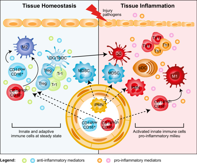
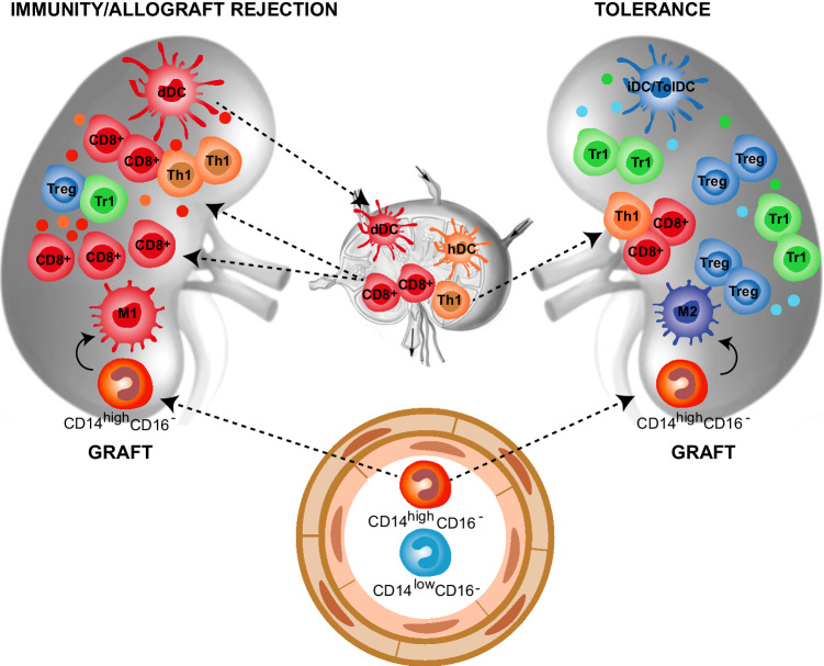
Myeloid cells play a pivotal role in regulating innate and adaptive immune responses. In inflammation, autoimmunity, and after transplantation, myeloid cells have contrasting roles: on the one hand they initiate the immune response, promoting activation and expansion of effector T-cells, and on the other, they counter-regulate inflammation, maintain tissue homeostasis, and promote tolerance. The latter activities are mediated by several myeloid cells including polymorphonuclear neutrophils, macrophages, myeloid-derived suppressor cells, and dendritic cells. Since these cells have been associated with immune suppression and tolerance, they will be further referred to as myeloid regulatory cells (MRCs). In recent years, MRCs have emerged as a therapeutic target or have been regarded as a potential cellular therapeutic product for tolerance induction. However, several open questions must be addressed to enable the therapeutic application of MRCs including: how do they function at the site of inflammation, how to best target these cells to modulate their activities, and how to isolate or to generate pure populations for adoptive cell therapies. In this review, we will give an overview of the current knowledge on MRCs in inflammation, autoimmunity, and transplantation. We will discuss current strategies to target MRCs and to exploit their tolerogenic potential as a cell-based therapy.

Keywords: Dendritic cells; Monocytes/macrophages; Mye-EUNITER; Myeloid regulatory cells (MRCs); Polymorphonuclear neutrophils; Tolerance.


Conflict of interest statement

The authors declare that they have no conflict of interests.

Figures

Fig. 1
Fig. 1
Myeloid regulatory cell contribution in tissue homeostasis and inflammation. Several subsets of myeloid regulatory cells (MRCs) are involved in preventing uncontrolled responses, in maintaining tissue homeostasis, and in promoting resolution of inflammation. Tissue homeostasis. Tissue-resident non-inflammatory M2 macrophages, immature and specialized DC subsets (iDC/TolDC), and MDSCs promote tissue homeostasis via different mechanisms: (1) secretion of anti-inflammatory mediators, such as IL-10 and TGF-β, and expression of IDO; (2) induction of T-regulatory cells, both FOXP3+ Tregs and Tr1 cells; (3) generation of a non-inflammatory milieu that leads to the differentiation of migrating classical inflammatory CD14highCD16 and not classical CD14lowCD16+ monocytes into anti-inflammatory M2 macrophages, which contribute to T-regulatory cell induction. Tissue inflammation. Upon tissue injury or pathogen entry, PMNs are recruited at the site of inflammation and, by secreting pro-inflammatory mediators, lead to the activation of plasmacytoid DC (pDCs), which consequently release IFN-α. The inflammatory milieu promotes the recruitment of classical inflammatory CD14highCD16 monocytes to the site of inflammation and their differentiation into pro-inflammatory M1 macrophages, and the activation and maturation of DCs. These cells in turn promote Th1 and Th17 cell responses via secretion of pro-inflammatory cytokines, such as IL-12 and IL-23. It still remains to be clarified whether MDSCs contribute to tissue inflammation
Fig. 2
Fig. 2
Myeloid cells in allograft rejection and tolerance. Myeloid cells play a central role in allograft rejection and tolerance induction after transplantation. Immunity/allograft rejection. Donor myeloid DCs (dDCs) migrate to the secondary lymphoid organs and activate recipient allo-reactive effector CD8+ and CD4+ (Th1) cells, which migrate back into the graft where they mediate rejection. Moreover, dying dDCs in the draining lymph nodes release alloAgs, host DCs (hDCs) uptake donor-derived alloAgs and contribute to the activation of alloAgs-specific effector CD8+ and CD4+ (Th1) cells. Within the graft, classical inflammatory CD14highCD16 monocytes are recruited from the circulation and differentiate into M1 macrophages that, by secreting pro-inflammatory mediators, contribute to the expansion of effector alloAg-specific T-cells. The limited number of T-regulatory cells (Tregs and Tr1 cells) present within the graft is not sufficient to control the massive infiltration of effector alloAg-specific T-cells. Tolerance. The graft microenvironment enriched of anti-inflammatory mediators, including IL-10, TGF-β, and CSF-1, leads to the differentiation of migrating classical inflammatory CD14highCD16 monocytes into anti-inflammatory M2 macrophages, which promote alloAgs-specific T-regulatory (Treg and Tr1) cells. In addition, the recruitment and/or induction of immature and tolerogenic DC subsets (iDC/TolDC) within the graft sustains the expansion/induction of alloAgs-specific T-regulatory (Treg and Tr1) cells, leading to long-term transplantation tolerance

References

    1. Majewski P, Majchrzak-Gorecka M, Grygier B, Skrzeczynska-Moncznik J, Osiecka O, Cichy J. Inhibitors of serine proteases in regulating the production and function of neutrophil extracellular traps. Front Immunol. 2016;7:261. doi: 10.3389/fimmu.2016.00261. - DOI - PMC - PubMed
    1. Diana J, Simoni Y, Furio L, Beaudoin L, Agerberth B, Barrat F, Lehuen A. Crosstalk between neutrophils, B-1a cells and plasmacytoid dendritic cells initiates autoimmune diabetes. Nat Med. 2013;19:65–73. doi: 10.1038/nm.3042. - DOI - PubMed
    1. Desnues B, Macedo AB, Ordonez-Rueda D, Roussel-Queval A, Malissen B, Bruhns P, Malissen M, Alexopoulou L. The transcriptional repressor Gfi1 prevents lupus autoimmunity by restraining TLR7 signaling. Eur J Immunol. 2016;46:2801–2811. doi: 10.1002/eji.201646573. - DOI - PubMed
    1. Kessenbrock K, Krumbholz M, Schonermarck U, Back W, Gross WL, Werb Z, Grone HJ, Brinkmann V, Jenne DE. Netting neutrophils in autoimmune small-vessel vasculitis. Nat Med. 2009;15:623–625. doi: 10.1038/nm.1959. - DOI - PMC - PubMed
    1. Vono M, Lin A, Norrby-Teglund A, Koup RA, Liang F, Lore K. Neutrophils acquire the capacity for antigen presentation to memory CD4(+) T cells in vitro and ex vivo. Blood. 2017;129:1991–2001. doi: 10.1182/blood-2016-10-744441. - DOI - PMC - PubMed

MeSH terms

LinkOut - more resources