. 2019 Jan 1;316(1):L216-L228.
doi: 10.1152/ajplung.00538.2017. Epub 2018 Oct 25.

Divergent changes of p53 in pulmonary arterial endothelial and smooth muscle cells involved in the development of pulmonary hypertension

Affiliations

Divergent changes of p53 in pulmonary arterial endothelial and smooth muscle cells involved in the development of pulmonary hypertension

Ziyi Wang et al. Am J Physiol Lung Cell Mol Physiol. .

Abstract

The tumor-suppressive role of p53, a transcription factor that regulates the expression of many genes, has been linked to cell cycle arrest, apoptosis, and senescence. The noncanonical function or the pathogenic role of p53 has more recently been implicated in pulmonary vascular disease. We previously reported that rapid nuclear accumulation of hypoxia-inducible factor (HIF)-1α in pulmonary arterial smooth muscle cells (PASMCs) upregulates transient receptor potential channels and enhances Ca2+ entry to increase cytosolic Ca2+ concentration ([Ca2+]cyt). Also, we observed differences in HIF-1α/2α expression in PASMCs and pulmonary arterial endothelial cells (PAECs). Here we report that p53 is increased in PAECs, but decreased in PASMCs, isolated from mice with hypoxia-induced pulmonary hypertension (PH) and rats with monocrotaline (MCT)-induced PH (MCT-PH). The increased p53 in PAECs from rats with MCT-PH is associated with an increased ratio of Bax/Bcl-2, while the decreased p53 in PASMCs is associated with an increased HIF-1α. Furthermore, p53 is downregulated in PASMCs isolated from patients with idiopathic pulmonary arterial hypertension compared with PASMCs from normal subjects. Overexpression of p53 in normal PASMCs inhibits store-operated Ca2+ entry (SOCE) induced by passive depletion of intracellularly stored Ca2+ in the sarcoplasmic reticulum, while downregulation of p53 enhances SOCE. These data indicate that differentially regulated expression of p53 and HIF-1α/2α in PASMCs and PAECs and the cross talk between p53 and HIF-1α/2α in PASMCs and PAECs may play an important role in the development of PH via, at least in part, induction of PAEC apoptosis and PASMC proliferation.

Keywords: Bcl-2 proteins; endothelial cell apoptosis; p53; smooth muscle cell proliferation; tumor-suppressor gene.


Conflict of interest statement

No conflicts of interest, financial or otherwise, are declared by the authors.

Figures

Fig. 1.
Fig. 1.
The protein level of p53 is downregulated in pulmonary arteries (PA) [mainly composed of PA smooth muscle cells (PASMCs)] but upregulated in lung tissue [mainly consisting of lung endothelial cells (ECs)] in mice with hypoxia-induced pulmonary hypertension (PH) and rats with monocrotaline-induced PH (MCT-PH). A: representative immunohistochemistry images (left) showing p53 level (shown in brown) in PA from normoxic (Nor) and chronically hypoxic (Hyp, 10% O2 for 14 days) mice. Right: summarized data (means ± SE) showing the percentage of p53-positive cells from Nor and Hyp mice. Scale bar = 50 µm. B: representative images (left) and summarized data (means ± SE, n = 5; right) showing Western blot analysis on p53 in isolated PA (PA, mainly containing PASMCs) and whole lung tissue (Lung, mainly containing ECs) in Nor and Hyp mice. *P < 0.05, **P < 0.01 and ***P < 0.001 vs. Nor. C and D: representative images (left) and summarized data (means ± SE, n = 5; right) showing Western blot analyses of CD31 (an EC marker) and α-smooth muscle actin (SMA; a SMC marker) in the whole lung tissue (Lung) and isolated PA (PA) from normal mice (C), as well as in mouse ECs and SMCs (D). E: representative images (left) and summarized data (means ± SE, n = 5; right) showing Western blot analysis on p53 in the isolated PA (PA) and whole lung tissue (Lung) from control (Cont) rats and rats with MCT-PH (MCT). **P < 0.01 and ***P < 0.001 vs. Cont. F: representative record of right ventricular pressure (RVP; left) and summarized data (means ± SE; right) showing right ventricular systolic pressure (RVSP) in Cont and MCT rats. ***P < 0.001 vs. Cont.
Fig. 2.
Fig. 2.
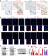
Chronic hypoxia selectively increases hypoxia-inducible factor-1α (HIF-1α) in pulmonary arterial smooth muscle cells (PASMCs) and HIF-2α in pulmonary arterial endothelial cells (PAECs). A: representative immunohistochemistry images showing HIF-1α (a) and HIF-2α (b) levels (shown in brown) in PA from normoxic (Nor) and chronically hypoxic (Hyp, 10% O2 for 21 days) mice. Scale bar = 100 µm. B: representative immunofluorescence images showing the colocalization of HIF-1α (a) and HIF-2α (b) levels (shown in red) together with the smooth muscle marker α-smooth muscle actin (SMA; shown in green) in PA from normoxic (Normoxia) and chronically hypoxic (Hypoxia, 10% O2 for 21 days) mice. Scale bar = 200 µm. C: representative images (a) and summarized data (means ± SE; n = 5; b) showing Western blot analysis on HIF-1α, HIF-2α, p21, Bax, and Bcl-2 in isolated PA (PA, mainly containing PASMCs) and whole lung tissue (Lung, mainly containing endothelial cells) in Nor and Hyp mice. *P < 0.05, **P < 0.01 and ***P < 0.001 vs. Nor.
Fig. 3.
Fig. 3.
Decreased p53 in isolated pulmonary arteries (PA) [mainly composed of PA smooth muscle cells (PASMCs)] is associated with the decreased Bax/Bcl-2 ratio, while increased p53 in whole lung tissue (mainly containing ECs) is associated with increased Bax/Bcl-2, in rats with monocrotaline-mediated PH (MCT-PH) compared with normal control (Cont) rats. A: Western blot analysis of Bax, Bcl-2, and p53 in isolated PAs from control and MCT rats (left). Summarized data (means ± SE; n = 5; right) showing the ratio of Bax to Bcl-2 and protein level of p53 in PA from control and MCT rats. B: Western blot analysis on Bax, Bcl-2, and p53 in whole lung tissues from control and MCT rats (left). Summarized data (means ± SE; right) showing the ratio of Bax to Bcl-2 and protein level of p53 in lung tissues from control and MCT rats. **P < 0.01 and ***P < 0.001 vs. Cont.
Fig. 4.
Fig. 4.
Hypoxia decreases p53 and Bax/Bcl-2 ratio in human pulmonary arterial smooth muscle cells (PASMCs) and increases p53 and Bax/Bcl-2 ratio in human pulmonary arterial endothelial cells (PAECs). A: representative images (a) and summarized data (means ± SE, n = 5; b–d) showing Western blot analysis of p53 (b), Bax/Bcl-2 (c), and hypoxia-inducible factor (HIF)-1α (d) in human PASMCs incubated under normoxic (Nor) and hypoxic (Hyp, 3% O2 for 4, 6, 8, 16, and 24 h) conditions. B: representative images (a) and summarized data (means ± SE; n = 5; b–d) showing Western blot analysis of p53 (b), Bax/Bcl-2 (c), and HIF-2α (d) in human PAECs incubated under normoxic (Nor) and hypoxic (Hyp, (3% O2 for 4, 6, 8, 16 and 24 h) conditions. C: representative images (a) and summarized data (means ± SE, n = 5; b–f) showing Western blot analyses of p53 (b), Bax/Bcl-2 (c), and HIF-1α (d) in human PASMCs incubated under normoxic (Nor) and hypoxic (Hyp, (3% O2 for 48 and 72 h) conditions. Summarized data on cell proliferation and cell apoptosis, determined by 5-ethynyl-2′-deoxyuridine (EdU) incorporation (e) and terminal deoxynucleotidyl transferase dUTP-mediated nick-end labeling (TUNEL) assay (f) in Nor and Hyp PASMCs is also shown. D: representative images (a) and summarized data (means ± SE, n = 5; b–f) showing Western blot analysis of p53 (b), Bax/Bcl-2 (c), and HIF-2α (d) in human PAECs incubated under normoxic (Nor) and hypoxic (Hyp, 3% O2 for 48 and 72 h) conditions. Summarized data on cell proliferation and cell apoptosis, determined by EdU incorporation (e) and TUNEL assay (f) in Nor and Hyp PAECs, are also shown. For these studies, all cells used were between 5 and 8 passages. We compare the same passage number of cells for each experiment. *P < 0.05, **P < 0.01 and ***P < 0.001 vs. Nor.
Fig. 5.
Fig. 5.
Decreased p53 in human pulmonary arterial smooth muscle cells (PASMCs) is associated with the decreased Bax/Bcl-2 ratio, while increased p53 in human pulmonary arterial endothelial cells (PAECs) is associated with increased Bax/Bcl-2, in patients with idiopathic pulmonary arterial hypertension (IPAH), compared with normal controls. A: Western blot analysis of p53, Bax, and Bcl-2 in normal and IPAH PASMCs (left). Summarized data (means ± SE, n = 5; right) showing the protein level of p53 and the ratio of Bax/Bcl-2 in normal and IPAH PASMCs. B: Western blot analysis of p53, Bax, and Bcl-2 in normal and IPAH PAECs (left). Summarized data (means ± SE; right) showing the protein levels of p53 and the ratio of Bax/Bcl-2 in normal and IPAH PAECs. For these studies, all cells used were between 5 and 8 passages. We compare the same passage number of cells for each experiment. **P < 0.01 and ***P < 0.001 vs. Normal.
Fig. 6.
Fig. 6.
Overexpression of p53 downregulates Transient receptor potential channel (TRPC) attenuates SOCE and inhibits pulmonary arterial smooth muscle cell (PASMC) proliferation and migration, while downregulation of p53 upregulates TRPC channels, enhances SOCE, and increases PASMC proliferation and migration. A: Western blot analysis of p53, TRPC1, and TRPC6 in human PASMC transfected with an empty vector and p53. B: representative record showing changes in cytosolic Ca2+ concentration ([Ca2+]cyt) before, during, and after application of 10 µM cyclopiazonic acid (CPA) in the absence (0Ca) or presence of extracellular Ca2+ in human PASMC transfected with an empty vector and p53. The bar graph shows the amplitude of [Ca2+]cyt increases due to CPA-induced SOCE (means ± SE; n = 5). C: cell proliferation [bromodeoxyuridine (BrdU) incorporation; means ± SE; n = 5] and migration (%cells migrated; means ± SE; n = 5) in PASMCs transfected with vector and p53. D: Western blot analyses of p53, TRPC1, and TRPC6 in human PASMCs transfected with siRNA-NT and siRNA-p53. E: representative record showing changes in [Ca2+]cyt before, during and after application of CPA in the absence (0Ca) or presence of extracellular Ca2+ in human PASMCs transfected with siRNA-NT and siRNA-p53. The bar graph shows the amplitude of [Ca2+]cyt increases due to CPA-induced SOCE (means ± SE; n = 5). F: cell proliferation (BrdU incorporation; means ± SE, n = 5) and migration (% cells migrated; means ± SE; n = 5) in human PASMCs transfected with siRNA-NT and siRNA-p53. G: Western blot analysis of p53, STIM1, and STIM2 in human PASMC transfected with siRNA-NT and siRNA-p53 (left). Summarized data (means ± SE; right) showing the protein levels of p53, STIM1, and STIM2 in control PASMCs and siRNA-p53 treated PASMCs. H: Western blot analysis of p53, Orai1, and Orai2 in human PASMC transfected with siRNA-NT and siRNA-p53 (left). Summarized data (means ± SE; n = 5; right) showing the protein levels of p53, Orai1, and Orai2 in control PASMCs and siRNA-p53-treated PASMCs. For these studies, all cells used were between 5 and 8 passages. We compared the same passage number of cells for each experiment. *P < 0.05 and ***P < 0.001 vs. vector or siRNA-NT.
Fig. 7.
Fig. 7.
Increasing hypoxia-inducible factor (Hif) by pharmacologically blocking prolyl hydroxylase domain proteins (PHDs) upregulates p53 in human pulmonary arterial endothelial cells (PAECs) and mRNA expression level of PHDs is comparable in lung tissues from normoxic and chronically hypoxic mice. A: Western blot analysis of HIF-1α and HIF-2α in normal human PAECs treated with vehicle (Cont) and FG-4592 (FG, 100 µM for 48 h), a selective blocker of PHDs. Summarized data (means ± SE; n = 5; right) showing protein levels of HIF-1α and HIF-2α in control PAECs and FG-treated PAECs. B: Western blot analysis of p53, Bax and Bcl-2 in control PAECs and FG-treated PAECs. Summarized data (means ± SE, n = 5; right) showing the protein levels of p53 and the ratio of Bax/Bcl-2 in control PAECs and FG-treated PAECs. C: Western blot analysis of HIF-1α and HIF-2α in normal human PASMCs treated with vehicle (Cont) and FG-4592 (FG, 100 µM for 48 h). Summarized data (means ± SE, n = 5; right) showing protein levels of HIF-1α and HIF-2α in control pulmonary arterial smooth muscle cells (PASMCs) and FG-treated PASMCs. D: Western blot analysis of p53, Bax and Bcl-2 in control PASMCs and FG-treated PASMCs. Summarized data (means ± SE, n = 5; right) showing the protein levels of p53, Bax, and Bcl-2 in control PASMCs and FG-treated PASMCs. E: RNA was extracted and regular quantitative RT-PCR was performed to determine mRNA expression levels for Phd1, Phd2, and Phd3 (left). Summarized data (means ± SE, n = 5; right) showing the mRNA levels of Phd1, Phd2, and Phd3 in whole lung tissues from normoxia and hypoxia (10% O2 for 21 days) mice. F: summarized data (means ± SE, n = 5) represented mRNA levels for Phd1, Phd2, and Phd3 in whole lung tissues from normoxia and hypoxia (10% O2 for 21 days) mice, using real-time quantitative RT-PCR. For these studies, all cells used were between 5 and 8 passages. We compared the same passage number of cells for each experiment. *P < 0.05, **P < 0.01, and ***P < 0.001 vs. Cont or Nor.

Similar articles

Cited by

References

    1. Abid S, Houssaïni A, Mouraret N, Marcos E, Amsellem V, Wan F, Dubois-Randé JL, Derumeaux G, Boczkowski J, Motterlini R, Adnot S. P21-dependent protective effects of a carbon monoxide-releasing molecule-3 in pulmonary hypertension. Arterioscler Thromb Vasc Biol 34: 304–312, 2014. doi:10.1161/ATVBAHA.113.302302. - DOI - PubMed
    1. Archer SL, Gomberg-Maitland M, Maitland ML, Rich S, Garcia JG, Weir EK. Mitochondrial metabolism, redox signaling, and fusion: a mitochondria-ROS-HIF-1α-Kv1.5 O2-sensing pathway at the intersection of pulmonary hypertension and cancer. Am J Physiol Heart Circ Physiol 294: H570–H578, 2008. doi:10.1152/ajpheart.01324.2007. - DOI - PubMed
    1. Archer SL, Weir EK, Wilkins MR. Basic science of pulmonary arterial hypertension for clinicians: new concepts and experimental therapies. Circulation 121: 2045–2066, 2010. doi:10.1161/CIRCULATIONAHA.108.847707. - DOI - PMC - PubMed
    1. Ball MK, Waypa GB, Mungai PT, Nielsen JM, Czech L, Dudley VJ, Beussink L, Dettman RW, Berkelhamer SK, Steinhorn RH, Shah SJ, Schumacker PT. Regulation of hypoxia-induced pulmonary hypertension by vascular smooth muscle hypoxia-inducible factor-1α. Am J Respir Crit Care Med 189: 314–324, 2014. doi:10.1164/rccm.201302-0302OC. - DOI - PMC - PubMed
    1. Berra E, Benizri E, Ginouvès A, Volmat V, Roux D, Pouysségur J. HIF prolyl-hydroxylase 2 is the key oxygen sensor setting low steady-state levels of HIF-1α in normoxia. EMBO J 22: 4082–4090, 2003. doi:10.1093/emboj/cdg392. - DOI - PMC - PubMed

Publication types

MeSH terms

Substances