. 2019 Jan 1;316(1):L94-L104.
doi: 10.1152/ajplung.00564.2017. Epub 2018 Oct 25.

Acute pulmonary effects of aerosolized nicotine

Affiliations

Acute pulmonary effects of aerosolized nicotine

Shama Ahmad et al. Am J Physiol Lung Cell Mol Physiol. .

Abstract

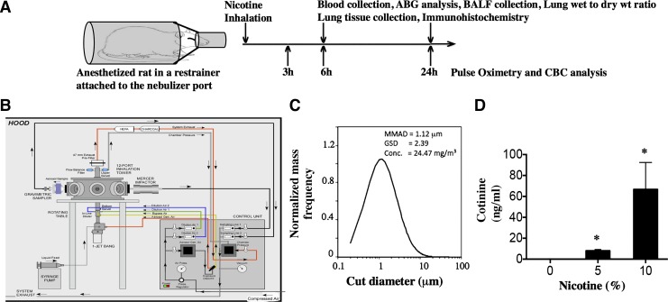
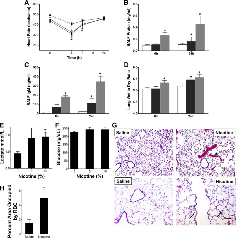
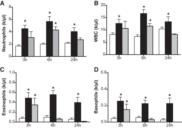
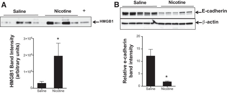
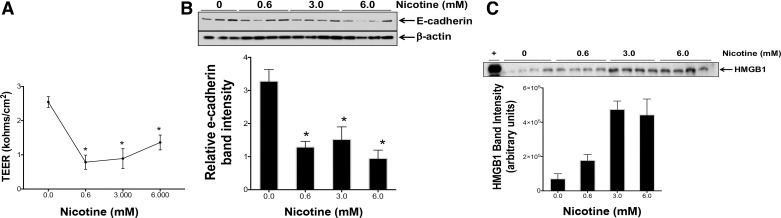
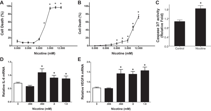
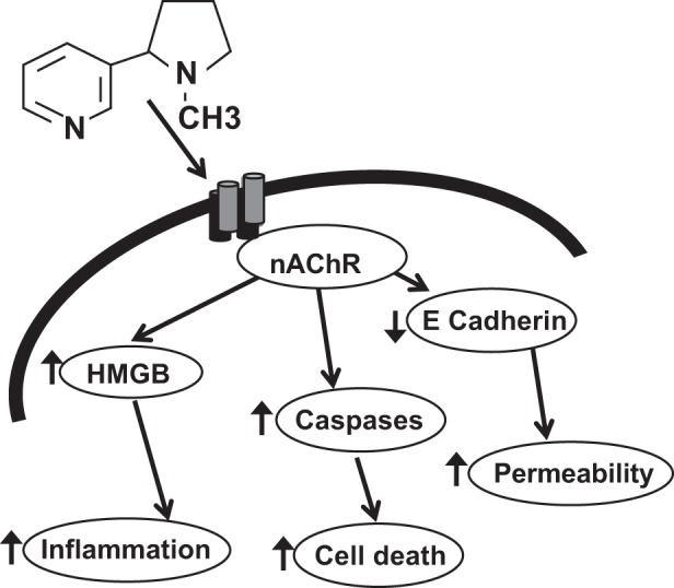
Nicotine is a highly addictive principal component of both tobacco and electronic cigarette that is readily absorbed in blood. Nicotine-containing electronic cigarettes are promoted as a safe alternative to cigarette smoking. However, the isolated effects of inhaled nicotine are largely unknown. Here we report a novel rat model of aerosolized nicotine with a particle size (~1 μm) in the respirable diameter range. Acute nicotine inhalation caused increased pulmonary edema and lung injury as measured by enhanced bronchoalveolar lavage fluid protein, IgM, lung wet-to-dry weight ratio, and high-mobility group box 1 (HMGB1) protein and decreased lung E-cadherin protein. Immunohistochemical analysis revealed congested blood vessels and increased neutrophil infiltration. Lung myeloperoxidase mRNA and protein increased in the nicotine-exposed rats. Complete blood counts also showed an increase in neutrophils, white blood cells, eosinophils, and basophils. Arterial blood gas measurements showed an increase in lactate. Lungs of nicotine-inhaling animals revealed increased mRNA levels of IL-1A and CXCL1. There was also an increase in IL-1α protein. In in vitro air-liquid interface cultures of airway epithelial cells, there was a dose dependent increase in HMGB1 release with nicotine treatment. Air-liquid cultures exposed to nicotine also resulted in a dose-dependent loss of barrier as measured by transepithelial electrical resistance and a decrease in E-cadherin expression. Nicotine also caused a dose-dependent increase in epithelial cell death and an increase in caspase-3/7 activities. These results show that the nicotine content of electronic cigarettes may have adverse pulmonary and systemic effects.

Keywords: aerosol; lung injury; nicotine; pulmonary edema; rats.


Conflict of interest statement

No conflicts of interest, financial or otherwise, are declared by the authors.

Figures

Fig. 1.
Fig. 1.
Nicotine aerosol generation, characterization, and exposure of rats. Sprague-Dawley rats were anesthetized and exposed to saline or nicotine aerosol for 15 min and transferred to room air. A: schematic representation of our exposure paradigm. Rats were monitored by pulse oximetry and clinical scoring after nose-only exposure nicotine aerosol. At the end of exposure rats were euthanized and necropsy was performed to evaluate parameters as shown in the experimental design (top). B: schematic of the CH-technology liquid particle generator and exposure system that utilizes Bio-Aerosol Nebulizing Generator (BANG) device to produce appropriate nicotine particles for alveolar delivery. C: plot showing particle size distribution of a 5% solution of nicotine in ethanol. The solution was injected into the BANG and an air flow of 6 l/min and a pressure of 60 psi was used to generate the aerosol. MMAD, mass median aerodynamic diameter; GSD, geometric standard deviation. D: cotinine, a nicotine metabolite was estimated in the plasma of rats exposed to aerosolized nicotine (15 min using 5 or 10% nicotine in normal saline). Plasma was collected and analyzed for cotinine 6 h after exposure. Data shown are means ± SE (n = 6). *P < 0.05 from the unexposed controls.
Fig. 2.
Fig. 2.
Acute nicotine inhalation causes depression in heart rate and pulmonary injury. Sprague-Dawley rats were exposed to aerosolized saline or nicotine for 15 min at 2 different concentrations (5 or 10%) of nicotine in normal saline. A: the heart rate (HR) was monitored at various time intervals by pulse oximetry (-■-: saline, -●-: 5% nicotine; -▲-: 10% nicotine group). P < 0.05 from HR before exposure of the same animal in A and from unexposed controls. B and C: animals were euthanized at 6 or 24 h postexposure and BALF was collected and protein (B) and IgM (C) content determined as described in the methods. D: lung wet-to-dry (W/D) weight ratios were also evaluated. The right upper lobe (RUL) was used to determine the weights. In BD, the open bars represent saline, black bars represent 5% nicotine and gray bars represent 10% saline. Data shown are means ± SE (n = 6). * P < 0.05 from saline controls. E and F: arterial blood gas measurements showing blood lactate (E) and glucose (F) are shown. G: representative hematoxylin and eosin-stained paraffin embedded lung sections from saline or nicotine (10%, 24 h)-exposed rats at ×10 (top) or ×20 (bottom) resolution are shown. Arrowhead demonstrates RBC congested areas that are quantified at bottom (H). The arrows demonstrate neutrophils. *P < 0.05 from saline controls.
Fig. 3.
Fig. 3.
Complete blood counts following nicotine inhalation. Rats were exposed to aerosols of saline or 5 or 10% solution of nicotine. Blood was collected from the tail vein at different time intervals and complete blood counts (CBCs) were determined using a Hemavet 950S differential cell counter. AD: neutrophils (A), white blood cells (WBC) (B), eosinophils (C), and basophils (D) were plotted. In AD, the open bars represent saline, black bars represent 5% nicotine and gray bars represent 10% nicotine. Data shown are means ± SE (n = 6). *P < 0.05 from saline control.
Fig. 4.
Fig. 4.
Inhaled nicotine caused increase in expression of genes in the inflammatory pathway. Rats were exposed to aerosols of saline or 10% solution of nicotine. Lungs were harvested after 24 h and total RNA was extracted. AC: mRNA encoding Myeloperoxidase (MPO; A) was estimated by using real-time RT-PCR analysis (B), MPO protein was measured by Western blot analysis and quantified (C). mRNA levels of IL-1a (D) and CXCL1 (E) were also estimated. F: lysates were prepared of the right middle lobe and analyzed by Luminex (IL-1α shown). Data shown are means ± SE (n = 5). *P < 0.05 from saline control.
Fig. 5.
Fig. 5.
Nicotine causes release of high-mobility group box 1 (HMGB1) and disruption of pulmonary epithelial barrier integrity. A: rats were exposed to saline or 10% nicotine aerosol following which BALF was collected at 24 h and immunoblot for HMGB1 was performed. B: 24 h following nicotine inhalation lungs were harvested and lysates were prepared and analyzed for E-cadherin by Western blots. (+) represents a positive HMGB1 control. The band intensity was measured for quantification, and the representative data are shown as means ± SE (n = 5). *P < 0.05 from saline control.
Fig. 6.
Fig. 6.
Nicotine causes loss of cell junction protein, disruption of epithelial barrier integrity and release of high-mobility group box 1 (HMGB1). A: air-liquid interface cultures of primary human bronchial epithelial cells were treated with varying concentrations of nicotine applied as thin layer (100 μl media) on the apical surface. B: transepithelial electrical resistance was measured using an epithelial voltohmmeter electrode after 24 h. C: supernatant media were collected 24 h later and cell lysates were prepared and analyzed for E-cadherin by Western blot. Released HMGB1 was measured using Western blot in the supernatant media. Data shown are means ± SE (n = 4). Figure represents experiments performed in airway epithelial cells isolated from at least 3 different donor tissues. (+) represents a positive HMGB1 control. *P < 0.05 from the values in the absence of nicotine.
Fig. 7.
Fig. 7.
Nicotine causes airway and alveolar epithelial cell death and expression of proinflammatory genes. A and B: to demonstrate effect of nicotine on airway and alveolar epithelial cells 16HBE (A) and A549 cells (B) were plated on 12 well plates (at 50 k/well) and exposed 24 h later to various concentrations of nicotine. Cell death was assessed 24 h later using Trypan blue dye. C: caspase-3/7 assay was carried out in supernatant media of A549 cells treated with nicotine (3 mM) for 6 h. Data shown are means ± SE (n = 8). *P < 0.05 from the values in the absence of nicotine. Nicotine-treated A549 cells were harvested after 24 h and total RNA was extracted. D and E: steady-state mRNA levels of IL-6 (D) and VEGFA (E) were estimated by real-time RT-PCR analysis using specific Taqman primers and probes. The expression level of each gene was normalized to 18S RNA and reported as fold change. Data shown are means ± SE (n = 6). *P < 0.05 from saline control.
Fig. 8.
Fig. 8.
Schematic representation of effects of inhaled nicotine aerosol on the lung. Nicotine acts via nicotinic acetylcholine receptor (nAChR) to cause release of damage associated molecular patterns (DAMPS) such as high-mobility group box 1 (HMGB1) causing increased inflamation, apoptotic cell death and increase cell permeability by decreasing cellular junction protens such as E-cadherin.

References

    1. Abraham E, Arcaroli J, Carmody A, Wang H, Tracey KJ. HMG-1 as a mediator of acute lung inflammation. J Immunol 165: 2950–2954, 2000. doi:10.4049/jimmunol.165.6.2950. - DOI - PubMed
    1. Adriani W, Macrì S, Pacifici R, Laviola G. Peculiar vulnerability to nicotine oral self-administration in mice during early adolescence. Neuropsychopharmacology 27: 212–224, 2002. doi:10.1016/S0893-133X(02)00295-6. - DOI - PubMed
    1. Ahmad A, Ahmad S, Glover L, Miller SM, Shannon JM, Guo X, Franklin WA, Bridges JP, Schaack JB, Colgan SP, White CW. Adenosine A2A receptor is a unique angiogenic target of HIF-2alpha in pulmonary endothelial cells. Proc Natl Acad Sci USA 106: 10684–10689, 2009. doi:10.1073/pnas.0901326106. - DOI - PMC - PubMed
    1. Ahmad S, Ahmad A, Dremina ES, Sharov VS, Guo X, Jones TN, Loader JE, Tatreau JR, Perraud AL, Schöneich C, Randell SH, White CW. Bcl-2 suppresses sarcoplasmic/endoplasmic reticulum Ca2+-ATPase expression in cystic fibrosis airways: role in oxidant-mediated cell death. Am J Respir Crit Care Med 179: 816–826, 2009. doi:10.1164/rccm.200807-1104OC. - DOI - PMC - PubMed
    1. Ahmad S, Nichols DP, Strand M, Rancourt RC, Randell SH, White CW, Ahmad A. SERCA2 regulates non-CF and CF airway epithelial cell response to ozone. PLoS One 6: e27451, 2011. doi:10.1371/journal.pone.0027451. - DOI - PMC - PubMed

Publication types

MeSH terms

LinkOut - more resources