. 2019 Jan 1;316(1):L175-L186.
doi: 10.1152/ajplung.00551.2017. Epub 2018 Oct 25.

HDAC8 inhibition ameliorates pulmonary fibrosis

Affiliations

HDAC8 inhibition ameliorates pulmonary fibrosis

Shigeki Saito et al. Am J Physiol Lung Cell Mol Physiol. .

Abstract

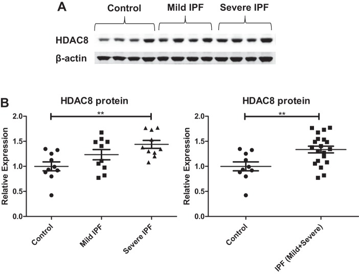
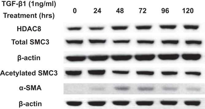
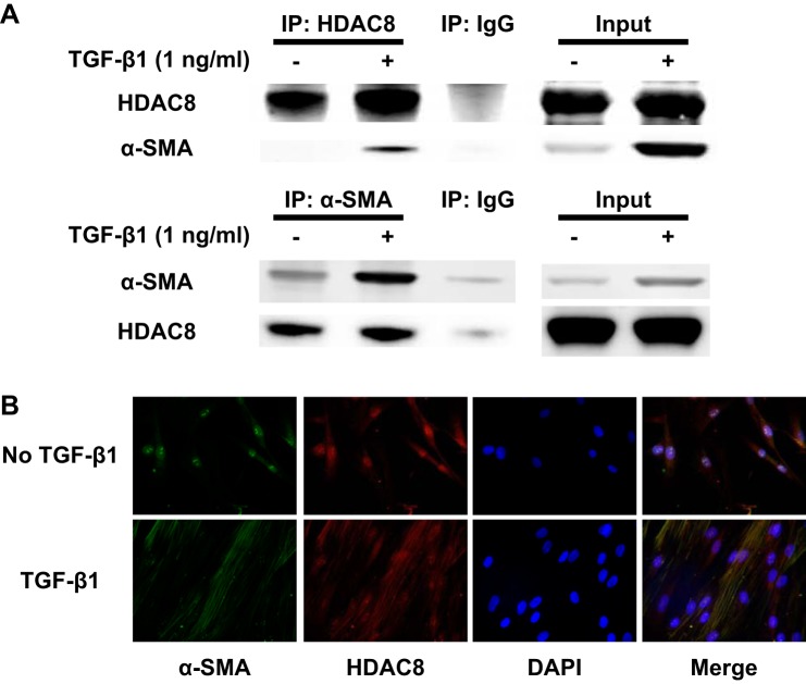
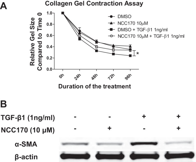
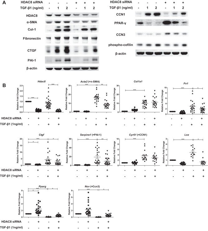
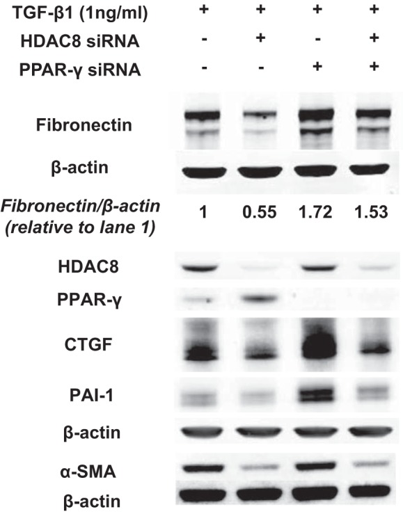
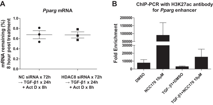
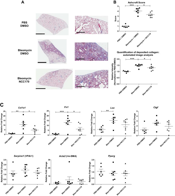
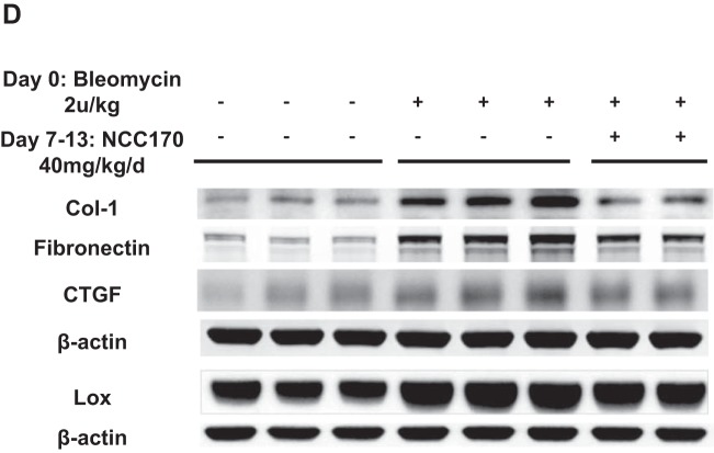
Idiopathic pulmonary fibrosis (IPF) is a fibroproliferative lung disease, and fibroblast-myofibroblast differentiation (FMD) is thought to be a key event in the pathogenesis of IPF. Histone deacetylase-8 (HDAC8) has been shown to associate with α-smooth muscle actin (α-SMA; a marker of FMD) and regulates cell contractility in vascular smooth muscle cells. However, the role of HDAC8 in FMD or pulmonary fibrosis has never been reported. This study investigated the role of HDAC8 in pulmonary fibrosis with a focus on FMD. We observed that HDAC8 expression was increased in IPF lung tissue as well as transforming growth factor (TGF)β1-treated normal human lung fibroblasts (NHLFs). Immunoprecipitation experiments revealed that HDAC8 was associated with α-SMA in TGFβ1-treated NHLFs. HDAC8 inhibition with NCC170 (HDAC8-selective inhibitor) repressed TGFβ1-induced fibroblast contraction and α-SMA protein expression in NHLFs cultured in collagen gels. HDAC8 inhibition with HDAC8 siRNA also repressed TGFβ1-induced expression of profibrotic molecules such as fibronectin and increased expression of antifibrotic molecules such as peroxisome proliferator-activated receptor-γ (PPARγ). Chromatin immunoprecipitation quantitative PCR using an antibody against H3K27ac (histone H3 acetylated at lysine 27; a known HDAC8 substrate and a marker for active enhancers) suggested that HDAC8 inhibition with NCC170 ameliorated TGFβ1-induced loss of H3K27ac at the PPARγ gene enhancer. Furthermore, NCC170 treatment significantly decreased fibrosis measured by Ashcroft score as well as expression of type 1 collagen and fibronectin in bleomycin-treated mouse lungs. These data suggest that HDAC8 contributes to pulmonary fibrosis and that there is a therapeutic potential for HDAC8 inhibitors to treat IPF as well as other fibrotic lung diseases.

Keywords: FMD; HDAC8; IPF; PPARγ; TGFβ1; bleomycin; fibronectin; type 1 collagen; α-SMA.


Conflict of interest statement

No conflicts of interest, financial or otherwise, are declared by the authors.

Figures

Fig. 1.
Fig. 1.
Histone deacetylase-8 (HDAC8) expression is increased in idiopathic pulmonary fibrosis (IPF) lungs. A: immunoblots of lung homogenates. β-Actin was used as a loading control. Control: forced vital capacity (FVC) ≥ 80% predicted; “Mild” IPF: FVC 50–79% predicted; “Severe” IPF: FVC < 50% predicted. FVC is a physiological parameter of lung function and a marker for disease severity in IPF. B: densitometry analyses of the immunoblots. HDAC8 protein expression is significantly increased in IPF lungs compared with control lungs. Left: HDAC8 protein expression is significantly increased in severe IPF lungs compared with controls (**P < 0.01, control vs. severe IPF). Although the comparison between control vs. mild IPF or mild IPF vs. severe IPF did not reach statistical significance, HDAC8 expression appeared to correlate with disease severity. Right: HDAC8 protein expression is significantly increased in IPF lungs compared with controls, when mild IPF and severe IPF were combined for analysis [**P < 0.01, control vs. IPF (mild + severe)].
Fig. 2.
Fig. 2.
Transforming growth factor-β1 (TGFβ1) mildly increases histone deacetylase-8 (HDAC8) protein expression and decreases structural maintenance of chromosomes protein-3 (SMC3) acetylation. Subconfluent normal human lung fibroblasts (NHLFs) were serum starved overnight and then treated with 1 ng/ml TGFβ1 for the designated durations. Immunoblots of HDAC8 and β-actin were performed on lysates prepared from NHLFs. TGFβ1 treatment mildly increased HDAC8 protein expression and decreased acetylation of SMC3 (a known HDAC8 substrate). Immunoblots of the extracts for α-smooth muscle actin (α-SMA) supported the occurrence of fibroblast-myofibroblast differentiation (FMD).
Fig. 3.
Fig. 3.
Histone deacetylase-8 (HDAC8) associates with α-smooth muscle actin (α-SMA) in transforming growth factor-β1 (TGFβ1)-treated normal human lung fibroblasts (NHLFs). Subconfluent NHLFs were serum starved overnight and then treated with TGFβ1 (1ng/ml) for 48 h. A: lysates from untreated and TGFβ1-treated cells were immunoprecipitated (IP) with an anti-HDAC8 antibody, anti-α-SMA antibody, or a nonspecific IgG control. Immunoprecipitated protein was immunoblotted for HDAC8 and α-SMA. The 2 right lanes (“Input”) represent an immunoblot of the lysates without IP. IP using anti-HDAC8 antibody suggested association between HDAC8 and α-SMA. Reverse IP using anti-α-SMA antibody also suggested association between HDAC8 and α-SMA, although HDAC8 was still weakly detected in IP using nonspecific IgG. This weak band was still detected even without nonspecific IgG (data not shown), which suggests that this weak band was due to nonspecific binding of α-SMA to beads. B: immunofluorescent staining revealed colocalization of HDAC8 and α-SMA within stress fibers in TGFβ1-treated NHLFs.
Fig. 4.
Fig. 4.
Histone deacetylase-8 (HDAC8) inhibitor NCC170 represses transforming growth factor-β1 (TGFβ1)-induced fibroblast contraction and α-smooth muscle actin (α-SMA) protein expression in collagen gels. normal human lung fibroblasts (NHLFs) were cultured in rat tail type I collagen gels (2 mg/ml) and treated with DMSO (vehicle) or NCC170, with or without TGFβ1 (1 ng/ml). A: NCC170 repressed TGFβ1-induced fibroblast contraction. The ratio of the area of the gel to culture well measured at 24 h intervals was used to assess gel contraction. Graph shows the area of the gel at indicated time points after treatment with DMSO (●), NCC170 (▲), DMSO+TGFβ1 (■), or NCC170+TGFβ1 (○). NCC170 ameliorated TGFβ1-induced collagen gel contraction (*P < 0.05; TGFβ1+DMSO vs. TGFβ1+NCC170). B: NCC-170 repressed TGFβ1-induced α-SMA protein expression. Gels were digested using type I collagenase (5 mg/ml), and cells were harvested in RIPA buffer at 48 h for detection of α-SMA by immunoblot. An immunoblot of β-actin was included as a loading control.
Fig. 5.
Fig. 5.
Histone deacetylase-8 (HDAC8) knockdown represses transforming growth factor-β1 (TGFβ1)-induced expression of several profibrotic proteins while inducing antifibrotic proteins in normal human lung fibroblasts (NHLFs). Subconfluent NHLFs were treated with negative control siRNA or HDAC8 siRNA for 72 h and then treated with TGFβ1 (1 or 2 ng/ml) for an additional 48 h before preparation of cell extracts in RIPA buffer (A) or an additional 24 h before preparation of total cellular RNA (B). A: immunoblots of the RIPA buffer extract revealed efficient knockdown of HDAC8 120 h posttransfection. HDAC8 knockdown repressed TGFβ1-induced expression of profibrotic proteins such as α-smooth muscle actin (α-SMA), type 1 collagen, fibronectin, connective tissue growth factor (CTGF), plasminogen activator inhibitor 1 (PAI-1), and CCN1, whereas HDAC8 knockdown increased expression of antifibrotic proteins such as peroxisome proliferator-activated receptor-γ (PPARγ) and CCN3. HDAC8 inhibition also repressed TGFβ1-induced phosphorylation of cofilin, an actin-binding protein essential for myofibroblast function. An immunoblot of β-actin is included as a loading control. B: quantitative RT-PCR analyses of total RNA isolated 96 h posttransfection of negative control siRNA or HDAC8 siRNA and 24 h after TGFβ1 treatment. Graphs show fold changes of the indicated mRNA after correction to the mRNA level of a housekeeping gene (36B4, a ribosomal protein) calculated by the 2−ΔΔCT method. Analyses confirmed efficient knockdown of HDAC8 mRNA (top left). HDAC8 knockdown repressed TGFβ1-induced mRNA expression of profibrotic genes such as α-SMA, fibronectin, CTGF, PAI-1, and lysyl oxidase (LOX), whereas HDAC8 knockdown increased mRNA expression of antifibrotic genes such as PPARγ and CCN3 (*P < 0.05, **P < 0.01, ***P < 0.001).
Fig. 6.
Fig. 6.
Histone deacetylase-8 (HDAC8) knockdown does not repress transforming growth factor-β1 (TGFβ1)-induced phosphorylation of Smad2/3 or Akt. Subconfluent normal human lung fibroblasts (NHLFs) were treated with negative control siRNA or HDAC8 siRNA for 72 h and then treated with TGFβ1 (1 ng/ml) for 30 min or 12 h. Immunoblots show that HDAC8 inhibition did not repress TGFβ1-induced phosphorylation of Smad2/3 or Akt.
Fig. 7.
Fig. 7.
Histone deacetylase-8 (HDAC8) knockdown represses transforming growth factor-β1 (TGFβ1)-induced expression of fibronectin, likely via peroxisome proliferator-activated receptor-γ (PPARγ)-dependent mechanism. Subconfluent normal human lung fibroblasts (NHLFs) were treated with negative control siRNA or HDAC8 siRNA, or PPARγ siRNA, or HDAC8 siRNA + PPARγ siRNA for 72 h and then treated with TGFβ1 (1 ng/ml) for an additional 48 h. Immunoblots were performed to probe for HDAC8, PPARγ, fibronectin, connective tissue growth factor (CTGF), plasminogen activator inhibitor 1 (PAI-1), and α-smooth muscle actin (α-SMA). An immunoblot of β-actin was included as a loading control. HDAC8 knockdown failed to significantly repress TGFβ1-induced expression of fibronectin when PPARγ was knocked down. This suggests that the repressive effect of HDAC8 inhibition on TGFβ1-induced expression of fibronectin was mostly PPARγ dependent. In contrast, HDAC8 knockdown was still able to repress TGFβ1-induced expression of CTGF, PAI-1, and α-SMA when PPARγ was knocked down. This suggests that the repressive effect of HDAC8 inhibition on TGFβ1-induced expression of CTGF/PAI-1/α-SMA was likely PPARγ independent and not due to upregulation of PPARγ. Fibronectin/β-actin denotes the ratio of fibronectin to β-actin expression, calculated using densitometry analyses of the immunoblots.
Fig. 8.
Fig. 8.
Histone deacetylase-8 (HDAC8) inhibition increases peroxisome proliferator-activated receptor-γ (PPARγ) expression, possibly by increasing H3K27 acetylation of the PPARγ gene enhancer. A: subconfluent normal human lung fibroblasts (NHLFs) were treated with negative control siRNA or HDAC8 siRNA for 72 h and then treated with transforming growth factor-β1 (TGFβ1, 1 ng/ml) for 24 h followed by addition of actinomycin D (Act D; 10 μg/ml) for 8 h to evaluate the effect of HDAC8 inhibition on PPARγ mRNA stability. Levels of PPARγ were determined by quantitative RT-PCR using 36B4 as an internal control. HDAC8 inhibition did not enhance PPARγ mRNA stability in NHLFs treated with TGFβ1 and actinomycin D. B: subconfluent NHLFs were pretreated with DMSO or NCC170 for 24 h and then treated with DMSO or NCC170 ± TGFβ1 for 24 h. Chromatin immunoprecipitation (ChIP)-PCR was performed using an antibody against acetylated histone 3 (H3)K27 (H3K27ac, a known HDAC8 substrate and marker for active enhancers) or IgG (as a control) and primers for the PPARγ gene enhancer region. For each treatment group, the ChIP signal was normalized to IgG control and expressed as “Fold Enrichment.” Averaged results of 3 independent experiments suggested that, in the enhancer region, 1) HDAC8 inhibition significantly increased the level of H3K27ac in the absence of TGFβ1; 2) TGFβ1 decreased the H3K27ac level; and 3) HDAC8 inhibition ameliorated TGFβ1-induced loss of H3K27ac.
Fig. 9.
Fig. 9.
NCC170-treated mice are protected against bleomycin-induced pulmonary fibrosis. Mice were treated with PBS or bleomycin (2 U/kg, by oropharyngeal aspiration) on day 0, followed by daily intraperitoneal injection of DMSO or NCC170 (40 mg·kg−1·day−1) on day 8 through day 13 (n = 6–7/group) and harvest of the lungs on day 14. A and B: collagen expression was assessed using Masson’s trichrome staining of lung sections. The entire lung tissue section on a single slide was scanned with Aperio Digital Pathology Whole Slide Scanner. A: representative images (taken at ×40 magnification) are shown. Scale bars, 3 mm (left), 300 μm (right). B, top: extent of lung fibrosis was quantified according to an 8-tier, modified Ashcroft scale. NCC170 treatment significantly ameliorated bleomycin-induced pulmonary fibrosis. Bottom: amount of deposited collagen was quantified using automated image analysis of the Masson trichrome staining. NCC170 treatment significantly repressed collagen deposition in bleomycin-induced pulmonary fibrosis. *P < 0.05, **P < 0.01, ***P < 0.001. C: mRNA expression levels were assessed using quantitative RT-PCR of lung homogenates. NCC170 treatment significantly diminished bleomycin-induced expression of type 1 collagen (Col1a1) and fibronectin (Fn1) mRNA. Lox, lysyl oxidase; Ctgf, connective tissue growth factor; PAI-1, plasminogen activator inhibitor 1; α-SMA, α-smooth muscle actin; Pparg, peroxisome proliferator-activated receptor-γ. *P < 0.05, **P < 0.01, ***P < 0.001. D: protein expression levels were assessed using immunoblots of lung homogenates. NCC170 treatment significantly diminished bleomycin-induced expression of type 1 collagen and fibronectin proteins.
Fig. 9.
Fig. 9.
Continued

Similar articles

Cited by

References

    1. Burgess HA, Daugherty LE, Thatcher TH, Lakatos HF, Ray DM, Redonnet M, Phipps RP, Sime PJ. PPARγ agonists inhibit TGF-β-induced pulmonary myofibroblast differentiation and collagen production: implications for therapy of lung fibrosis. Am J Physiol Lung Cell Mol Physiol 288: L1146–L1153, 2005. doi:10.1152/ajplung.00383.2004. - DOI - PubMed
    1. Conforti F, Davies ER, Calderwood CJ, Thatcher TH, Jones MG, Smart DE, Mahajan S, Alzetani A, Havelock T, Maher TM, Molyneaux PL, Thorley AJ, Tetley TD, Warner JA, Packham G, Ganesan A, Skipp PJ, Marshall BJ, Richeldi L, Sime PJ, O’Reilly KMA, Davies DE. The histone deacetylase inhibitor, romidepsin, as a potential treatment for pulmonary fibrosis. Oncotarget 8: 48737–48754, 2017. doi:10.18632/oncotarget.17114. - DOI - PMC - PubMed
    1. Davies ER, Haitchi HM, Thatcher TH, Sime PJ, Kottmann RM, Ganesan A, Packham G, O’Reilly KM, Davies DE. Spiruchostatin A inhibits proliferation and differentiation of fibroblasts from patients with pulmonary fibrosis. Am J Respir Cell Mol Biol 46: 687–694, 2012. doi:10.1165/rcmb.2011-0040OC. - DOI - PMC - PubMed
    1. Deardorff MA, Bando M, Nakato R, Watrin E, Itoh T, Minamino M, Saitoh K, Komata M, Katou Y, Clark D, Cole KE, De Baere E, Decroos C, Di Donato N, Ernst S, Francey LJ, Gyftodimou Y, Hirashima K, Hullings M, Ishikawa Y, Jaulin C, Kaur M, Kiyono T, Lombardi PM, Magnaghi-Jaulin L, Mortier GR, Nozaki N, Petersen MB, Seimiya H, Siu VM, Suzuki Y, Takagaki K, Wilde JJ, Willems PJ, Prigent C, Gillessen-Kaesbach G, Christianson DW, Kaiser FJ, Jackson LG, Hirota T, Krantz ID, Shirahige K. HDAC8 mutations in Cornelia de Lange syndrome affect the cohesin acetylation cycle. Nature 489: 313–317, 2012. doi:10.1038/nature11316. - DOI - PMC - PubMed
    1. El Agha E, Moiseenko A, Kheirollahi V, De Langhe S, Crnkovic S, Kwapiszewska G, Szibor M, Kosanovic D, Schwind F, Schermuly RT, Henneke I, MacKenzie B, Quantius J, Herold S, Ntokou A, Ahlbrecht K, Braun T, Morty RE, Günther A, Seeger W, Bellusci S. Two-way conversion between lipogenic and myogenic fibroblastic phenotypes marks the progression and resolution of lung fibrosis. Cell Stem Cell 20: 261–273, .e3, 2017. doi:10.1016/j.stem.2016.10.004. - DOI - PMC - PubMed

Publication types

MeSH terms

LinkOut - more resources