

doi: 10.1093/brain/awy273.
Brodmann: a pioneer of human brain mapping-his impact on concepts of cortical organization
Affiliations
- PMID: 30358817
- PMCID: PMC6202576
- DOI: 10.1093/brain/awy273
Item in Clipboard
Brodmann: a pioneer of human brain mapping-his impact on concepts of cortical organization
Brain.
.
Abstract
On the 150th anniversary of Korbinian Brodmann’s birth, and the 100th anniversary of his death, Zilles celebrates his pioneering role in brain mapping. With the aid of hitherto unpublished documents and figures, he explains the concepts behind Brodmann’s cytoarchitectonic maps and considers their impact on current neuroimaging approaches.
Figures
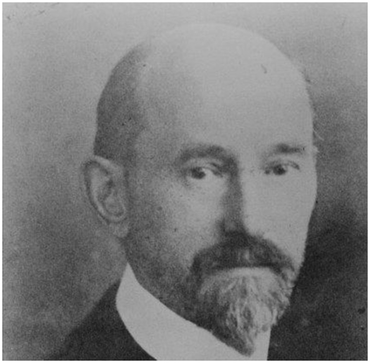
Korbinian Brodmann (17 November 1868–22 August 1918). With permission of the C. & O. Vogt Archive, Institute of Brain Research, University Düsseldorf.
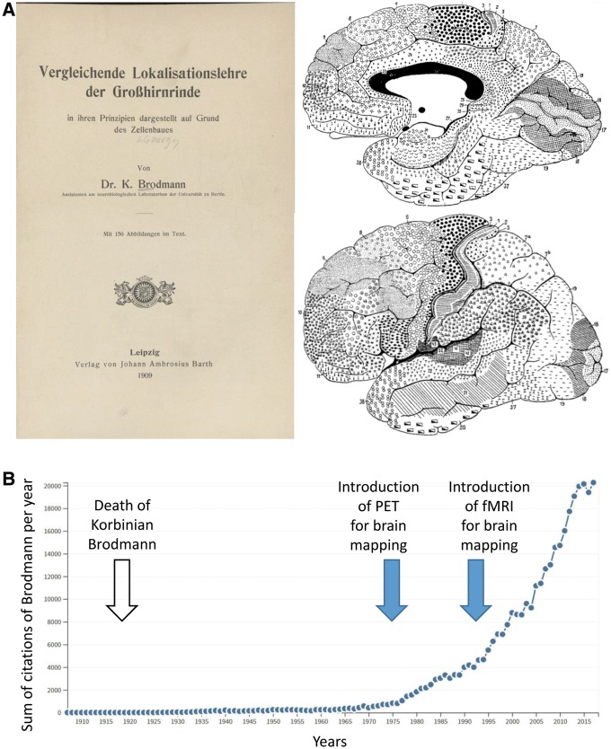
Influence of Brodmann's monography and map on modern day neuroimaging studies. (A) Title page of the famous monography (Brodmann, 1909) and the final version of the map of the entire human cerebral cortex (Brodmann, 1910). (B) Development of the citations of Brodmann’s work up to the year 2017. Source: Web of Science.
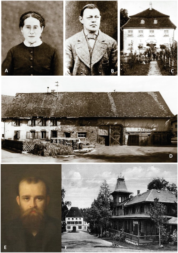
Photographs portraying Brodmann's life as a child and a young adult. Korbinian Brodmann’s mother Sophie Benkler (A) and his father Josef Brodmann (B). Hofgut of the Brodmann family (C) and his birthplace, the farming house of the Benkler family (D). With permission of the Brodmann Museum, Hohenfels-Liggersdorf. Oskar Vogt (E) at the age of 33 years. Painting by W. Döring, 1905. With permission of the C. & O. Vogt Archive, Institute of Brain Research, University Düsseldorf. Alexandersbad (F) around the time of the first encounter of Vogt and Brodmann. With permission of the Brodmann Museum, Hohenfels-Liggersdorf.
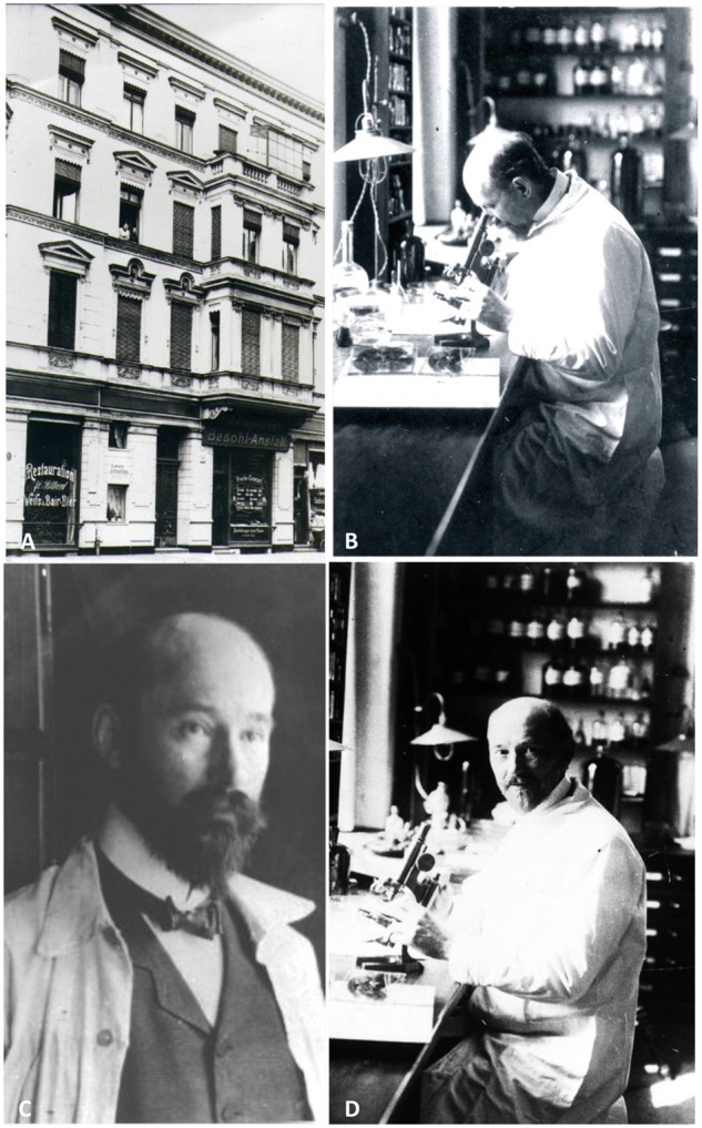
Photographs portraying Brodmann's time in Berlin. (A) ‘Neurobiological Central Station’, Berlin, Magdeburgerstr. 16. (B–D) Korbinian Brodmann during his time in Vogt’s ‘Neurobiologischem Laboratorium’ in Berlin. With permission of the C. & O. Vogt Archive, Institute of Brain Research, University Düsseldorf.
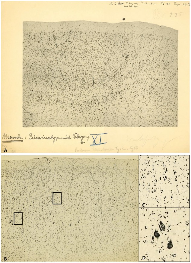
Micrographs of the human visual cortex with annotations by Brodmann. (A) Border (asterisk) between the primary (BA17, ‘Calcarinatypus’, right) and secondary (BA18, left) visual cortex of a human brain. Original micrograph with hand-written notes by K. Brodmann. (B) Brodmann’s area 4 (primary motor cortex) with Betz giant pyramidal cells (left). Note the excellent quality of the photographic technique in the magnified details (inserts C and D on the right; their positions are indicated as rectangles in the original micrograph). With permission of the C. & O. Vogt Archive, Institute of Brain Research, University Düsseldorf.
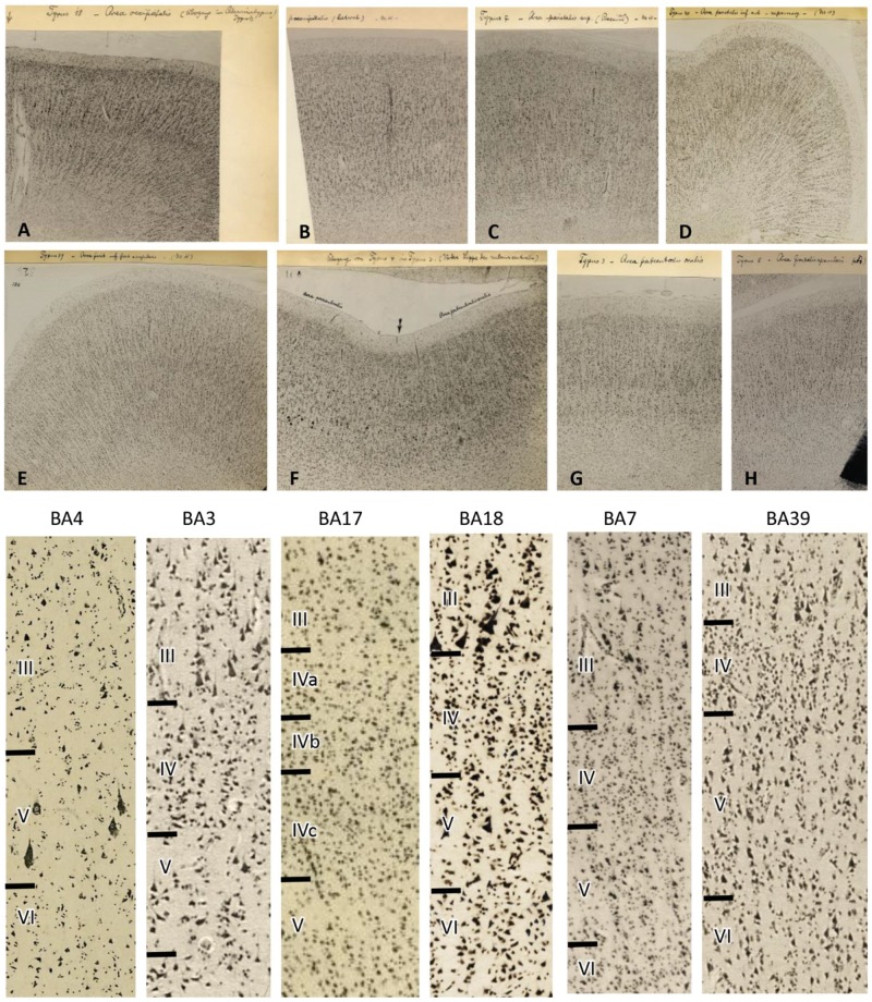
Original micrographs of human cortical areas by K. Brodmann (specimen M15). Transcriptions of Brodmann’s hand writing: (A) Area occipitalis BA18, secondary visual cortex at the transition to BA17. (B) Praeoccipitalis BA19 (higher visual cortex). (C) Area parietalis sup. (precuneus ant) BA7. (D) Area parietalis inf. ant = supramarg BA40, inferior parietal cortex. (E) Area pariet. post. inf = angularis BA39, temporo-occipital cortex. (F) Transition from BA4 to BA3, (primary motor to primary somatosensory cortex). (G) Area postcentralis oralis BA3, primary somatosensory cortex. (H) Area frontalis agranularis BA6, premotor cortex. Bottom: Higher magnifications of the original micrographs (A–H) demonstrate the larger (or at least equally large) pyramidal cells in layer III compared with those cells in layer V. This cytoarchitectonic feature is called ‘externopyramidization’ and typical for higher unimodal (BA18 as an example in the visual system) and multimodal (examples BA7 and BA39) areas. In contrast, the primary motor (BA4), somatosensory (BA3), and visual (BA17) cortices display larger pyramidal cells in layer V than III. Note the wider layer IV in primary sensory (BA3 and BA17) than multimodal (BA7 and BA39) areas, and the lack of a clearly visible layer IV in the primary motor area (BA4). With permission of the C. & O. Vogt Archive, Institute of Brain Research, University Düsseldorf.
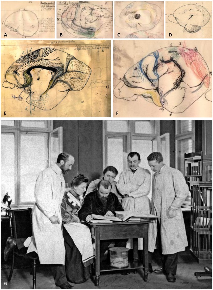
Hand drawings of Brodmann (with permission of the Brodmann Museum, Hohenfels-Liggersdorf, Germany). (A) Lateral view of the left hemisphere of a platypus (Ornithorhynchus paradoxus, Monotremata). (B) Lateral view of the right hemisphere of a spiny anteater (Echidna, Monotremata). (C) Mesial surface of the right hemisphere of a common brushtail possum (Trichosurus vulpecula). ‘Am’ in the yellow circle depicts the Corpus amygdaloideum. Large black dot commissura anterior. The commissura anterior is far smaller in the human than in the marsupial brain, since it preferentially connects allocortical regions, which are large in marsupials but belong to the relatively small (relative to the isocortex) human allocortex (archi- and palaeocortex). (D) Mesial surface of the right hemisphere of the Kinkajou (Cercoleptes caudivolvolus). Fine dots label the primary visual cortex (BA17), other symbols mark the olfactory and entorhinal cortex. (E) Mesial surface of the right hemisphere of a prosimian (Lemur niger). Large black pyramids mark the area praeparietalis (BA5), filled circles the primary motor cortex (BA4), open circles the premotor cortex (BA6), dots the primary visual cortex (BA17), and the large and small crosses the cingulate cortex (par of BA24 and the whole BA23, respectively). This sketch is an early stage of Fig. 99 in the monography (Brodmann, 1909). (F) Lateral view of the left hemisphere of a kangaroo (Macropus pennicillatus). Blue contour primary motor cortex (BA4), red primary visual cortex (BA17), yellow olfactory cortex. (G) Brodmann together with Cécile and Oskar Vogt in the ‘Neurobiological Laboratory’ of the University of Berlin. From left to right Korbinian Brodmann, Cécile and Oskar Vogt, the technician Louise Bosse, and the scientific collaborators Max Lewandowski and Max Borchert. Photo taken around 1905. With permission of the C. & O. Vogt Archive, Institute of Brain Research, University Düsseldorf, Photograph No. 272.
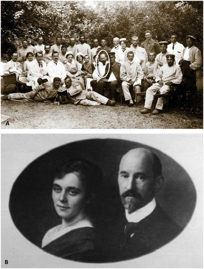
Photographs portraying Brodmann's life as an established MD. (A) Brodmann as a physician in a hospital for soldiers. Photo taken between 1914 and 1916. With permission of the Brodmann Museum, Hohenfels-Liggersdorf. (B) Korbinian Brodmann and his wife Margarete Francke in the year 1917. With permission of the C. & O. Vogt Archive, Institute of Brain Research, University Düsseldorf.
References
-
- Amunts K, Zilles K. A multimodal analysis of structure and function in Broca’s region. In: Grodzinski Y, Amunts K, editors, Broca’s Region. New York, NY: Oxford University Press; 2006. p. 17–30.
-
- Amunts K, Zilles K. Architectonic mapping of the human brain beyond Brodmann. Neuron 2015; 88: 1086–107. - PubMed
-
- Barbas H. Pattern in the laminar origin of corticocortical connections. J Comp Neurol 1986; 1252: 415–22. - PubMed
-
- Barbas H. General cortical and special prefrontal connections: principles from structure to function. Annu Rev Neurosci 2015; 38: 269–89. - PubMed
-
- Bailey P, von Bonin G. The isocortex of man. Urbana, IL: University of Illinois Press; 1951.
Publication types
MeSH terms
Personal name as subject
- Actions
