

Imaging Sodium Flux during Action Potentials in Neurons with Fluorescent Nanosensors and Transparent Microelectrodes
- PMID: 30358986
- PMCID: PMC6958690
- DOI: 10.1021/acssensors.8b00903
Imaging Sodium Flux during Action Potentials in Neurons with Fluorescent Nanosensors and Transparent Microelectrodes
Abstract
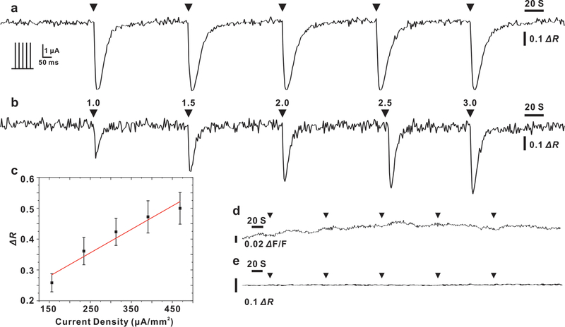
Sodium flux plays a pivotal role in neurobiological processes including initiation of action potentials and regulation of neuronal cell excitability. However, unlike the wide range of fluorescent calcium indicators used extensively for cellular studies, the choice of sodium probes remains limited. We have previously demonstrated optode-based nanosensors (OBNs) for detecting sodium ions with advantageous modular properties such as tunable physiological sensing range, full reversibility, and superb selectivity against key physiological interfering ion potassium. (1) Motivated by bridging the gap between the great interest in sodium imaging of neuronal cell activity as an alternative to patch clamp and limited choices of optical sodium indicators, in this Letter we report the application of nanosensors capable of detecting intracellular sodium flux in isolated rat dorsal root ganglion neurons during electrical stimulation using transparent microelectrodes. Taking advantage of the ratiometric detection scheme offered by this fluorescent modular sensing platform, we performed dual color imaging of the sensor to monitor the intracellular sodium currents underlying trains of action potentials in real time. The combination of nanosensors and microelectrodes for monitoring neuronal sodium dynamics is a novel tool for investigating the regulatory role of sodium ions involved during neural activities.
Keywords: dorsal root ganglion; ion sensing; nanosensor; optode; ratiometric imaging; sodium; transparent microelectrode.
Conflict of interest statement
The authors declare no competing financial interest
Figures


Similar articles
-
Optical Probes for Neurobiological Sensing and Imaging.Acc Chem Res. 2018 May 15;51(5):1023-1032. doi: 10.1021/acs.accounts.7b00564. Epub 2018 Apr 13. Acc Chem Res. 2018. PMID: 29652127 Free PMC article. Review.
-
Polymer-free optode nanosensors for dynamic, reversible, and ratiometric sodium imaging in the physiological range.Sci Rep. 2013 Nov 28;3:3366. doi: 10.1038/srep03366. Sci Rep. 2013. PMID: 24284431 Free PMC article.
-
Transparent arrays of bilayer-nanomesh microelectrodes for simultaneous electrophysiology and two-photon imaging in the brain.Sci Adv. 2018 Sep 5;4(9):eaat0626. doi: 10.1126/sciadv.aat0626. eCollection 2018 Sep. Sci Adv. 2018. PMID: 30191176 Free PMC article.
-
Electrophysiological properties of neurons in intact rat dorsal root ganglia classified by conduction velocity and action potential duration.J Neurophysiol. 1996 Sep;76(3):1924-41. doi: 10.1152/jn.1996.76.3.1924. J Neurophysiol. 1996. PMID: 8890304
-
Microelectrodes for studying neurobiology.Curr Opin Chem Biol. 2008 Oct;12(5):491-6. doi: 10.1016/j.cbpa.2008.06.035. Curr Opin Chem Biol. 2008. PMID: 18675377 Free PMC article. Review.
Cited by
-
Recent Developments in Nanosensors for Imaging Applications in Biological Systems.Annu Rev Anal Chem (Palo Alto Calif). 2019 Jun 12;12(1):109-128. doi: 10.1146/annurev-anchem-061417-125747. Epub 2019 Mar 11. Annu Rev Anal Chem (Palo Alto Calif). 2019. PMID: 30857408 Free PMC article. Review.
-
Nanotechnology and quantum science enabled advances in neurological medical applications: diagnostics and treatments.Med Biol Eng Comput. 2022 Dec;60(12):3341-3356. doi: 10.1007/s11517-022-02664-3. Epub 2022 Oct 8. Med Biol Eng Comput. 2022. PMID: 36207564 Review.
-
Real-time particle-by-particle detection of erythrocyte-camouflaged microsensor with extended circulation time in the bloodstream.Proc Natl Acad Sci U S A. 2020 Feb 18;117(7):3509-3517. doi: 10.1073/pnas.1914913117. Epub 2020 Feb 4. Proc Natl Acad Sci U S A. 2020. PMID: 32019879 Free PMC article.
-
Optical Nanosensors for in vivo Physiological Chloride Detection for Monitoring Cystic Fibrosis Treatment.Anal Methods. 2020 Mar 21;12(11):1441-1448. doi: 10.1039/C9AY02717C. Epub 2020 Feb 26. Anal Methods. 2020. PMID: 32226484 Free PMC article.
-
Oxygen-Sensitive Optical Nanosensors: Current Advances and Future Perspectives.ACS Sens. 2025 May 23;10(5):3194-3206. doi: 10.1021/acssensors.5c00180. Epub 2025 Apr 24. ACS Sens. 2025. PMID: 40272943 Review.
References
-
- Amorino GP; Fox MH Intracellular Na+ Measurements Using Sodium Green Tetraacetate with Flow Cytometry. Cytometry 1995, 21, 248–256. - PubMed
-
- Meier SD; Kovalchuk Y; Rose CR Properties of the New Fluorescent Na+ Indicator CoroNa Green: Comparison with SBFI and Confocal Na+ Imaging. J. Neurosci. Methods 2006, 155, 251–259. - PubMed
-
- Kim MK; Lim CS; Hong JT; Han JH; Jang HY; Kim HM; Cho BR Sodium-Ion-Selective Two-Photon Fluorescent Probe for in vivo Imaging. Angew. Chem., Int. Ed 2010, 49, 364–367. - PubMed
Publication types
MeSH terms
Substances
Grants and funding
LinkOut - more resources
Full Text Sources
Miscellaneous
