

Tolerance and Innate Immunity Shape the Development of Postpartum Uterine Disease and the Impact of Endometritis in Dairy Cattle
- PMID: 30359085
- PMCID: PMC6450715
- DOI: 10.1146/annurev-animal-020518-115227
Tolerance and Innate Immunity Shape the Development of Postpartum Uterine Disease and the Impact of Endometritis in Dairy Cattle
Abstract
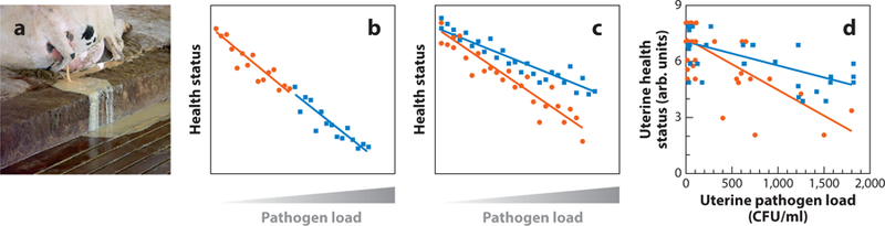
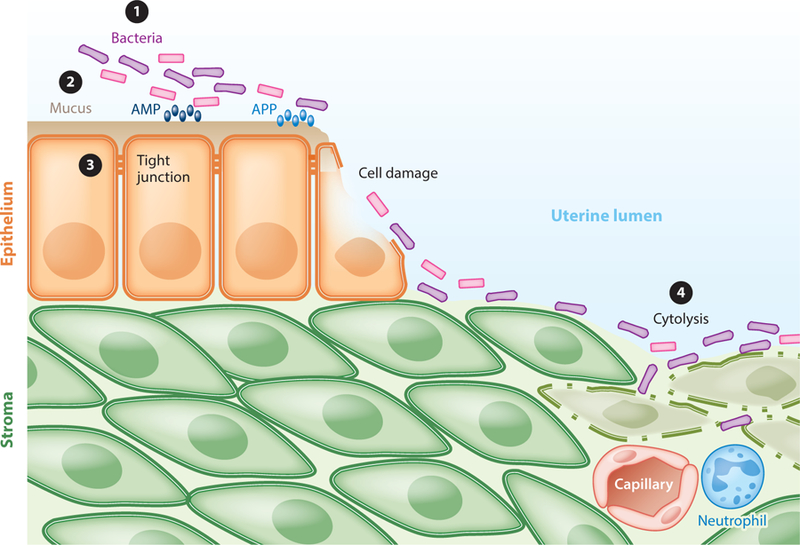
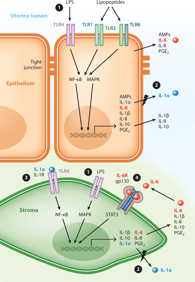
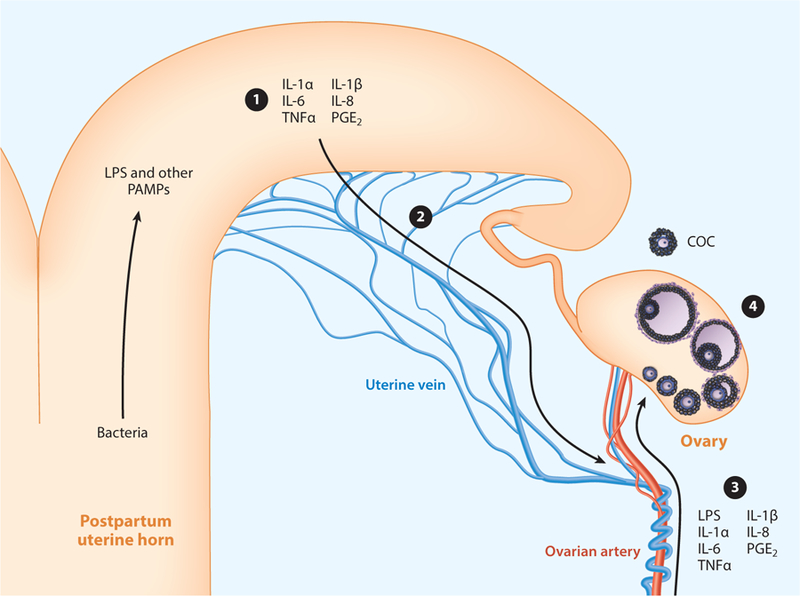
Bacteria are ubiquitous in the bovine uterus after parturition, but 50 years ago, cows tolerated these bacteria and few animals developed uterine disease. Now, up to 40% of dairy cattle develop postpartum uterine disease. Uterine disease causes infertility by compromising the function of not only the endometrium but also the ovary. Animals defend themselves against pathogens using tolerance and resistance mechanisms. Tolerance is the ability to limit the disease severity induced by a given pathogen burden. Resistance is the ability to limit the pathogen burden and is usually the function of immunity. Endometrial cells contribute to tolerance and have roles in innate immunity and the inflammatory response to pathogens. However, failures in endometrial tolerance and the character of the inflammatory response shape postpartum uterine disease. We propose that uterine health is more dependent on the ability of the endometrium to tolerate pathogens than the ability to resist invading bacteria.
Keywords: bovine; fertility; infection; inflammation; ovary; uterus.
Figures
Similar articles
-
Preventing postpartum uterine disease in dairy cattle depends on avoiding, tolerating and resisting pathogenic bacteria.Theriogenology. 2020 Jul 1;150:158-165. doi: 10.1016/j.theriogenology.2020.01.017. Epub 2020 Jan 11. Theriogenology. 2020. PMID: 31973964 Free PMC article. Review.
-
Mechanisms linking bacterial infections of the bovine endometrium to disease and infertility.Reprod Biol. 2016 Mar;16(1):1-7. doi: 10.1016/j.repbio.2015.12.002. Epub 2015 Dec 23. Reprod Biol. 2016. PMID: 26952747 Review.
-
PHYSIOLOGY AND ENDOCRINOLOGY SYMPOSIUM: Uterine infection: linking infection and innate immunity with infertility in the high-producing dairy cow.J Anim Sci. 2015 May;93(5):2021-33. doi: 10.2527/jas.2014-8496. J Anim Sci. 2015. PMID: 26020298 Review.
-
Expression of genes associated with immunity in the endometrium of cattle with disparate postpartum uterine disease and fertility.Reprod Biol Endocrinol. 2009 May 29;7:55. doi: 10.1186/1477-7827-7-55. Reprod Biol Endocrinol. 2009. PMID: 19476661 Free PMC article.
-
Dynamics of uterine infections with Escherichia coli, Streptococcus uberis and Trueperella pyogenes in post-partum dairy cows and their association with clinical endometritis.Vet J. 2014 Dec;202(3):527-32. doi: 10.1016/j.tvjl.2014.08.023. Epub 2014 Aug 27. Vet J. 2014. PMID: 25439441
Cited by
-
Effects of Resveratrol on Receptor Expression and Serum Levels of Estrogen and Progesterone in the Rat Endometritis Model.Reprod Sci. 2021 Sep;28(9):2610-2622. doi: 10.1007/s43032-021-00586-3. Epub 2021 May 8. Reprod Sci. 2021. PMID: 33966185
-
Modulation of Bovine Endometrial Cell Receptors and Signaling Pathways as a Nanotherapeutic Exploration against Dairy Cow Postpartum Endometritis.Animals (Basel). 2021 May 23;11(6):1516. doi: 10.3390/ani11061516. Animals (Basel). 2021. PMID: 34071093 Free PMC article. Review.
-
The Effect of Housing System on Disease Prevalence and Productive Lifespan of Dairy Herds-A Case Study.Animals (Basel). 2022 Jun 22;12(13):1610. doi: 10.3390/ani12131610. Animals (Basel). 2022. PMID: 35804508 Free PMC article.
-
Molecular Mechanisms Associated with the Development of the Metritis Complex in Dairy Cattle.Genes (Basel). 2024 Mar 30;15(4):439. doi: 10.3390/genes15040439. Genes (Basel). 2024. PMID: 38674374 Free PMC article.
-
Dynamics of uterine microbiota in postpartum dairy cows with clinical or subclinical endometritis.Sci Rep. 2020 Jul 23;10(1):12353. doi: 10.1038/s41598-020-69317-z. Sci Rep. 2020. PMID: 32704012 Free PMC article.
References
-
- Britt JH, Cushman RA, Dechow CD, Dobson H, Humblot P, et al. 2018. Invited review: learning from the future: a vision for dairy farms and cows in 2067. J. Dairy Sci 101:3722–41 - PubMed
-
-
Raberg L, Sim D, Read AF. 2007. Disentangling genetic variation for resistance and tolerance to infectious diseases in animals. Science 318:812–14.
Provides a framework for exploring resistance and tolerance in animals.
-
Publication types
MeSH terms
Grants and funding
LinkOut - more resources
Full Text Sources
Medical