Review
. 2019 Feb;92(1094):20180492.
doi: 10.1259/bjr.20180492. Epub 2018 Nov 1.

Clinical 7 T MRI: Are we there yet? A review about magnetic resonance imaging at ultra-high field

Affiliations
Review

Clinical 7 T MRI: Are we there yet? A review about magnetic resonance imaging at ultra-high field

Giuseppe Barisano et al. Br J Radiol. 2019 Feb.

Abstract

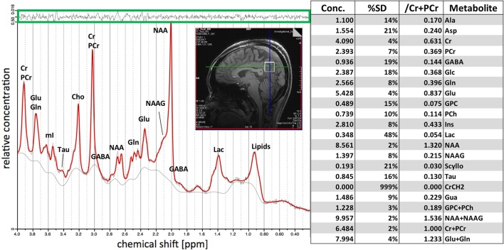
In recent years, ultra-high field MRI (7 T and above) has received more interest for clinical imaging. Indeed, a number of studies have shown the benefits from the application of this powerful tool not only for research purposes, but also in realms of improved diagnostics and patient management. The increased signal-to-noise ratio and higher spatial resolution compared with conventional and high-field clinical scanners allow imaging of small anatomical detail and subtle pathological findings. Furthermore, greater spectral resolution achieved at ultra-high field allows the resolution of metabolites for MR spectroscopic imaging. All these advantages have a significant impact on many neurological diseases, including multiple sclerosis, cerebrovascular disease, brain tumors, epilepsy and neurodegenerative diseases, in part because the pathology can be subtle and lesions small in these diseases, therefore having higher signal and resolution will help lesion detection. In this review, we discuss the main clinical neurological applications and some technical challenges which remain with ultra-high field MRI.


Figures

Figure 1.
Figure 1.
Axial SWI minIP Images at 7 T (left) and 3 T (right) of a healthy volunteer. The depiction of vessels is noticeably superior at UHF. The higher susceptibility and spatial resolution at 7 T allows detection of smaller vessels and the deoxyhenoglobin in veins which may be important for the detection of the “CVS” in the differential diagnoses of multiple sclerosis compare to other diseases which can cause demyelination such as neuromyelitis optica spectrum disorder, systemic autoimmune diseases, cerebral small vessel disease, Susac syndrome, and migraine, which typically woud not have the CVS. Resolution 7 T: 0.2 x 0.2 x 1.5 mm3, scanning time = 5 min. Resolution 3 T: 0.9 x 0.9 x 1.2 mm3, scanning time = 5 min. CVS, central vein sign; SWI, susceptibility-weighted imaging.
Figure 2.
Figure 2.
7 T MR spectroscopy with metabolite peaks. The single voxel spectrum (2 cm isotropic) is located in the posterior cingulate cortex. The green box on top demonstrates the residual of the LCmodel fit .Reliable concentrations have been demonstrated, with a standard deviation (%SD) inferior to 20% for most metabolites (Table). The improved sensitivity and specificity of 7 T MRS allow identification of metabolites with low concentration and discrimination of peaks of metabolites that overlap at lower field strength, such as glutamate (Glu), glutamine (Gln), and myo-inositol (mI). SD, standard deviation.
Figure 3.
Figure 3.
Axial TOF-MRA MIP images at 7 T (left) and 3 T (right) of a healthy volunteer. An increased number of vessels is visible at UHF. Please note the increase conspicuity of the lenticulostriate arteries (arrows) arising from the M1 segement of the MCAs and the higher contrast seen in the insular branches of the MCAs (arrowheads). Resolution 7 T: 0.3 × 0.3 × 0.3 mm3, 4 × 72 slices, scanning time = 4 × 2.23 min. MCAs, middle cerebral arteries; MRA, MR Angiography; MIP, maximum intensity projection; TOF,time of flight.
Figure 4.
Figure 4.
Thin minimum intensity projection across 10 mm slices from a 22-years-old healthy female subject at 3 and 7 T. Improved delineation of LSAs (white arrows) and reduced blurring, especially in the more distal vessels (arrowheads), can be appreciated at 7 T compared to 3 T. The visualization of these LSAs allows for visualization of normal vessels as well as the detection of possible pathologies such as arterial dissection and small LSA aneurysms (so called Charcot Bouchard microaneurysms). LSA, Lenticulostriate artery.
Figure 5.
Figure 5.
Postcontrast T1-weighted MR images at 7 T (left), 3 T (middle), and 1.5 T (right). 7 T imaging demonstrates what appears to be an 8-mm right-sided hypoenhancing pituitary microadenoma (white arrow), not visible at 3 and 1.5 T. From Law et al., JNS 2018 (https://doi.org/10.3171/2017.9.JNS171969), permission from Elsevier.
Figure 6.
Figure 6.
Hippocampal subfield imaging with high resolution coronal T2 weighted contrast at 3 and 7 T. 3 T image: in-plane resolution: 400 μm, slice thickness: 2 mm, sequence: BLADE , no repetition, total acquisition time: 10 mins. 7 T image: in-plane resolution: 300 μm, slice thickness: 2 mm, sequence: TSE, 4 averages and 2 concatenations, total acquisition time: 11 mins. Note that Fimbria (white arrow), Alveus (arrowhead), and Stratum layers were resolved at 7 T. SE, turbo spin echo.
Figure 7.
Figure 7.
Color fractional anisotropy (A, B, D, E) and DTI principal direction of diffusion (C, F) maps from the Human Connectome Project. 7 T (A, B, and C, 1.05 mm3 isotropic resolution) and 3 T (D, E, and F, 1.25 mm3 isotropic resolution) from the same subject. Red, green and blue indicate regions with diffusivities oriented primarily laterolateral, ventrodorsal, and rostrocaudal, respectively. Improved resolution allows identification of fiber tracks (red arrows and yellow arrows in B) which are faintly visible at 3 T. Note increased B1 + inhomogeneity resulting in signal loss in temporal lobe regions at 7 T (white arrows in A).
Figure 8.
Figure 8.
Fiber orientation glyphs (A, B) and tractography reconstructions (C, D) from the Human Connectome Project. 7 T (A, C: 1.05 mm3 isotropic resolution) and 3 T (B, D: 1.25 mm3 isotropic resolution) from the same subject. Fiber orientations were estimated using FSL BEDPOSTX and visualized using the Quantitative Imaging Toolkit (QIT); the 7 T data shows improved modeling of cortical fiber orientations (blue arrow). Tractography models of the superior longitudinal fasciculus I were created using deterministic streamline tractography in QIT; the 7 T data shows improved reconstruction of orbitofrontal connections of the pathway in both hemispheres (white arrow).

References

    1. Alvarez-Linera J. 3T MRI: Advances in brain imaging. Eur J Radiol 2008; 67: 415–26. doi: 10.1016/j.ejrad.2008.02.045 - DOI - PubMed
    1. Robitaille P-ML, Abduljalil AM, Kangarlu A, Zhang X, Yu Y, Burgess R, et al. . Human magnetic resonance imaging at 8 T. NMR Biomed . 1998; 11: 263–5. doi: 10.1002/(SICI)1099-1492(199810)11:6<263::AID-NBM549>3.0.CO;2-0 - DOI - PubMed
    1. Kraff O, Fischer A, Nagel AM, Mönninghoff C, Ladd ME. MRI at 7 Tesla and above: demonstrated and potential capabilities. J Magn Reson Imaging 2015; 41: 13–33. doi: 10.1002/jmri.24573 - DOI - PubMed
    1. Park C-A, Kang C-K, Kim Y-B, Cho Z-H. Advances in MR angiography with 7T MRI: From microvascular imaging to functional angiography. Neuroimage . 2018; 168(March): 269–78. doi: 10.1016/j.neuroimage.2017.01.019 - DOI - PubMed
    1. US Food and Drug Administration (FDA) Criteria for significant risk investigations of magnetic resonance diagnostic devices. 2014. Available from: https://www.fda.gov/RegulatoryInformation/Guidances/ucm072686.htm [October 2018].

MeSH terms