

Imaging appearance following surgical decompression of the ulnar nerve
- PMID: 30359100
- PMCID: PMC6404839
- DOI: 10.1259/bjr.20180757
Imaging appearance following surgical decompression of the ulnar nerve
Abstract
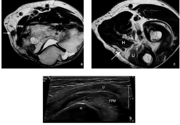
Ulnar neuropathy at the elbow is the second most common entrapment neuropathy of the upper extremity. Yet, there is a paucity of literature focusing on the imaging appearance following surgical decompression of the ulnar nerve at the elbow. Diagnostic imaging studies obtained after surgical decompression at The University of Michigan were reviewed and imaging findings were documented. We aim to describe the various techniques of ulnar nerve decompression and corresponding post-operative appearance on imaging. Potential complications following decompression will also be described with imaging and clinical correlation of recalcitrant ulnar neuropathy. It is important for the radiologist who performs MRI or ultrasound of the elbow to be aware of the various ulnar nerve decompression procedures. This knowledge will facilitate rapid and accurate diagnosis of normal and abnormal appearance of the ulnar nerve in this context.
Figures


Similar articles
-
Ulnar nerve entrapment neuropathy at the elbow: simple decompression.Neurosurgery. 2004 Nov;55(5):1150-3. doi: 10.1227/01.neu.0000140841.28007.f2. Neurosurgery. 2004. PMID: 15509321 Review.
-
Ulnar neuropathy at the elbow: follow-up and prognostic factors determining outcome.Neurology. 2005 May 10;64(9):1664-5; author reply 1664-5. doi: 10.1212/wnl.64.9.1664-b. Neurology. 2005. PMID: 15883347 No abstract available.
-
Ultrasonography Detects Ulnar Nerve Dislocation Despite Normal Electrophysiology and Magnetic Resonance Imaging.World Neurosurg. 2017 Mar;99:809.e1-809.e5. doi: 10.1016/j.wneu.2017.01.007. Epub 2017 Jan 12. World Neurosurg. 2017. PMID: 28089807
-
Imaging the intermuscular septum in the context of ulnar neuropathy.Skeletal Radiol. 2022 Mar;51(3):505-511. doi: 10.1007/s00256-021-03835-3. Epub 2021 Jul 10. Skeletal Radiol. 2022. PMID: 34245322 Review.
-
Ultrasound imaging of the ulnar nerve: correlation of preoperative and intraoperative dimensions.Clin Neurol Neurosurg. 2008 Jul;110(7):687-90. doi: 10.1016/j.clineuro.2008.04.003. Epub 2008 May 19. Clin Neurol Neurosurg. 2008. PMID: 18486322
Cited by
-
Comparison of 68Ga-FAPI PET/CT and 18FDG PET/CT Modalities in Gastrointestinal System Malignancies with Peritoneal Involvement.Mol Imaging Biol. 2022 Oct;24(5):789-797. doi: 10.1007/s11307-022-01729-x. Epub 2022 Apr 11. Mol Imaging Biol. 2022. PMID: 35411447
-
Ulnar neuropathy at the elbow: associations of pre-operative DTI parameters with clinical outcomes after cubital tunnel decompression.Eur Radiol. 2023 Sep;33(9):6351-6358. doi: 10.1007/s00330-023-09562-8. Epub 2023 Apr 4. Eur Radiol. 2023. PMID: 37014404
-
Cubital tunnel syndrome: anatomy, pathology, and imaging.Skeletal Radiol. 2025 Jan;54(1):1-15. doi: 10.1007/s00256-024-04705-4. Epub 2024 May 18. Skeletal Radiol. 2025. PMID: 38760642 Review.
-
Qualitative and quantitative analysis of 3D T1 Silent imaging.Radiol Med. 2021 Sep;126(9):1207-1215. doi: 10.1007/s11547-021-01380-6. Epub 2021 Jun 15. Radiol Med. 2021. PMID: 34131844
-
Diagnostic ultrasonography of upper extremity dynamic compressive neuropathies in athletes: A narrative review.Int Orthop. 2025 Apr;49(4):925-933. doi: 10.1007/s00264-025-06417-3. Epub 2025 Jan 30. Int Orthop. 2025. PMID: 39883178 Review.
References
Publication types
MeSH terms
LinkOut - more resources
Full Text Sources
